. 2000 May;105(9):1317-25.
doi: 10.1172/JCI9918.

IL-10-producing T cells suppress immune responses in anergic tuberculosis patients

Affiliations

IL-10-producing T cells suppress immune responses in anergic tuberculosis patients

V A Boussiotis et al. J Clin Invest. 2000 May.

Abstract

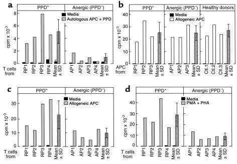
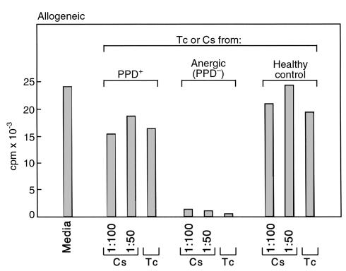
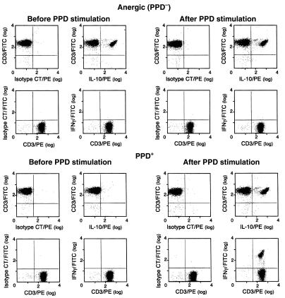
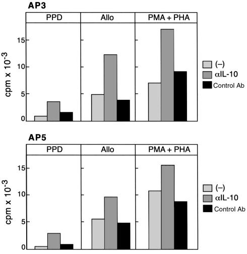
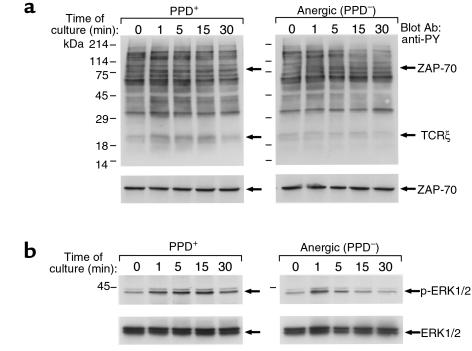
The lethality of Mycobacterium tuberculosis remains the highest among infectious organisms and is linked to inadequate immune response of the host. Containment and cure of tuberculosis requires an effective cell-mediated immune response, and the absence, during active tuberculosis infection, of delayed-type hypersensitivity (DTH) responses to mycobacterial antigens, defined as anergy, is associated with poor clinical outcome. To investigate the biochemical events associated with this anergy, we screened 206 patients with pulmonary tuberculosis and identified anergic patients by their lack of dermal reactivity to tuberculin purified protein derivative (PPD). In vitro stimulation of T cells with PPD induced production of IL-10, IFN-gamma, and proliferation in PPD(+) patients, whereas cells from anergic patients produced IL-10 but not IFN-gamma and failed to proliferate in response to this treatment. Moreover, in anergic patients IL-10-producing T cells were constitutively present, and T-cell receptor-mediated (TCR-mediated) stimulation resulted in defective phosphorylation of TCRzeta and defective activation of ZAP-70 and MAPK. These results show that T-cell anergy can be induced by antigen in vivo in the intact human host and provide new insights into mechanisms by which M. tuberculosis escapes immune surveillance.


Figures

Figure 1
Figure 1
APC from anergic TB patients do not generate a PPD-specific proliferative response of autologous T cells but can efficiently generate allo-MLR. (a) CD4+ T cells from PPD+ (responding) patients (RP) and anergic patients (AP) were stimulated with autologous APC loaded with PPD, and 3H-thymidine incorporation was determined. Results of four representative patients among 10 studied in each group are shown. (b) T cells from the same healthy volunteer donor were stimulated with APC from RP, AP, and healthy control individuals (Ctl) for 7 days, and DNA synthesis was determined by 3H-thymidine incorporation. Results of three representative individuals among eight studied in each group are shown. T cells from anergic TB patients have diminished responses to alloantigen and nonspecific mitogens. (c) CD4+ T cells from anergic and PPD+ patients were used as responders, and APC from the same healthy volunteer donor were used as stimulators. Cultures were continued for 7 days and response was assessed by 3H-thymidine incorporation. (d) CD4+ T cells from TB patients were cultured with PMA and PHA and response was examined by 3H-thimindine incorporation. Results of four representative patients among eight studied in each group are shown in c and d.
Figure 2
Figure 2
Culture supernatants after PPD stimulation and CD4+ T cells from anergic TB patients inhibit allogeneic MLR. CD4+ T cells and APC from HLA-disparate healthy individuals were used as responders and stimulators, respectively. Incubation was continued for 7 days with either media alone or in the presence of the indicated concentrations of culture supernatants (Cs) collected at 36 hours after PPD stimulation of CD4+ T cells from PPD+ TB patients, anergic (PPD) TB patients, and control healthy individuals, or with CD4+ T cells (Tc) from the same individuals. Response was examined by 3H-thymidine incorporation. Results from experiments with one representative patient among three tested in each group are shown.
Figure 3
Figure 3
IL-10+ T cells are constitutively present but IFN-γ+ T cells are not induced by PPD in anergic TB patients, whereas both IL-10+ and IFN-γ+ T cells are induced in PPD+ patients by PPD. Before stimulation and after culture with PPD, T cells were stained with anti-CD3 mAb. Cells were fixed, permeabilized, and stained for detection of intracellular cytokines using directly conjugated mAb’s specific for human IFN-γ and IL-10 (IFN-γ-FITC and IL-10-PE) or isotype-matched control as indicated. Results of one PPD+ and one anergic patient are shown and are representative of three patients examined in each group.
Figure 4
Figure 4
Anti–IL-10 neutralizing mAb increases proliferation of T cells from anergic TB patients in response to PPD, alloantigen, and PMA plus PHA. CD4+ T cells from two anergic patients (AP3 and AP5) were stimulated with PPD, allogeneic PBMCs, or PMA plus PHA in the presence of either media, neutralizing anti–IL-10 mAb, or isotype-matched control antibody. After the indicated time intervals of culture as described in Methods, response was determined by 3H-thymidine incorporation.
Figure 5
Figure 5
Defective TCR-mediated phosphorylation of TCRζ and ZAP-70 and defective activation of MAPK in anergic TB patients. (a) T cells from PPD+ and anergic TB patients (top panel) were stimulated in vitro with anti-CD3 and anti-CD28 mAb for the indicated time intervals. Cell lysates were prepared and analyzed by SDS-PAGE, transferred to nitrocellulose membrane, and blotted with anti-phosphotyrosine mAb. Blots were stripped and reblotted with ZAP-70 mAb (bottom panel). (b) Phosphorylation of ERK1 and ERK2 MAPK (top panel) was examined by immunoblotting with phospho-ERK–specific antibody, which recognizes only the activated phosphorylated form of ERK1 and ERK2. The blot was stripped and reprobed with ERK-specific antibody (bottom panel), which recognizes total ERK. The blots presented are representative of five PPD+ and five PPD individuals.

References

    1. Daley CL, et al. An outbreak of tuberculosis with accelerated progression among persons infected with the human immunodeficiency virus. An analysis using restriction-fragment-length polymorphisms. N Engl J Med. 1992;326:231–235. - PubMed
    1. Cisneros JR, Murray KM. Corticosteroids in tuberculosis. Ann Pharmacother. 1996;30:1298–1303. - PubMed
    1. Bloom BR, Small PM. The evolving relation between humans and Mycobacterium tuberculosis. N Engl J Med. 1998;338:677–678. - PubMed
    1. Karim, M., and Elner, J.J. 1995. Mechanisms of anergy. In Tuberculosis. W.N. Rom and S.M. Garay, editors. Little, Brown & Co. New York, New York, USA. 343–351.
    1. Daniel, T. 1991. Tuberculosis. In Harrison’s principles of internal medicine. J.D. Wilson et al., editors. McGraw-Hill. New York, New York, USA. 637–645.

Publication types