

Epithelial and endothelial expression of the green fluorescent protein reporter gene under the control of bovine prion protein (PrP) gene regulatory sequences in transgenic mice
- PMID: 10792029
- PMCID: PMC25844
- DOI: 10.1073/pnas.080081197
Epithelial and endothelial expression of the green fluorescent protein reporter gene under the control of bovine prion protein (PrP) gene regulatory sequences in transgenic mice
Abstract
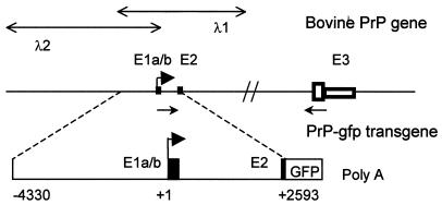
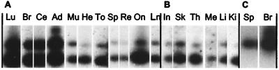
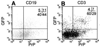
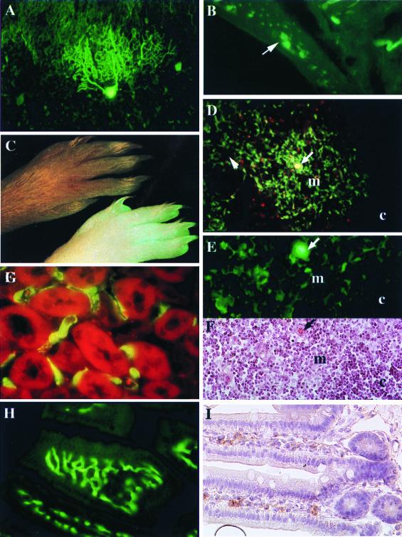
The expression of the cellular form of the prion protein (PrP(c)) gene is required for prion replication and neuroinvasion in transmissible spongiform encephalopathies. The identification of the cell types expressing PrP(c) is necessary to understanding how the agent replicates and spreads from peripheral sites to the central nervous system. To determine the nature of the cell types expressing PrP(c), a green fluorescent protein reporter gene was expressed in transgenic mice under the control of 6.9 kb of the bovine PrP gene regulatory sequences. It was shown that the bovine PrP gene is expressed as two populations of mRNA differing by alternative splicing of one 115-bp 5' untranslated exon in 17 different bovine tissues. The analysis of transgenic mice showed reporter gene expression in some cells that have been identified as expressing PrP, such as cerebellar Purkinje cells, lymphocytes, and keratinocytes. In addition, expression of green fluorescent protein was observed in the plexus of the enteric nervous system and in a restricted subset of cells not yet clearly identified as expressing PrP: the epithelial cells of the thymic medullary and the endothelial cells of both the mucosal capillaries of the intestine and the renal capillaries. These data provide valuable information on the distribution of PrP(c) at the cellular level and argue for roles of the epithelial and endothelial cells in the spread of infection from the periphery to the brain. Moreover, the transgenic mice described in this paper provide a model that will allow for the study of the transcriptional activity of the PrP gene promoter in response to scrapie infection.
Figures
Similar articles
-
Prion protein (PrPc) immunocytochemistry and expression of the green fluorescent protein reporter gene under control of the bovine PrP gene promoter in the mouse brain.J Comp Neurol. 2004 May 24;473(2):244-69. doi: 10.1002/cne.20117. J Comp Neurol. 2004. PMID: 15101092
-
[Mechanisms of neuroinvasion by prions: molecular principles and present state of research].Schweiz Med Wochenschr. 2000 Mar 25;130(12):435-42. Schweiz Med Wochenschr. 2000. PMID: 10780058 Review. German.
-
Lentivector-mediated RNAi efficiently suppresses prion protein and prolongs survival of scrapie-infected mice.J Clin Invest. 2006 Dec;116(12):3204-10. doi: 10.1172/JCI29236. J Clin Invest. 2006. PMID: 17143329 Free PMC article.
-
Expression of prion protein in the gut of mice infected orally with the 301V murine strain of the bovine spongiform encephalopathy agent.J Comp Pathol. 2005 May;132(4):273-82. doi: 10.1016/j.jcpa.2004.10.004. J Comp Pathol. 2005. PMID: 15893985
-
New insights into cellular prion protein (PrPc) functions: the "ying and yang" of a relevant protein.Brain Res Rev. 2009 Oct;61(2):170-84. doi: 10.1016/j.brainresrev.2009.06.002. Epub 2009 Jun 10. Brain Res Rev. 2009. PMID: 19523487 Review.
Cited by
-
Role of hypoxia‑mediated cellular prion protein functional change in stem cells and potential application in angiogenesis (Review).Mol Med Rep. 2017 Nov;16(5):5747-5751. doi: 10.3892/mmr.2017.7387. Epub 2017 Aug 29. Mol Med Rep. 2017. PMID: 28901450 Free PMC article.
-
Transgenesis applied to transmissible spongiform encephalopathies.Transgenic Res. 2002 Dec;11(6):547-64. doi: 10.1023/a:1021125510220. Transgenic Res. 2002. PMID: 12509129 Review.
-
Roles of the cellular prion protein in the regulation of cell-cell junctions and barrier function.Tissue Barriers. 2013 Apr 1;1(2):e24377. doi: 10.4161/tisb.24377. Tissue Barriers. 2013. PMID: 24665391 Free PMC article. Review.
-
Immune hyporesponsiveness to amyloid beta-peptide in amyloid precursor protein transgenic mice: implications for the pathogenesis and treatment of Alzheimer's disease.Proc Natl Acad Sci U S A. 2001 Aug 28;98(18):10273-8. doi: 10.1073/pnas.191118298. Epub 2001 Aug 21. Proc Natl Acad Sci U S A. 2001. PMID: 11517335 Free PMC article.
-
Translation of the prion protein mRNA is robust in astrocytes but does not amplify during reactive astrocytosis in the mouse brain.PLoS One. 2014 Apr 21;9(4):e95958. doi: 10.1371/journal.pone.0095958. eCollection 2014. PLoS One. 2014. PMID: 24752288 Free PMC article.
References
-
- Prusiner S B, Scott M, Foster D, Pan K M, Groth D, Mirenda C, Torchia M, Yang S L, Serban D, Carlson G A, et al. Cell. 1990;63:673–686. - PubMed
-
- Caughey B W, Dong A, Bhat K S, Ernst D, Hayes S F, Caughey W S. Biochemistry. 1991;30:7672–7680. - PubMed
-
- Bolton D C, McKinley M P, Prusiner S B. Science. 1982;218:1309–1311. - PubMed
Publication types
MeSH terms
Substances
Associated data
- Actions
LinkOut - more resources
Full Text Sources
Other Literature Sources
Research Materials
