. 2000 May 9;97(10):5416-21.
doi: 10.1073/pnas.100501197.

Myogenic stem cell function is impaired in mice lacking the forkhead/winged helix protein MNF

Affiliations

Myogenic stem cell function is impaired in mice lacking the forkhead/winged helix protein MNF

D J Garry et al. Proc Natl Acad Sci U S A. .

Abstract

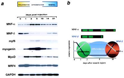
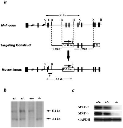
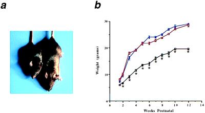
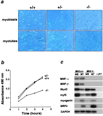
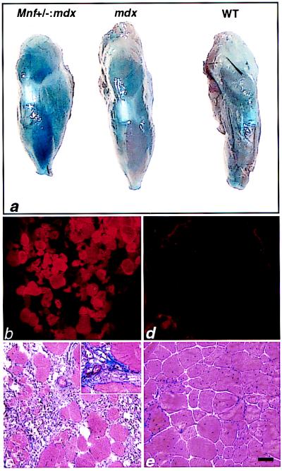
Myocyte nuclear factor (MNF) is a winged helix transcription factor that is expressed selectively in myogenic stem cells (satellite cells) of adult animals. Using a gene knockout strategy to generate a functional null allele at the Mnf locus, we observed that mice lacking MNF are viable, but severely runted. Skeletal muscles of Mnf-/- animals are atrophic, and satellite cell function is impaired. Muscle regeneration after injury is delayed and incomplete, and the normal timing of expression of cell cycle regulators and myogenic determination genes is dysregulated. Mnf mutant mice were intercrossed with mdx mice that lack dystrophin and exhibit only a subtle myopathic phenotype. In contrast, mdx mice that also lack MNF die in the first few weeks of life with a severe myopathy. Haploinsufficiency at the Mnf locus (Mnf+/-) also exacerbates the mdx phenotype to more closely resemble Duchenne's muscular dystrophy in humans. We conclude that MNF acts to regulate genes that coordinate the proliferation and differentiation of myogenic stem cells after muscle injury. Animals deficient in MNF may prove useful for evaluation of potential therapeutic interventions to promote muscle regeneration for patients having Duchenne's muscular dystrophy.


Figures

Figure 1
Figure 1
Skeletal muscle gene expression after cardiotoxin injury. (a) RT-PCR/Southern blot assay of the expression of MNF-α, MNF-β, and basic helix–loop–helix proteins of the MyoD family in regenerating muscles after cardiotoxin injury. MNF-α and MNF-β are reciprocally expressed in myogenic cells during regeneration, with MNF-β expressed in the quiescent stem cell and MNF-α expressed during stem cell proliferation. (b) Schematic of MNF-α and MNF-β expression during muscle regeneration.
Figure 2
Figure 2
Targeted disruption of Mnf. (a) Restriction map of the Mnf locus and gene targeting strategy. The targeting vector was constructed by replacing a BamHI–HindIII genomic fragment containing exons encoding the winged helix domain of MNF with a neomycin expression cassette, driven by an RNA polymerase II promoter. The targeting vector included a 1.0-kb 5′ Mnf sequence, 7.1 kb of 3′ Mnf sequence, and two thymidine kinase drug-resistance cassettes. Homologous recombinants were identified by Southern blot analysis using an 0.8-kb EcoRI/BamHI genomic fragment as the probe (hybridizing to the Mnf gene 5′ to the targeted region). Exons are represented as filled boxes. E, EcoRI; B, BamHI; H, HindIII; X, XhoI. (b) Offspring were genotyped by Southern blot analysis. (c) RT-PCR analysis of adult skeletal muscle reveals an absence of MNF-α and MNF-β transcripts in mutant mice.
Figure 3
Figure 3
Growth retardation of Mnf mutant mice. (a) The Mnf−/− mouse (Left) is approximately 60% the size of its wild-type, gender-matched (male) littermate (Right) at 6 weeks of age. (b) Mice without MNF are significantly smaller at each age up to a 1-year period compared with gender-matched littermates that are heterozygous for the targeted allele or wild type (Mnf+/+, blue line; Mnf+/−, red line; Mnf−/−, black line; n = 6 male mice at each age; *, P < 0.05). There were no significant differences noted between the heterozygotes and wild-type mice at 6 months of age, but significant differences were observed at 12 months of age (Mnf−/−, 26.4 ± 1.3 g vs. Mnf+/−, 36.2 ± 1.9 g vs. Mnf+/+, 41.7 ± 2.5 g; n = 6 pair, P < 0.05).
Figure 4
Figure 4
Morphological and molecular assessment of the skeletal muscle after cardiotoxin delivery in Mnf+/+ and Mnf−/− mice. (a) In the Mnf+/+ mouse after cardiotoxin delivery, edematous myofibers are evident by day 1, an extensive cellular proliferative response by day 2, regenerating myofibers with central nuclei (arrows) by day 5, and restoration of the hind limb architecture by 10 days (12, 15). (b) In contrast, the Mnf−/− skeletal muscle is characterized by a persistent hypercellular response that remains 10 days after injury, the delayed appearance of regenerating myofibers at 21 days, and restoration of muscle architecture only after 28 days (n = 3). Note focal regions of myofiber replacement by fat (days 10 and 21) in regenerating Mnf mutant skeletal muscle. (Bar = 100 μ.) RT-PCR/Southern analysis using primers specific for MyoD, c-myc, MRF4, myogenin, or GAPDH sequences from total RNA (10 μg) isolated from hind limbs of control (c) or Mnf-null (d) mice at the indicated times after injection of cardiotoxin. Peak expression of MyoD and c-myc mRNA after cardiotoxin injury is delayed compared with the wild-type controls. Myogenin and MRF4 also are dysregulated in the Mnf−/− mouse (n = 3).
Figure 5
Figure 5
Growth inhibition and differential gene expression of cultured muscle cells lacking MNF. (a) Muscle cells isolated from 3-day-old wild-type (+/+), heterozygous (+/−), and Mnf null (−/−) neonates were cultured in growth medium (myoblasts) and differentiation medium (myotubes). Mnf mutant myoblasts are capable of forming differentiated myotubes under low serum conditions. (b) Cell proliferation assays were performed on muscle cells isolated from 3-day-old neonatal wild-type (+/+), Mnf-heterologous (+/−), and Mnf-null (−/−) mice (performed in triplicate). (c) Semiquantitative RT-PCR/Southern blot analysis of MNF isoforms and MyoD family members in wild-type myoblasts (MB) and myotubes (MT) compared with Mnf−/− myoblasts (MB) and myotubes (MT) (performed in quadruplicate).
Figure 6
Figure 6
F2 progeny of the Mnf null and mdx intercross. (a) Four-day-old littermates including the Mnf-mdx double knockout (Left), Mnf null (Middle), and mdx (Right). (b–e) Two-week-old Mnf-mdx double knockout and mdx littermates were injected with Evans blue dye, which accumulates in cells with damaged membranes (–23) (n = 3 for each group). (b) Extensive dye uptake in the double mutant chest wall skeletal muscle and diaphragm (Inset) is apparent when using fluorescent microscopy (to identify Evan's blue dye positive myofibers) with an absence of uptake in the chest wall of mdx mice at this age (d). (c) Adjacent sections of chest wall musculature and diaphragm (Inset) were stained with hematoxylin and eosin. Note evidence of necrosis and inflammation in the double mutant chest wall/diaphragm (Inset) skeletal muscle (c) compared with a lack of inflammation associated with the chest wall/diaphragm of the 2-week-old mdx littermates (e). (Bar = 100 μ.)
Figure 7
Figure 7
Increased myonecrosis in the tibialis anterior (TA) muscle of Mnf+/−:mdx mice compared with mdx mice. (a) Macroscopic evaluation of Evans blue staining after i.v. dye injection into 6-month-old Mnf+/−:mdx, mdx, and wild-type mice. Note extensive blue dye incorporation in the double mutant TA compared with the mdx or wild-type TA (n = 6 mice in each group). (b and c) Fluorescent microscopy of cryosectioned TA muscles revealing extensive dye-filled myofibers in damaged myofibers of the Mnf+/−:mdx mouse (b) compared with the mdx skeletal muscle (d). (c) Adjacent sections of double mutant TA stained with Masson trichrome reveals extensive ongoing myonecrosis and fibrosis (Inset). (e) Adjacent sections of mdx TA stained with Masson trichrome reveals an absence of inflammation and a relative absence of fibrosis at 6 months of age. (Bar = 100 μ.)

References

    1. Gussoni E, Blau H M, Kunkel L M. Nat Med. 1997;3:970–977. - PubMed
    1. Engel A G, Yamamoto M, Fischbeck K H. In: Myology. Engel A G, Franzini-Armstrong C, editors. New York: McGraw–Hill; 1994. pp. 1130–1188.
    1. Monaco A, Neve R, Colletti-Feener C, Bertelson C, Kurnit D, Kunkel L M. Nature (London) 1986;323:646–650. - PubMed
    1. Burghes A, Logan C, Hu X, Belfall B, Worton R, Ray P. Nature (London) 1987;328:434–437. - PubMed
    1. Hoffman E P, Brown R H, Kunkel L M. Cell. 1987;51:919–928. - PubMed

Publication types

MeSH terms