Clinical Trial
. 2000 May;120(2):324-32.
doi: 10.1046/j.1365-2249.2000.01223.x.

Decreased CD95 expression on naive T cells from HIV-infected persons undergoing highly active anti-retroviral therapy (HAART) and the influence of IL-2 low dose administration. Irhan Study Group

Affiliations
Clinical Trial

Decreased CD95 expression on naive T cells from HIV-infected persons undergoing highly active anti-retroviral therapy (HAART) and the influence of IL-2 low dose administration. Irhan Study Group

A Amendola et al. Clin Exp Immunol. 2000 May.

Abstract

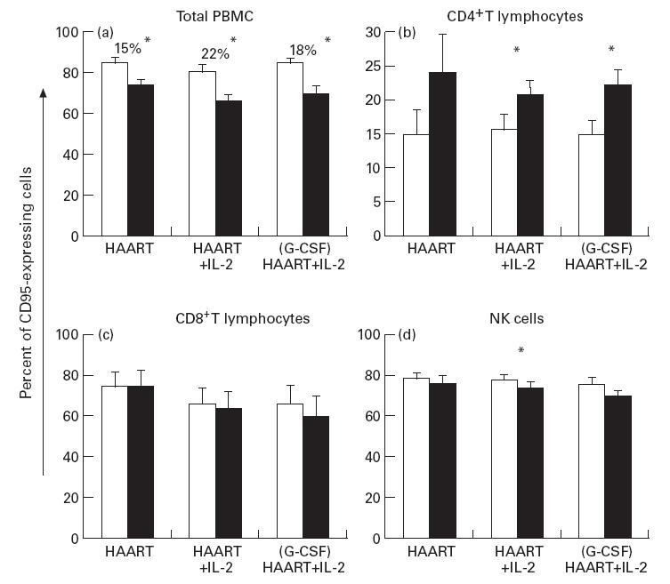
The functional recovery of the immune system in HIV-infected persons receiving HAART and the role of adjuvant immune therapy are still matters of intensive investigation. We analysed the effects of HAART combined with cytokines in 22 naive asymptomatic individuals, randomized to receive HAART (n = 6), HAART plus a low dose (1000 000 U/daily) of rIL-2 (n = 8), and HAART plus rIL-2 after previous administration of granulocyte colony-stimulating factor (n = 8). After 3 months of therapy, increased CD4+ T cell counts and diminished viral loads were observed in all patients, independently of cytokine addition. A decreased expression of CD95 (Apo 1/Fas) was evident in all groups when compared with values before therapy. The percentages of peripheral blood mononuclear cells (PBMC) expressing CD95 after therapy decreased by 15%, 22% and 18% in the three treatment groups, respectively (P < 0.05). Analysis of PBMC subsets demonstrated that CD95 expression was significantly reduced on CD45RA+CD62L+ naive T cells (25.3%, 22.4%, and 18.6%, respectively; P < 0.05) in each group, after therapy. Accordingly, all patients showed a reduced rate of in vitro spontaneous apoptosis (P < 0.05). Another effect induced by HAART was a significant increase in IL-2Ralpha expression on total PBMC (P < 0.05), independently of cytokine addition. Altogether, our results suggest that very low dose administration of rIL-2 (1000 000 U/daily) may be not enough to induce a significant improvement in the immune system as regards HAART alone. The employment of higher doses of recombinant cytokines and/or different administration protocols in clinical trials might however contribute to ameliorate the immune reconstitution in patients undergoing HAART.


Figures

Fig. 1
Fig. 1
Frequencies of naive cells in HIV-infected individuals undergoing anti-retroviral therapy. Detection of peripheral blood mononuclear cells (PBMC) with a naive phenotype (identified as expression of CD45RA+ and CD62L+ receptors) was performed by single-cell analysis using a 4-fluorescence cytometric protocol at t = 0 (before therapy; □) and at t = 12 weeks (after 12 weeks of therapy; ▪). The percentage of naive cells analysed in each patient is shown in (a) (group 1), (b) (group 2), and (c) (group 3). In parallel, CD4+ T cells with naive phenotype were also analysed and relative percentages are shown in (d) (group 1), (e) (group 2), and (f) (group 3).
Fig. 2
Fig. 2
Analysis of CD95 expression on peripheral blood mononuclear cells (PBMC) obtained from HIV-infected individuals undergoing therapy. The expression of the CD95 receptor on total PBMC was investigated by single-cell analysis at t = 0 (□) and at t = 12 weeks (▪). (a) Changes in the percentage of total PBMC expressing the CD95 receptor in each group, before and after differentiated therapies. Percentages of CD95+ cell decrease, relative to each group following therapy, are shown. Results of phenotypical single-cell analysis of CD95+ PBMC subsets are shown in (b) (percentage of CD4+CD95+ T cells), (c) (percentage of CD8+CD95+ T cells), and (d) (percentage of natural killer (NK) CD16+CD95+ cells). Data represent the arithmetic mean percentage; error bars indicate s.e.m. P values are indicated: *P < 0·05.
Fig. 3
Fig. 3
Decrease of CD95 death receptor expression on naive subset of total peripheral blood mononuclear cells (PBMC) after differentiated therapy in HIV-infected individuals. (a) Changes in the percentage of naive (CD45RA+CD62L+) cells expressing the CD95 receptor, before (□) and after 12 weeks of therapy (▪) in each group of patients. Mean percentages of decrease, relative to each group following therapy, are reported in the text. Data represent the arithmetic mean percentages; error bars indicate s.e.m. P values are indicated: *P < 0·05. (b–e) Representative flow cytometric images of a patient undergoing (G-CSF) HAART +IL-2, before (b,d) and after (c,e) therapy. Naive cells, shown in (b,c) by gating CD45RA+CD62L+ cells (R2), were then analysed for the expression of CD95 at t = 0 (d) and t = 12 weeks (e). Percentages of CD95+ naive cells were 81% at starting therapy (d) and 28·7% after 3 months (e).
Fig. 4
Fig. 4
rIL-2 administration during HAART affects the expression of CD25 (IL-2Rα) in HIV-infected individuals undergoing therapy. (a) Changes in the percentage of CD25-expressing total peripheral blood mononuclear cells (PBMC) in all groups, investigated by single-cell analysis at t = 0 (□) and at t = 12 weeks (▪). Mean percentages of CD25+ cell increase, relative to each group following therapy, are reported in the text. Analysis of different subsets of PBMC expressing CD25 was carried out by flow cytometry on the three groups of patients, and percentages reported in (b,c,d). A significant rIL-2 treatment-induced increase in the percentages of CD3+, CD8+, CD16+ and CD19+ lymphocytes was observed after therapy in groups 2 and 3, when compared with baseline values at starting treatment. Data represent the arithmetic mean percentage; error bars indicate s.e.m. P values are indicated: *P < 0·05.
Fig. 5
Fig. 5
rIL-2 administration during HAART does not affect the expression of CD122 (IL-2Rβ) in HIV-infected individuals undergoing therapy. (a) Mean percentages of CD122-expressing total peripheral blood mononuclear cells (PBMC) in all groups, investigated by single-cell analysis at t = 0 (□) and at t = 12 weeks (▪). Mean percentages of CD122+ cells did not change significantly after therapy in all groups. Different subsets of PBMC expressing CD122 were then analysed and are reported in (b,c,d). No significant treatment-induced changes in the percentage of CD122+ lymphocytes were observed after therapy, when compared with baseline values at starting treatment. Data represent the arithmetic mean percentage; error bars indicate s.e.m. P values are indicated: *P < 0·05.

References

    1. Collier AC, Coombs RW, Schoenfeld DA, et al. Treatment of human immunodeficiency virus infection with saquinavir, zidovudine and zalcitabine. N Engl J Med. 1996;334:1011–7. - PubMed
    1. Autran B, Carcelain G, Li TS, et al. Positive effects of combined antiretroviral therapy on CD4+ T cell homeostasis and function in advanced HIV disease. Science. 1997;277:112–6. - PubMed
    1. Mathez D, Bagnarelli P, Gorin I, et al. Reduction in viral load and increases in T lymphocyte numbers in treatment-naive patients with advanced HIV-1 infection treated with ritonavir, zidovudine, zalcitabine triple therapy. Antiviral Ther. 1997;2:175–83. - PubMed
    1. Pontesilli O, Kerkhof-Garde S, Pakker NG, et al. Antigen-specific T-lymphocyte proliferative responses during highly active antiretroviral therapy (HAART) of HIV-1 infection. Immunol Letters. 1999;66:213–7. - PubMed
    1. Foudraine NA, Hovenkamp E, Notermans DW, et al. Immunopathology as a result of highly active antiretroviral therapy in HIV-1-infected patients. AIDS. 1999;13:177–84. - PubMed

Publication types