

Granulocyte-macrophage colony-stimulating factor (GM-CSF) but not granulocyte colony-stimulating factor (G-CSF) induces plasma membrane expression of proteinase 3 (PR3) on neutrophils in vitro
- PMID: 10792393
- PMCID: PMC1905642
- DOI: 10.1046/j.1365-2249.2000.01205.x
Granulocyte-macrophage colony-stimulating factor (GM-CSF) but not granulocyte colony-stimulating factor (G-CSF) induces plasma membrane expression of proteinase 3 (PR3) on neutrophils in vitro
Abstract
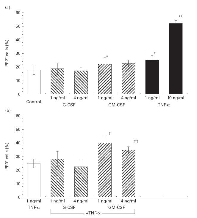
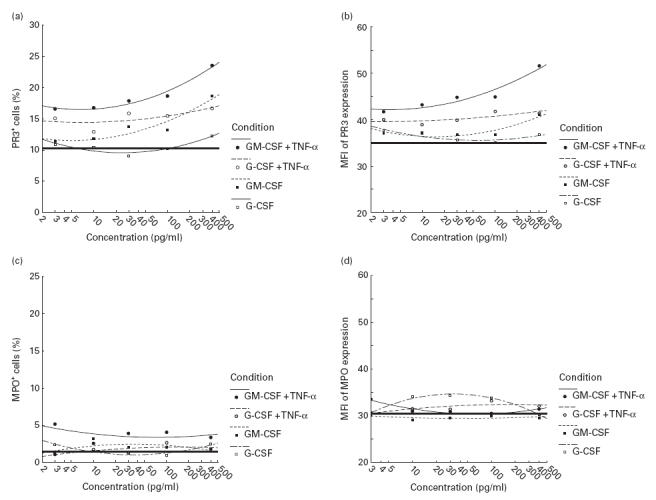
The theoretical risk of triggering vasculitis resulting from administration of G-CSF and GM-CSF to patients with anti-neutrophil cytoplasmic antibody (ANCA)-associated vasculitides (AAV), such as Wegener's granulomatosis (WG), who develop agranulocytosis due to cytotoxic therapy, is unknown. Since there is strong evidence that activation of polymorphonuclear neutrophils (PMN) induced by binding of ANCA to PR3 or myeloperoxidase (MPO) expressed on their plasma membrane is involved in the pathogenesis of systemic vasculitides (SV), we studied the surface expression of PR3 and MPO on PMN from healthy donors in response to G-CSF and GM-CSF in vitro by flow cytometric analysis. Increasing doses of G-CSF did not alter PR3 expression on either untreated or tumour necrosis factor-alpha (TNF-alpha)-primed donor PMN significantly. In contrast, GM-CSF significantly increased PR3 membrane expression on both intact PMN and neutrophils primed with TNF-alpha. MPO expression was not significantly altered by either G-CSF or GM-CSF. In summary, these data demonstrate that GM-CSF, but not G-CSF, induces plasma membrane expression of PR3 on PMN in vitro. Since in AAV accessibility of the antigen (PR3 or MPO) to the antibody (ANCA) on the plasma membrane of PMN is thought to be essential for neutrophil activation by ANCA, the results of the present study suggest that administration of GM-CSF to patients with WG with neutropenia implies a definite theoretical risk of deterioration of vasculitis via this mechanism.
Figures
Similar articles
-
IgG from myeloperoxidase-antineutrophil cytoplasmic antibody-positive patients stimulates greater activation of primed neutrophils than IgG from proteinase 3-antineutrophil cytosplasmic antibody-positive patients.Arthritis Rheum. 2001 Apr;44(4):921-30. doi: 10.1002/1529-0131(200104)44:4<921::AID-ANR149>3.0.CO;2-4. Arthritis Rheum. 2001. PMID: 11315931
-
Human anti-neutrophil cytoplasm autoantibodies to proteinase 3 (PR3-ANCA) bind to neutrophils.Kidney Int. 2005 Aug;68(2):537-41. doi: 10.1111/j.1523-1755.2005.00431.x. Kidney Int. 2005. PMID: 16014030
-
Neutrophil priming and apoptosis in anti-neutrophil cytoplasmic autoantibody-associated vasculitis.Kidney Int. 2001 May;59(5):1729-38. doi: 10.1046/j.1523-1755.2001.0590051729.x. Kidney Int. 2001. PMID: 11318943
-
Pathogenesis of PR3-ANCA associated vasculitis.J Autoimmun. 2008 Feb-Mar;30(1-2):29-36. doi: 10.1016/j.jaut.2007.11.005. Epub 2007 Dec 26. J Autoimmun. 2008. PMID: 18162369 Review.
-
'Classic' anti-neutrophil cytoplasmic autoantibodies (cANCA), 'Wegener's autoantigen' and their immunopathogenic role in Wegener's granulomatosis.J Autoimmun. 1993 Apr;6(2):171-84. doi: 10.1006/jaut.1993.1015. J Autoimmun. 1993. PMID: 8388690 Review.
Cited by
-
Aberrant cytokine pattern of the nasal mucosa in granulomatosis with polyangiitis.Arthritis Res Ther. 2012 Oct 17;14(5):R203. doi: 10.1186/ar4041. Arthritis Res Ther. 2012. PMID: 23031229 Free PMC article.
-
New Insights into Pathogenesis and Treatment of ANCA-Associated Vasculitis: Autoantibodies and Beyond.Antibodies (Basel). 2023 Mar 21;12(1):25. doi: 10.3390/antib12010025. Antibodies (Basel). 2023. PMID: 36975372 Free PMC article. Review.
-
Anti-neutrophil cytoplasm antibody IgG subclasses in Wegener's granulomatosis: a possible pathogenic role for the IgG4 subclass.Clin Exp Immunol. 2004 Oct;138(1):183-92. doi: 10.1111/j.1365-2249.2004.02566.x. Clin Exp Immunol. 2004. PMID: 15373923 Free PMC article.
-
The use of small molecule high-throughput screening to identify inhibitors of the proteinase 3-NB1 interaction.Clin Exp Immunol. 2010 Aug;161(2):389-96. doi: 10.1111/j.1365-2249.2010.04174.x. Epub 2010 May 7. Clin Exp Immunol. 2010. PMID: 20456416 Free PMC article.
-
Anti-endothelial cell antibodies (AECA) in patients with propylthiouracil (PTU)-induced ANCA positive vasculitis are associated with disease activity.Clin Exp Immunol. 2005 Mar;139(3):569-74. doi: 10.1111/j.1365-2249.2005.02725.x. Clin Exp Immunol. 2005. PMID: 15730404 Free PMC article.
References
-
- Welte K, Gabrilove J, Bronchud MH, Platzer E, Morstyn G. Filgrastim (r-metHuG-CSF): the first 10 years. Blood. 1996;88:1907–29. - PubMed
-
- Spiekermann K, Rosler J, Emmendorfer A, Elsner J, Welte K. Functional features of neutrophils induced by G-CSF and GM-CSF treatment: differential effects and clinical implications. Leukemia. 1997;11:466–78. - PubMed
-
- Frampton JE, Lee CR, Faulds D. Filgrastim. A review of its pharmacological properties and therapeutic efficacy in neutropenia. Drugs. 1994;48:731–60. - PubMed
-
- Anderlini P, Przepiorka D, Champlin R, Körbling M. Biological and clinical effects of granulocyte colony-stimulating factor in normal individuals. Blood. 1996;88:2819–25. - PubMed
-
- Lieschke GJ, Burgess AW. Granulocyte colony-stimulating factor and granulocyte-macrophage colony-stimulating factor (first of two parts) N Engl J Med. 1991;327:28–35. - PubMed
MeSH terms
Substances
LinkOut - more resources
Full Text Sources
Research Materials
Miscellaneous
