

Immunohistochemical analysis of cyclin D1 shows deregulated expression in multiple myeloma with the t(11;14)
- PMID: 10793062
- PMCID: PMC1876932
- DOI: 10.1016/S0002-9440(10)65022-5
Immunohistochemical analysis of cyclin D1 shows deregulated expression in multiple myeloma with the t(11;14)
Abstract
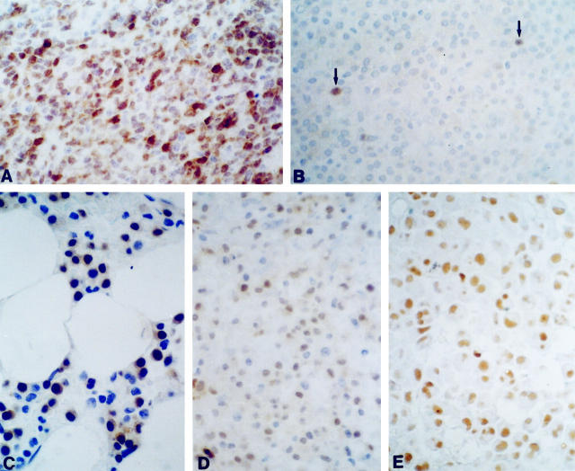
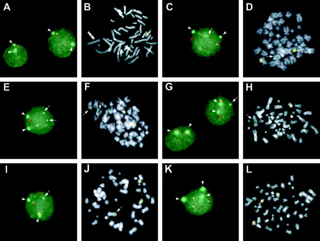
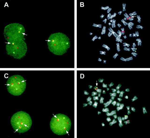
The t(11;14)(q13;q32) chromosomal translocation, the hallmark of mantle cell lymphoma (MCL), is recurrently found in multiple myelomas (MM) by means of conventional cytogenetics. Unlike MCL, recent molecular studies of MM-derived cell lines with t(11;14) have indicated that the breakpoints are highly dispersed over the 11q13 region; however, the fact that cyclin D1 is generally overexpressed in these cell lines suggests that this gene is the target of the translocation. To evaluate further the involvement of cyclin D1 in MM, we used immunohistochemistry and fluorescence in situ hybridization to investigate cyclin D1 expression and the presence of chromosome 11 abnormalities in a representative panel of 48 MM patients (40 at diagnosis and 8 at relapse). Cyclin D1 overexpression occurred in 12/48 (25%) of cases; combined immunohistochemistry and fluorescence in situ hybridization analyses in 39 patients showed cyclin D1 positivity in all of the cases (7/7) bearing the t(11;14), in two of the 13 cases with trisomy 11, and in one of the 19 cases with no apparent abnormalities of chromosome 11. Our data indicate that the t(11;14) translocation in MM leads to cyclin D1 overexpression and that immunohistochemical analysis may represent a reliable means of identifying this lesion in MM.
Figures
Similar articles
-
Frequency and distribution of trisomy 11 in multiple myeloma patients: relation with overexpression of CCND1 and t(11;14).Cancer Genet Cytogenet. 2007 Feb;173(1):51-6. doi: 10.1016/j.cancergencyto.2006.09.017. Cancer Genet Cytogenet. 2007. PMID: 17284370
-
The (11;14)(q13;q32) translocation in multiple myeloma. A morphologic and immunohistochemical study.Am J Clin Pathol. 2000 Jun;113(6):831-7. doi: 10.1309/4W8E-8F4K-BHUP-UBE7. Am J Clin Pathol. 2000. PMID: 10874884
-
Identification of cyclin D1 mRNA overexpression in B-cell neoplasias by real-time reverse transcription-PCR of microdissected paraffin sections.Clin Cancer Res. 2002 Sep;8(9):2902-11. Clin Cancer Res. 2002. PMID: 12231535
-
B-prolymphocytic leukaemia with t(11;14) revisited: a splenomegalic form of mantle cell lymphoma evolving with leukaemia.Br J Haematol. 2004 May;125(3):330-6. doi: 10.1111/j.1365-2141.2004.04913.x. Br J Haematol. 2004. PMID: 15086413 Review.
-
Mantle cell lymphoma lacking the t(11;14) translocation: a case report and brief review of the literature.J Clin Pathol. 2008 Jul;61(7):869-70. doi: 10.1136/jcp.2007.048629. J Clin Pathol. 2008. PMID: 18587018 Review.
Cited by
-
Overlapping morphologic and immunophenotypic profiles in small B-cell lymphoma. A report of two cases.Virchows Arch. 2006 Sep;449(3):320-7. doi: 10.1007/s00428-006-0242-1. Epub 2006 Jul 18. Virchows Arch. 2006. PMID: 16847683
-
Immunohistochemical analysis of cyclin D1 and p53 in multiple myeloma: relationship to proliferative activity and prognostic significance.Med Oncol. 2004;21(1):73-80. doi: 10.1385/MO:21:1:73. Med Oncol. 2004. PMID: 15034217
-
One Patient, Two Uncommon B-Cell Neoplasms: Solitary Plasmacytoma following Complete Remission from Intravascular Large B-Cell Lymphoma Involving Central Nervous System.Case Rep Med. 2014;2014:620423. doi: 10.1155/2014/620423. Epub 2014 Feb 18. Case Rep Med. 2014. PMID: 24715915 Free PMC article.
-
Cyclin D1 expression in multiple myeloma by immunohistochemistry: Case series of 14 patients and literature review.Indian J Med Paediatr Oncol. 2013 Oct;34(4):283-91. doi: 10.4103/0971-5851.125246. Indian J Med Paediatr Oncol. 2013. PMID: 24604959 Free PMC article.
-
Immunophenotypic expression profile of multiple myeloma cases at a tertiary hospital in Nairobi Kenya.Front Med (Lausanne). 2023 May 12;10:1177775. doi: 10.3389/fmed.2023.1177775. eCollection 2023. Front Med (Lausanne). 2023. PMID: 37250623 Free PMC article.
References
-
- Heim S, Mitelman F: Cancer Cytogenetics. 1995. Wiley-Liss, New York
-
- Dewald GW, Jenkins RB: Cytogenetic studies of patients with monoclonal gammopathies. Wiernik PH Canellos GP Dutcher JP Kyle RA eds. Neoplastic Diseases of the Blood. 1996, :pp 515-523 Churchill Livingstone, New York
-
- Nishida K, Tamura A, Nakazawa N, Ueda Y, Abe T, Matsuda F, Kashima K, Taniwaki M: The Ig heavy chain gene is frequently involved in chromosomal translocations in multiple myeloma and plasma cell leukemia as detected by in situ hybridization. Blood 1997, 90:526-534 - PubMed
Publication types
MeSH terms
Substances
LinkOut - more resources
Full Text Sources
Other Literature Sources
Medical
Research Materials
