. 2000 May;156(5):1515-25.
doi: 10.1016/S0002-9440(10)65023-7.

E-cadherin expression in melanoma cells restores keratinocyte-mediated growth control and down-regulates expression of invasion-related adhesion receptors

Affiliations

E-cadherin expression in melanoma cells restores keratinocyte-mediated growth control and down-regulates expression of invasion-related adhesion receptors

M Y Hsu et al. Am J Pathol. 2000 May.

Abstract

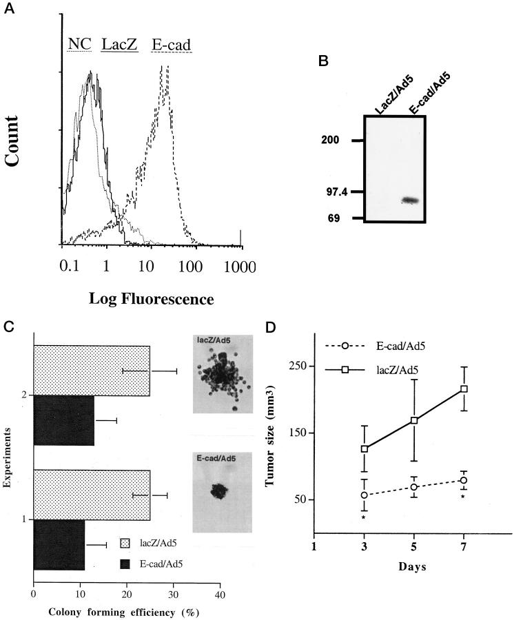
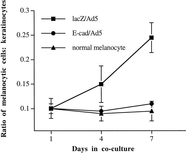
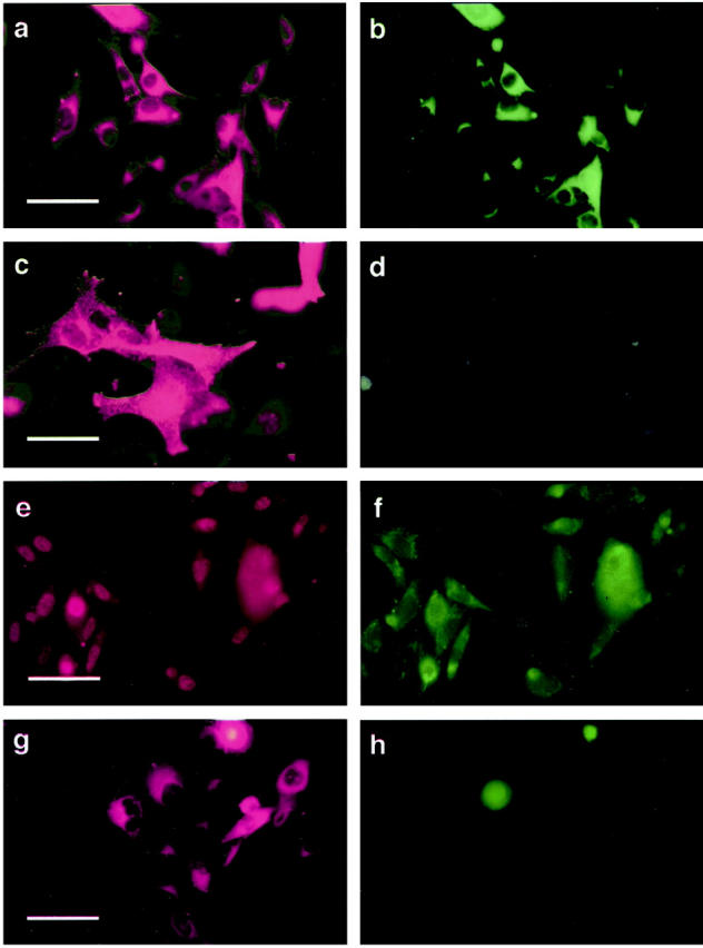
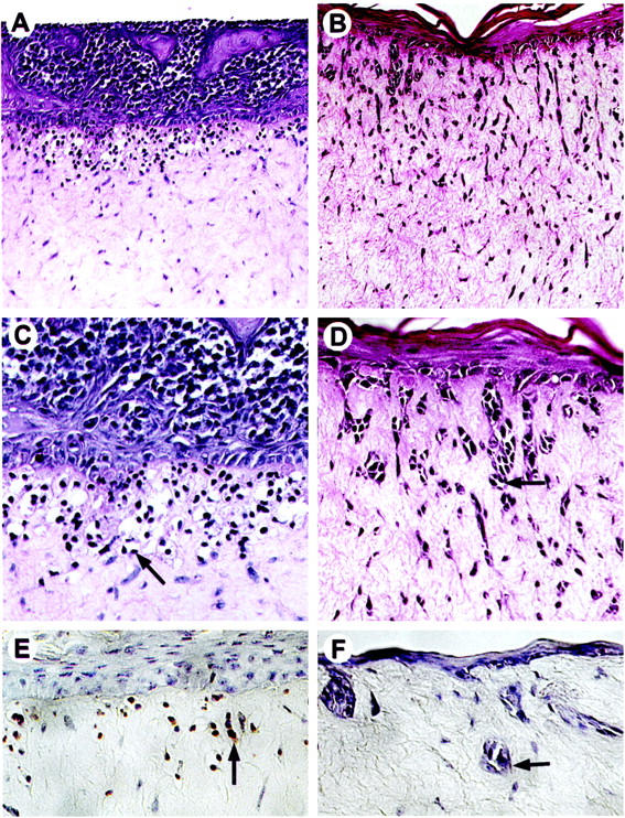
In human epidermis, functional symbiosis requires homeostatic balance between keratinocytes and melanocytes. Compelling evidence from co-culture studies demonstrated a sophisticated, multileveled regulation of normal melanocytic phenotype orchestrated by undifferentiated, basal-type keratinocytes. Keratinocytes control cell growth and dendricity, as well as expression of melanoma-associated cell surface molecules of normal melanocytes. In contrast, melanoma cells are refractory to the keratinocyte-mediated regulation. The loss of regulatory dominance by keratinocytes occurs in concert with down-regulation of E-cadherin expression in melanoma cells. To investigate the potential role of E-cadherin in melanoma-keratinocyte interaction, we transduced E-cadherin-negative melanoma cells with full-length E-cadherin cDNA using an adenoviral vector. Our results show that functional E-cadherin expression in melanoma cells leads to cell adhesion to keratinocytes rendering them susceptible for keratinocyte-mediated control. In a skin reconstruction model, ectopic E-cadherin expression inhibits invasion of melanoma cells into dermis by down-regulating invasion-related adhesion receptors, MelCAM/MUC18 and beta3 integrin subunit, and by induction of apoptosis. Thus, disruption of the E-cadherin-mediated, normal regulatory control from keratinocytes may represent one of the mechanisms accounting for melanocyte transformation.


Figures

Figure 1.
Figure 1.
Characterization of E-cad/Ad5-transduced melanoma cells. A: Expression of exogenous E-cadherin in E-cad/Ad5-transduced WM115 melanoma cells (E-cad, broken line) as determined by fluorescence-activated cell sorting analysis with an anti-E-cadherin mAb 48 hours after transduction. The solid line represents lacZ/Ad5-transduced cells and the dotted line represents the negative control (NC) cells stained with an isotype-matched negative control antibody. Similar results were obtained with three other melanoma cell lines. B: Detection of E-cadherin/catenin complex in E-cad/Ad5-transduced WM115 melanoma cells by immunoprecipitation. Forty-eight hours after transduction, cell lysates were immunoprecipitated with an anti-E-cadherin mAb. Samples were subjected to electrophoresis and probed with an anti-β-catenin mAb. A 92-kd band representing β-catenin co-immunoprecipitated with E-cadherin in E-cad/Ad5-transduced cells but not in lacZ-transduced cells. C: Soft agar growth of 1205Lu melanoma cells overexpressing E-cadherin. Cells were transduced with lacZ/Ad5 or E-cad/Ad5, and 24 hours later, resuspended in 0.25% agar and seeded in triplicate 35-mm wells at 6 × 10 cells/well. After 3 weeks, colony-forming efficiency was determined as the percentage of cells forming colonies containing four or more cells. Ten random fields were examined for each condition. Data represent mean ± SD from two independent experiments. Asterisks indicate significant difference by Student’s t-test (experiment 1: P < 0.001; experiment 2: P = 0.001). Inserts show the micrographs of representative colonies. D: Tumorigenicity of melanoma cells after induction of E-cadherin. One day after transduction, 1205Lu melanoma cells in subconfluent cultures were trypsinized and resuspended in growth medium at 5 × 10 cells/ml. Suspended cells (100 μl) were injected subcutaneously onto the back of SCID mice (3 mice/condition). Tumor size was monitored on days 3, 5, and 7. Asterisks indicate significant differences between groups (day 3, P = 0.045; day 5, P = 0.052; and day 7, P = 0.003).
Figure 2.
Figure 2.
Growth control and regulation of cell surface molecule expression. Keratinocytes control growth of human melanoma cells expressing E-cadherin. Melanoma cells WM115, transduced with either E-cad/Ad5 (•) for overexpression of E-cadherin or lacZ/Ad5 control vector (▪), and normal human melanocytes as positive control (▴) were co-cultured with keratinocytes at an initial seeding ratio of 1:10. On days 1, 4, and 7, co-cultures were stained with mAb Mel5, an anti-TRP-1 antibody, to identify melanocytic cells and then counterstained with Höechst dye to visualize all cells. Cell ratios were determined by counting under a fluorescence microscope. E-cadherin- and lacZ-transduced melanoma cells showed significant differences in cell ratios (P < 0.05 on days 4 and 7 by Satterthwaite’s method for unequal variances).
Figure 3.
Figure 3.
Keratinocytes down-regulate cell surface expression of Mel-CAM (a–d) and β3 integrin subunit (e–h) by E-cadherin-expressing melanoma cells. Melanoma cells were transduced with either E-cad/Ad5 or lacZ/Ad5. After 24 hours, cells were mixed with normal human keratinocytes at a 1:5 ratio and the co-cultures were stained 7 days later. a: Identification of lacZ-transduced WM115 melanoma cells in co-cultures using mAb Mel-5, a melanosomal marker, followed by Cy3-conjugated secondary antibody. b: lacZ-transduced melanoma cells double-stained with mAb A32 defining Mel-CAM. c: E-cadherin-transduced melanoma cells identified in the co-cultures using mAb Mel-5. d: E-cadherin-transduced melanoma cells double-stained with anti-Mel-CAM mAb A32. Similar down-regulation was observed using SAP mAb against β3 subunit of the vitronectin receptor (e–h): lacZ-transduced 1205Lu melanoma cells in co-culture double-stained with Mel5 (e) and SAP (f); E-cadherin-transduced 1205Lu melanoma cells in co-culture double-stained with Mel5 (g) and SAP (h). Scale bars: a, e, and g, 40 μm; c, 20 μm.
Figure 5.
Figure 5.
Keratinocytes inhibit invasion of E-cadherin-expressing melanoma cells into the dermis in human skin reconstructs. 1205Lu metastatic melanoma cells were transduced with either E-cad/Ad5 or lacZ/Ad5 and incorporated into the epidermal compartments of skin reconstructs. At maturation, reconstructs were harvested, fixed, and embedded in paraffin for hematoxylin and eosin staining. E-cadherin-expressing melanoma cells grew exclusively in the epidermis, at the epidermal/dermal junction, and in the upper dermis (A and C), displaying typical signs of apoptosis (C). These cells stained positive by the ApopTag in situ apoptosis detection kit (Oncor) (E). Arrows in C and E indicate a melanoma cell with apoptotic bodies. In contrast, lacZ-transduced cells (B, D, and F) formed strands of cell nests (arrows) that invaded deep into the dermis and were not apoptotic (F). Magnification: A and B, ×100; C–F, ×250.
Figure 4.
Figure 4.
Western blot analyses of β3 integrin expression in melanoma cells. Melanoma cells were transduced with either E-cad/Ad5 or lacZ/Ad5. After 24 hours, cell lysates were prepared, quantified, resolved by electrophoresis, and immunoblotted with mAb SAP. Lane 1: lacZ/Ad5-transduced normal melanocytes; lane 2: E-cad/Ad5-transduced normal melanocytes; lane 3: lacZ/Ad5-transduced 1205Lu melanoma cells; lane 4: E-cad/Ad5-transduced 1205Lu melanoma cells; lane 5: lacZ/Ad5-transduced WM115 melanoma cells; lane 6: E-cad/Ad5-transduced WM115 melanoma cells; lane 7: lacZ/Ad5-transduced WM164 melanoma cells; lane 8: E-cad/Ad5-transduced WM164. Expression of β3 integrin subunit of melanoma cells after transduction of E-cadherin is not altered in the absence of keratinocytes. Total protein loaded per lane: 100 μg.

References

    1. Tang A, Eller MS, Hara M, Yaar M, Hirohashi S, Gilchrest BA: E-cadherin is the major mediator of human melanocyte adhesion to keratinocytes in vitro. J Cell Sci 1994, 107:983-992 - PubMed
    1. Hsu M-Y, Wheelock MJ, Johnson KR, Herlyn M: Shifts in cadherin profiles between human normal melanocytes and melanomas. J Invest Dermatol Symp Proc 1996, 1:188-194 - PubMed
    1. Herlyn M, Rodeck U, Mancinati ML, Cardillo FM, Lang A, Ross AH, Jambrosi J, Koprowski H: Expression of melanoma-associated antigens in rapidly dividing human melanocytes in culture. Cancer Res 1987, 47:3057-3061 - PubMed
    1. Elder DE, Rodeck U, Thurin J, Cardillo F, Clark WH, Stewart R: Antigenic profile of tumor progression in human melanocytic nevi and melanomas. Cancer Res 1989, 49:5091-5096 - PubMed
    1. Valyi-Nagy IT, Hirka G, Jensen PJ, Shih I-M, Juhasz I, Herlyn M: Undifferentiated keratinocytes control growth, morphology, and antigen expression of normal melanocytes through cell-cell contact. Lab Invest 1993, 69:152-159 - PubMed

Publication types

MeSH terms