. 2000 May;156(5):1549-56.
doi: 10.1016/S0002-9440(10)65026-2.

Complement activation after oxidative stress: role of the lectin complement pathway

Affiliations

Complement activation after oxidative stress: role of the lectin complement pathway

C D Collard et al. Am J Pathol. 2000 May.

Abstract

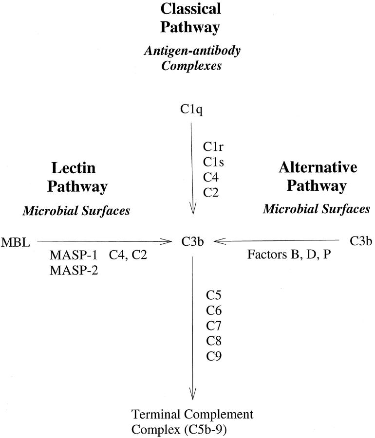
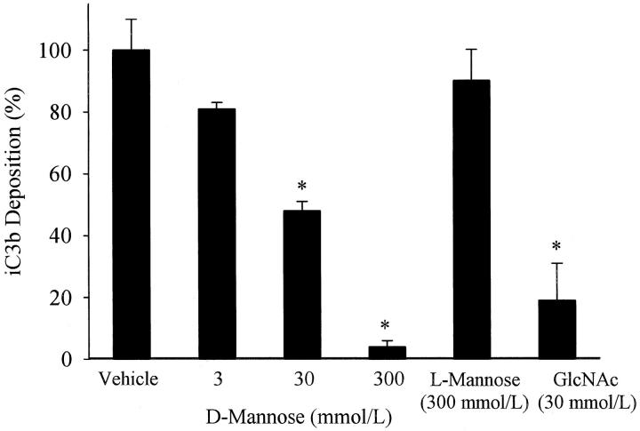
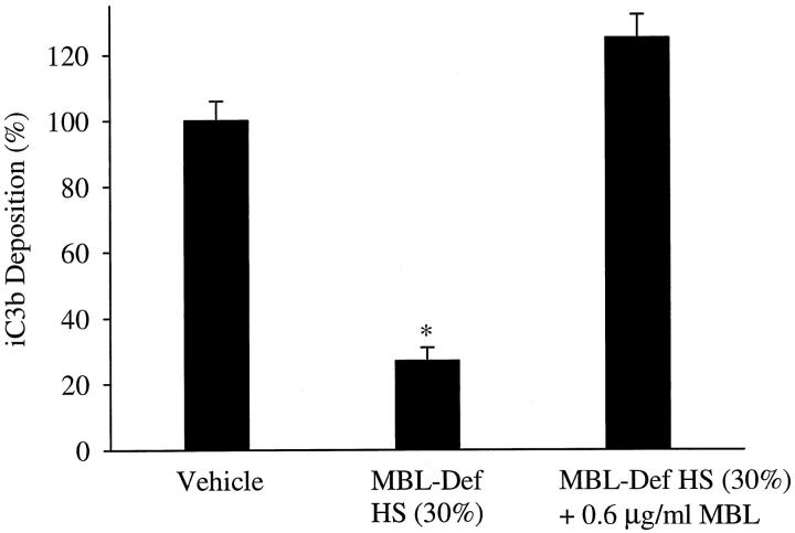
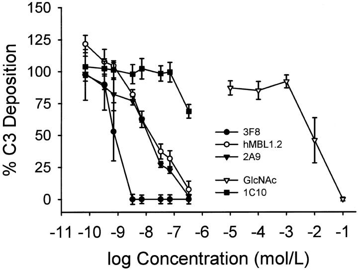
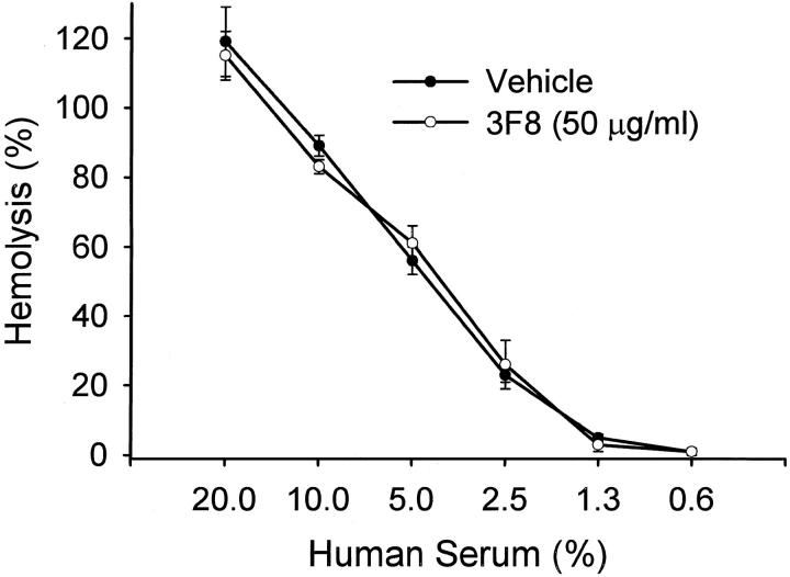
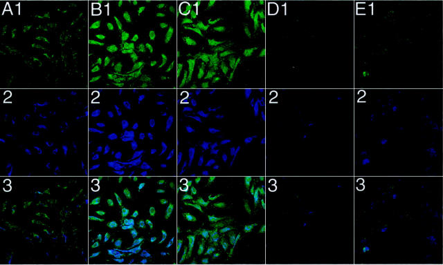
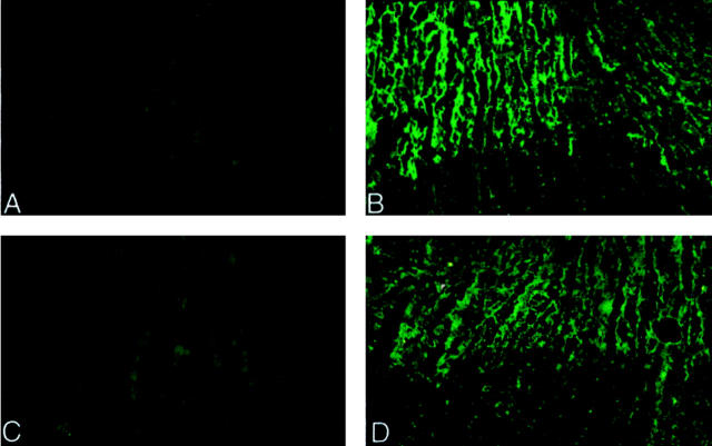
The complement system plays an important role in mediating tissue injury after oxidative stress. The role of mannose-binding lectin (MBL) and the lectin complement pathway (LCP) in mediating complement activation after endothelial oxidative stress was investigated. iC3b deposition on hypoxic (24 hours; 1% O(2))/reoxygenated (3 hours; 21% O(2)) human endothelial cells was attenuated by N-acetyl-D-glucosamine or D-mannose, but not L-mannose, in a dose-dependent manner. Endothelial iC3b deposition after oxidative stress was also attenuated in MBL-deficient serum. Novel, functionally inhibitory, anti-human MBL monoclonal antibodies attenuated MBL-dependent C3 deposition on mannan-coated plates in a dose-dependent manner. Treatment of human serum with anti-MBL monoclonal antibodies inhibited MBL and C3 deposition after endothelial oxidative stress. Consistent with our in vitro findings, C3 and MBL immunostaining throughout the ischemic area at risk increased during rat myocardial reperfusion in vivo. These data suggest that the LCP mediates complement activation after tissue oxidative stress. Inhibition of MBL may represent a novel therapeutic strategy for ischemia/reperfusion injury and other complement-mediated disease states.


Figures

Figure 1.
Figure 1.
Three pathways of complement activation. A simplified schematic figure depicting the antigen/antibody-dependent classical complement pathway and the antibody-independent alternative and lectin complement pathways. Note that activation of the classical or LCP is C2-dependent.
Figure 2.
Figure 2.
Mannose inhibition of endothelial iC3b deposition. iC3b deposition on hypoxic (24 hours) HUVECs reoxygenated (3 hours) in the presence of 30% HS treated with 0, 3, 30, or 300 mmol/L D-mannose or L-mannose (300 mmol/L) was measured by ELISA. D-mannose, but not L-mannose, reduced endothelial iC3b deposition in a dose-dependent manner. Similarly, GlcNAC (30 mmol/L) inhibited iC3b deposition. Data are normalized to hypoxic HUVECs reoxygenated in 30% HS (vehicle; OD405 = 0.14 ± 0.01). n = 3; error bars = SE; *P < 0.05 compared to vehicle.
Figure 3.
Figure 3.
Endothelial iC3b deposition is attenuated in MBL-deficient serum. iC3b deposition on hypoxic HUVECs reoxygenated in 30% MBL-deficient HS was significantly less (P < 0.05) than hypoxic HUVECs reoxygenated in 30% HS (vehicle). In contrast, iC3b deposition on hypoxic HUVECs reoxygenated in 30% MBL-deficient HS reconstituted with MBL did not significantly differ from 30% HS (vehicle). Data are normalized to hypoxic HUVECs reoxygenated in 30% HS (vehicle; OD405 = 0.16 ± 0.01). n = 3; error bars = SE; *P < 0.05 compared to vehicle.
Figure 4.
Figure 4.
Western blot analysis of human MBL. Monoclonal anti-human MBL antibodies 3F8, hMBL1.2, 2A9, or 1C10 were used for Western blot analysis of reduced human MBL. Lanes 1–4 represent staining of reduced human MBL with 10 μg/ml of mAb 2A9, hMBL1.2, 1C10, or 3F8, respectively. A single band with an approximate molecular weight of 32 kd (ie, consistent with reduced MBL) was observed with each mAb. This figure is representative of 3 separate experiments.
Figure 5.
Figure 5.
C3 deposition on mannan-coated plates. Monoclonal anti-human MBL antibodies 3F8, hMBL1.2 and 2A9, as well as N-acetylglucosamine (GlcNAc) inhibited C3 deposition on mannan coated plates in a dose-dependent manner. The control anti-human MBL mAb, 1C10, did not inhibit C3 deposition. Each symbol represents the mean of 4 to 5 individual experiments. Concentrations of antibodies (shown in mol/L) represent 0.01, 0.05, 0.1, 0.5, 1, 5, 10, and 50 μg/ml, respectively. Brackets represent SE.
Figure 6.
Figure 6.
Anti-human MBL mAbs do not attenuate HS hemolytic activity. HS treated with vehicle (PBS) or mAb 3F8 (50 μg/ml) demonstrated similar hemolytic activity to sensitized chicken red blood cells. Similar data were obtained with clones 2A9, hMBL1.2 and 1C10 (data not presented). Thus, these mAbs do not inhibit classical complement pathway activation. Each symbol represents the mean of 3 individual experiments. Brackets represent SE.
Figure 7.
Figure 7.
Colocalization of C3 and MBL on HUVECs after oxidative stress. Immunofluorescent confocal microscopic demonstration of MBL (blue) and C3 (green) deposition on normoxic and hypoxic (24 hours) HUVECs reoxygenated (3 hours) in 30% HS with and without the functionally inhibitory anti-human MBL mAbs 3F8 or 2A9 (5 μg/ml), or the non-functionally inhibitory anti-human MBL mAb, 1C10 (50 μg/ml). Columns A-E represent normoxic untreated, hypoxic/reoxygenated untreated, 1C10-treated, 3F8-treated, and 2A9-treated cells, respectively. Rows 1–3 represent staining for C3, MBL, and C3 + MBL, respectively. C3 (B1) and MBL (B2) staining on hypoxic/reoxygenated HUVECs was significantly increased compared to normoxic HUVECs (A1 and A2, respectively). Treatment of HS with clone 1C10 (50 μg/ml) did not attenuate C3 or MBL deposition (C1 and C2, respectively) after oxidative stress. Treatment of HS with clone 3F8 or 2A9 (5 μg/ml) significantly decreased C3 and MBL deposition (D1 or E1, and D2 or E2, respectively). Row 3 represents colocalization of MBL and C3 under normoxic untreated, hypoxic/reoxygenated untreated, 1C10-treated, 3F8-treated, and 2A9-treated conditions, respectively. These panels are representative of 3 different experiments. Original magnification, ×40.
Figure 8.
Figure 8.
Immunofluorescence analysis of rat C3 and MBL. The left ventricular free wall of rats undergoing 60 minutes of ischemia and no reperfusion (A and C) or 30 minutes of ischemia and 30 minutes of reperfusion (B and D) were processed for immunohistochemical staining for rat C3 (A and B) or MBL (clone 14C3.74; C and D). Positive staining for C3 (B) and MBL (D) is observed in the ischemic/reperfused hearts compared to the ischemia-only hearts (A and C, respectively). Furthermore, the straining is localized to the ischemic area at risk. No staining for C3 or MBL was observed in the sham-operated rat hearts (data not shown). Original magnification, ×100.

References

    1. Weisman HF, Bartow T, Leppo MK, Marsh HC, Jr, Carson GR, Concino MF, Boyle MP, Roux KH, Weisfeldt ML, Fearon DT: Soluble human complement receptor type 1: in vivo inhibitor of complement suppressing post-ischemic myocardial inflammation and necrosis. Science 1990, 249:146-151 - PubMed
    1. Collard CD, Vakeva A, Bukusoglu C, Zünd G, Sperati CJ, Colgan SP, Stahl GL: Reoxygenation of hypoxic human umbilical vein endothelial cells activates the classic complement pathway. Circulation 1997, 96:326-333 - PubMed
    1. Collard CD, Agah A, Stahl GL: Complement activation following reoxygenation of hypoxic human endothelial cells: role of intracellular reactive oxygen species, NF-κB and new protein synthesis. Immunopharmacology 1998, 39:39-50 - PubMed
    1. Collard CD, Agah A, Reenstra WR, Buras JA, Stahl GL: Endothelial nuclear factor-κB translocation and vascular cell adhesion molecule-1 induction by complement: Inhibition with anti-C5 therapy or cGMP analogues. Arterioscler Thromb Vasc Biol 1999, 19:2623-2629 - PubMed
    1. Buerke M, Murohara T, Lefer AM: Cardioprotective effects of a C1 esterase inhibitor in myocardial ischemia and reperfusion. Circulation 1995, 91:393-402 - PubMed

Publication types

MeSH terms