. 2000 May;156(5):1557-64.
doi: 10.1016/S0002-9440(10)65027-4.

Severe destructive autoimmune lesions with aging in murine Sjögren's syndrome through Fas-mediated apoptosis

Affiliations

Severe destructive autoimmune lesions with aging in murine Sjögren's syndrome through Fas-mediated apoptosis

N Ishimaru et al. Am J Pathol. 2000 May.

Abstract

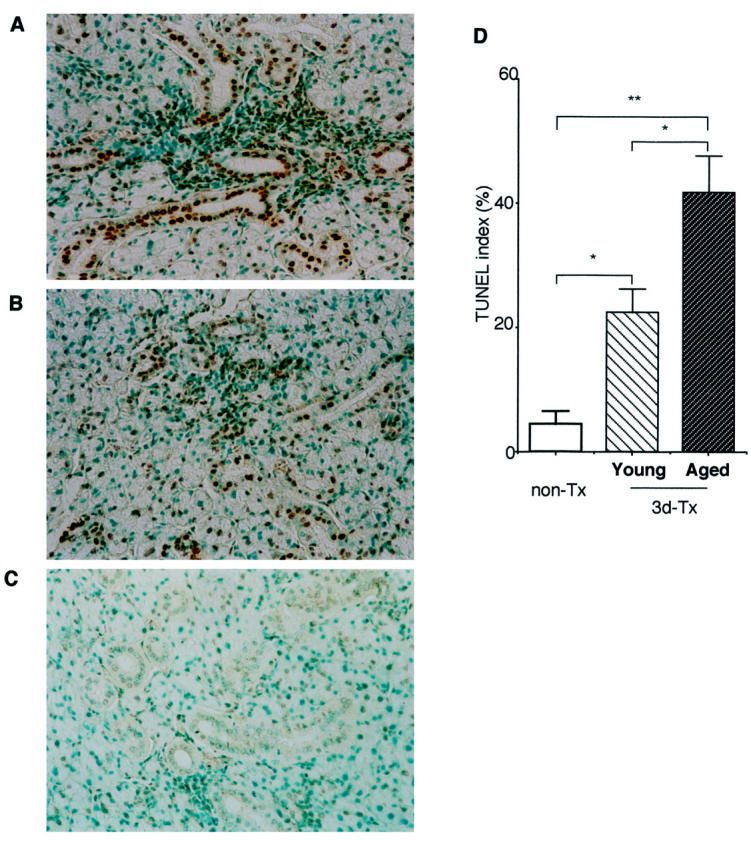
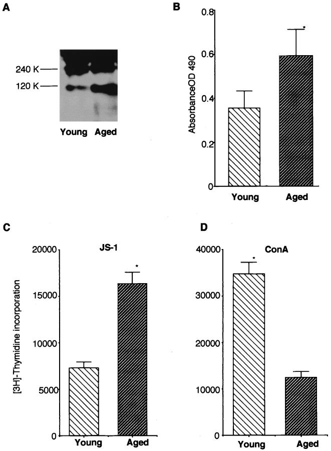
When we evaluated the age-associated changes in autoimmune exocrinopathy in a NFS/sld murine model for primary Sjögren's syndrome (SS), severe destructive autoimmune lesions developed in the salivary and lacrimal glands in the aged mice, compared with those observed in the younger model. We detected a decreased secretion of saliva and tear flow in the aged group. A significant increase of TUNEL(+)-apoptotic epithelial duct cells in the salivary glands was detected in the aged SS animal model. A higher proportion of mouse salivary gland cells bearing Fas was found in the aged group, whereas no significant changes were seen on tissue-infiltrating CD4(+) T cells bearing FasL in the salivary glands from young and aged mice. We detected an increased cleavage product of organ-specific autoantigen, 120-kd alpha-fodrin, in the aged salivary gland tissues on immunoblotting, and an increase in serum autoantibody production against 120-kd alpha-fodrin by enzyme-linked immunosorbent assay. An increase in the proliferative response of splenic T cells against organ-specific autoantigen was observed, whereas nonspecific concanavalin A responsiveness was decreased in the aged mice. In addition, a decrease in Fas expression was found on splenic CD4(+) T cells in the aged mice, and anti-Fas mAb-stimulated apoptosis was down-regulated on CD4(+) T cells. These results indicate that age-associated dysregulation of CD4(+) T cells may play a crucial role on acceleration of organ-specific autoimmune lesions in a murine model for primary SS through Fas-mediated apoptosis.


Figures

Figure 1.
Figure 1.
Mean grade of autoimmune lesions in the salivary and lacrimal glands in aging NFS/sld animal model of both sexes. We observed a significant increase in mean grade of autoimmune lesions with aging, and the predominance in female at all ages. Grading of inflammatory lesions was classified according to the modified method of White and Casarett (*, P < 0.01; **, P < 0.001, Mann-Whitney U test).
Figure 2.
Figure 2.
Representative severe destructive autoimmune lesions in the aged parotid (A) and lacrimal (B) glands of 20-month-old NFS/sld animal model, showing focal fibrosis, ductal dilatation, and fatty infiltration. H&E, ×120.
Figure 3.
Figure 3.
Secretion of saliva and tears. The average saliva and tear volume of the aged SS model mice (18 to 20 months of age) was significantly lower than that of the young mice (2 to 4 months of age) and the total amount of fluid decreased with advancing age. Results are expressed as mean ± SEM in five mice examined per each group (*, P < 0.05; **, P < 0.005, Student’s t-test).
Figure 4.
Figure 4.
Detection of TUNEL+-apoptotic duct cells in the salivary gland sections from aged (18 to 20 months of age) (A) and young (2 to 4 months of age) 3d-Tx NFS/sld mice (B) and non-Tx NFS/sld control mice (6 months of age) (C). D: A significant increase of apoptotic epithelial duct cells was observed in the aged salivary gland tissues from 3d-Tx NFS/sld mice. The percentage of duct cells staining positively with TUNEL was enumerated using a 10 × 20-grid net micrometer disk covering an objective of area 0.16 mm2. Data were analyzed in 10 fields per section, and were expressed as mean percentage ± SD in five mice examined in each group (*, P < 0.01; **, P < 0.001, Student’s t-test).
Figure 5.
Figure 5.
Flow cytometric analysis of Fas expression on MSG cells from aged (18 to 20 months of age) and young (2 to 4 months of age) 3d-Tx NFS/sld and FasL expression on the tissue-infiltrating lymphocytes (TIL) purified from salivary glands gated on CD4. A higher proportion of MSG cells expressing Fas was observed in the aged than that in the young mice. No significant changes were found on the tissue-infiltrating CD4+ T cells bearing FasL both from the aged and the young SS model mice. Five mice in each group were analyzed, and similar results were obtained.
Figure 6.
Figure 6.
A: Detection of organ-specific autoantigen, 120-kd α-fodrin, in the salivary gland tissues from aged (18 to 20 months of age) and young (2 to 4 months of age) SS model mice on Western blot analysis. Representative experiment demonstrates a more intense band of anti-120-kd α-fodrin in the salivary gland tissues from aged mice than those in young mice. Five tissue samples were examined for each group. B: A high titer of serum autoantibodies against 120-kd α-fodrin was detected in aged mice (18 to 20 months of age) compared with that in young SS model mice (2 to 4 months of age) by ELISA. Results indicate mean ± SEM values for five samples examined in each group (*, P < 0.01, Student’s t-test). C: A significant increase in proliferative response of splenic T cells against organ-specific autoantigen, 120-kd α-fodrin was detected in the aged (18 to 20 months of age), as compared with that in the young mice (2 to 4 months of age) (*, P < 0.01, Student’s t-test). D: A significant decrease in nonspecific concanavalin A responsiveness of splenic T cells was detected in the aged SS model mice (18 to 20 months of age) (*, P < 0.001, Student’s t-test). Five mice in each age group were analyzed. Data are expressed as counts per minute per culture ± SD in triplicate.
Figure 7.
Figure 7.
A: Flow cytometric analysis of Fas-expressing CD4+ and CD8+ T cells freshly isolated from the spleens in young (2 to 4 months of age), and aged (18 to 20 months of age) mice. A significant decrease of CD4+ T cells expressing Fas in the aged mice was observed as compared with those in the young mice, whereas no difference was found in CD8+ T cells expressing Fas. Five samples were analyzed in each group. B: Anti-Fas mAb-stimulated apoptosis was significantly decreased in the aged CD4+ T cells in spleen than those in the young mice, but not in CD8+ T cells. Isolated CD4+ T cells were stimulated with anti-Fas mAb and analyzed for apoptotic nuclei with PI and Anexin V. Five mice in each age group were analyzed.

Similar articles

Cited by

References

    1. Miller RA: The aging immune system: primers and prospectus. Science 1996, 273:70-74 - PubMed
    1. Pawelec G, Adibadeh M, Pohla H, Schaudt K: Immunosenescence: aging of the immune system. Immunol Today 1995, 16:420-422 - PubMed
    1. Hodes RJ: Molecular alterations in the aging immune system. J Exp Med 1995, 182:1-3 - PMC - PubMed
    1. Nagel JE, Chopra RK, Chrest FJ, McCoy MT, Schneider EL, Holbrook NJ, Adler WH: Decreased proliferation, interleukin 2 synthesis, and interleukin 2 receptor expression are accompanied by decreased mRNA expression in phytohemagglutinin-stimulated cells from elderly donors. J Clin Invest 1988, 81:1096-1102 - PMC - PubMed
    1. Proust JJ, Filburn CR, Harrison SA, Buchholz MA, Nordin AA: Age-related defect in signal transduction during lectin activation of murine T lymphocytes. J Immunol 1987, 139:1472-1478 - PubMed

Publication types