. 2000 May;156(5):1589-98.
doi: 10.1016/S0002-9440(10)65031-6.

Contribution of apoptosis and apoptosis-related proteins to the malformation of the primitive intrahepatic biliary system in Meckel syndrome

Affiliations

Contribution of apoptosis and apoptosis-related proteins to the malformation of the primitive intrahepatic biliary system in Meckel syndrome

C Sergi et al. Am J Pathol. 2000 May.

Abstract

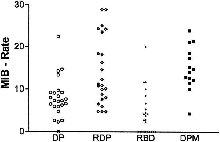
In the developing liver, the complete or partial persistence of the primitive double-layered cylinder of biliary-type cells that surrounds the branches of portal vein and its mesenchyme gives origin to portal tracts with an increased number of bile duct structures. The term "ductal plate malformation of the liver" was coined to label the insufficient remodeling of the primitive intrahepatic biliary system. Meckel syndrome is an autosomal recessive inherited disease characterized by occipital encephalocele, postaxial polydactyly, diffuse cystic renal dysplasia, and malformation of the ductal plate of the liver. We studied 52 fetuses with Meckel syndrome from five German centers (Berlin, Freiburg, Heidelberg, Mainz, and Marburg). Analysis of apoptosis and cell proliferation (Ki-67) was performed by terminal deoxynucleotide transferase-mediated dUTP nick-end labeling (TUNEL) and immunohistochemistry in the liver of 24 normal fetuses of different gestational ages (14-38 weeks of gestation) and in 14 fetuses with Meckel syndrome (17-38 weeks of gestation). The expression of two apoptosis-related proteins, Fas (a transmembrane cell surface protein involved in the apoptosis) and Bcl-2 (an anti-apoptotic protein), was studied by immunohistochemistry in the liver of 11 normal fetuses of different gestational ages (14-40 weeks of gestation) and in 40 fetuses with Meckel syndrome (16-38 weeks of gestation). In control fetuses, apoptosis rate and cell proliferation were high in the remodeling ductal plate and moderate in the ductal plate and in remodeled bile ducts. During gestation, expression of Fas and Bcl-2 decreased and increased, respectively. The malformed ductal plates in the fetal livers with Meckel syndrome showed a marked decrease in the apoptotic rate and Fas expression and an increase in proliferative activity and Bcl-2 expression in comparison with control fetuses. Furthermore, by linear regression analysis, we found that both proliferation activity and apoptosis rate in the ductal plate malformation of fetuses with Meckel syndrome were practically constant along the gestation. These results, which represent the first systematic study of apoptosis in ductal plate malformation of the liver, indicate that 1) animals harboring the gene defect of Meckel syndrome could be a good model for the study of the abnormal development of the primitive intrahepatic biliary system, 2) a decreased cell turnover occurs in the ductal plate malformation of fetuses with Meckel syndrome, and 3) the increase of Bcl-2 expression contributes to the pathogenesis of the lack of remodeling of ductal plate of the liver in Meckel syndrome.


Figures

Figure 1.
Figure 1.
Malformation of the ductal plate of the liver of a fetus affected with Meckel syndrome with dilatation of the primitive ductal plate structure and portal tract fibrosis. In small portal tracts the dilatation in the portal tract suggested a “polypoid”-like image. AE 1 + 3. Original magnifications: a, ×62.5; b, ×200.
Figure 2.
Figure 2.
Proliferation activity (MIB − Rate) expressed as the percentage of MIB-positive cells on all counted cells in a determinate structure during the stages of human fetal liver development (ductal plate (DP), remodeling ductal plate (RDP), and remodeled bile ducts (RBD)) and in the malformation of the primitive embryonic ductal plate or ductal plate malformation (DPM). For statistical significance see text.
Figure 3.
Figure 3.
Proliferation activity (Ki-67) during fetal development of intrahepatic bile ducts and in ductal plate malformation of the liver of fetuses with Meckel syndrome. Cells of remodeling ductal plate in a normal fetus (a, arrow) and in several biliary cells of malformed ductal plates in Meckel syndrome (b, c–d, arrows). Original magnifications: a, ×200; b, ×160; c, ×220; d, ×320.
Figure 4.
Figure 4.
Results of linear regression analysis in all three stages of normal bile duct development (ductal plate (DP), remodeling ductal plate (RDP), remodeled bile ducts (RBD)) of 24 normal fetuses and in the ductal plate malformation (DPM) of 14 fetuses with Meckel syndrome. The best-fit lines are shown as a solid line for DP, a dashed line for RDP, a dotted line for RBD, and a mixed broken line for DPM. The proliferative activity (MIB − Rate) was expressed as the percentage of MIB-positive cells on all counted cells in a determinate structure during the stages of human fetal liver development. Two or more of these developmental stages may be present in the same liver specimen of normal fetuses. Replicate values were averaged and treated as single data points. For statistical significance and r see text.
Figure 5.
Figure 5.
Apoptosis rate expressed as the percentage of TUNEL-positive cells on all counted cells in a determinate structure during the stages of human fetal liver development (ductal plate (DP), remodeling ductal plate (RDP), and remodeled bile ducts (RBD)) and in the malformation of the primitive embryonic ductal plate or ductal plate malformation (DPM). For statistical significance see text.
Figure 6.
Figure 6.
Apoptotic cells during fetal development of intrahepatic bile ducts and in ductal plate malformation of the liver of fetuses with Meckel syndrome are recognizable as nuclei stained by the TUNEL method. Shown are cells of remodeling ductal plate in a normal fetus (a, arrows) and in very few biliary cells of a malformed ductal plate in Meckel syndrome (b, arrow). Original magnifications: a, ×320; b, ×200.
Figure 7.
Figure 7.
Results of linear regression analysis in all three stages of normal bile duct development (ductal plate (DP), remodeling ductal plate (RDP), remodeled bile ducts (RBD)) of 24 normal fetuses and in the ductal plate malformation (DPM) of 14 fetuses with Meckel syndrome. The best-fit lines are shown as a solid line for DP, a dashed line for RDP, a dotted line for RBD, and a mixed broken line for DPM. The apoptosis rate was expressed as the percentage of TUNEL-positive cells on all counted cells in a determinate structure during the stages of human fetal liver development. Two or more of these developmental stages may be present in the same liver specimen of normal fetuses. Replicate values were averaged and treated as single data points. For statistical significance and r see text.
Figure 8.
Figure 8.
Expression of Fas and Bcl-2 antigens during fetal development of intrahepatic bile ducts and in ductal plate malformation of the liver of fetuses with Meckel syndrome. Moderate Fas expression in cells of remodeling ductal plate stage (a, ×320), and negative to faintly positive Fas expression in the malformed ductal plates of Meckel syndrome (b, ×200). Bcl-2 is faintly positive in the remodeling ductal plate stage (c, ×200), whereas it is strongly positive in the malformed ductal plates of Meckel syndrome (d, ×200).
Figure 9.
Figure 9.
Fas score during human fetal bile duct development (x axis: weeks of gestation) of 14 normal fetuses (○) and 40 fetuses with malformation of the primitive embryonic ductal plate (•). In normal fetuses the Fas score comprises the analysis in all three stages of bile duct development (DP, RDP, RBD). Replicate values for the same week of gestation are averaged and treated as single data points. For statistical significance and linear regression analysis see text.
Figure 10.
Figure 10.
Bcl-2 score during human fetal bile duct development (x axis: weeks of gestation) of 14 normal fetuses (○) and 40 fetuses with malformation of the primitive embryonic ductal plate (•). In normal fetuses the Bcl-2 score comprises the analysis in all three stages of bile duct development (DP, RDP, RBD). Replicate values for the same week of gestation are averaged and treated as single data points. For statistical significance and linear regression analysis see text.

Similar articles

Cited by

References

    1. Zou H, Niswander L: Requirement for BMP signaling in interdigital apoptosis and scale formation. Science 1996, 272:738-741 - PubMed
    1. Koseki C, Herzlinger D, al-Awqati Q: Apoptosis in metanephric development. J Cell Biol 1992, 119:1327-1333 - PMC - PubMed
    1. Terada T, Nakanuma Y: Detection of apoptosis and expression of apoptosis-related proteins during human intrahepatic bile duct development. Am J Pathol 1995, 146:67-74 - PMC - PubMed
    1. Desmet VJ: Intrahepatic bile ducts under the lens. J Hepatol 1985, 1:545-559 - PubMed
    1. Blankenberg TA, Lund JK, Reubner BH: Normal and abnormal development of human intrahepatic bile ducts: an immunohistochemical perspective. Perspect Pediatr Pathol 1991, 14:143-167 - PubMed