. 2000 May;156(5):1599-612.
doi: 10.1016/S0002-9440(10)65032-8.

Characterization and isolation of ductular cells coexpressing neural cell adhesion molecule and Bcl-2 from primary cholangiopathies and ductal plate malformations

Affiliations

Characterization and isolation of ductular cells coexpressing neural cell adhesion molecule and Bcl-2 from primary cholangiopathies and ductal plate malformations

L Fabris et al. Am J Pathol. 2000 May.

Abstract

It has recently been shown that reactive bile ductules display neuroendocrine features, including immunoreactivity for the neural cell adhesion molecule (NCAM). In this study we have compared the immunohistochemical expression of NCAM with that of HEA-125 (biliary specific) and LKM-1 (hepatocyte specific) and other markers relevant to morphogenesis (Bcl-2, EMA) and cell proliferation (Ki-67) in cryostat sections from different chronic liver diseases and from fetal livers at different gestational ages. In parallel, viable NCAM-positive ductular cells were purified from collagenase digests of cirrhotic livers by immunomagnetic separation and characterized by immunocytochemistry and transmission electron microscopy. We demonstrated that reactive ductules with atypical morphology coexpressed NCAM and Bcl-2 and were found mainly in congenital diseases associated with ductal plate malformation and in primary cholangiopathies. On the contrary, reactive ductules with typical morphology were negative for NCAM/Bcl-2 and positive for EMA. Reactive ductules coexpressing NCAM/Bcl-2 were negative for the proliferation marker Ki-67 and appeared to be directly connected with periportal hepatocytes. In fetal livers NCAM/Bcl-2 was transiently expressed during the early developmental stages of ductal plate (10-16 weeks) and started to disappear as the ductal plate began duplicating. NCAM-positive ductal plate cells were Ki-67 negative, becoming positive in duplicated segments. Thus the histogenesis of ductular reactive cells seems to recapitulate the early stages of biliary ontogenesis. In primary cholangiopathies and ductal plate malformations, these cells do not appear to maturate further, and thus abundant ductular structures coexist with vanishing mature ducts. These NCAM-positive ductular cells were immunopurified from patients with chronic cholestatic liver diseases and showed ultrastructural features consistent with a less differentiated phenotype than mature cholangiocytes. These isolated cells represent a useful model for in vitro studies.


Figures

Figure 1.
Figure 1.
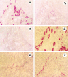
Light micrograph of single staining (immunoperoxidase method) showing the distribution of NCAM during the early steps in bile duct morphogenesis (a–c). An indirect three-step immunoperoxidase technique was used with peroxidase-labeled rabbit anti-mouse and swine anti-rabbit immunoglobulin (Dako) and diaminobenzidine substrate. NCAM is expressed on a single-layer ductal plate surrounding a large portal vein branch, as seen in fetal liver at 12 weeks’ gestation (a: original magnification, ×60). However, NCAM is disappearing as duplicated segments are formed with staining still present in residual ductal plate remnants, as seen in fetal liver at 14 weeks’ gestation (b: original magnification, ×40). Faint patchy NCAM staining is observed in bile ducts with an easily recognizable lumen incorporating into mesenchyma, as seen in fetal liver at 16 weeks’ gestation (c: original magnification, ×120). In d the high level of colocalization between NCAM (red) and Bcl-2 (green) is demonstrated in developing ductal plate by dismerged images of double staining at confocal analysis.
Figure 2.
Figure 2.
Light micrograph of single staining (APAAP method) showing the inverse relationship between EMA and NCAM expression on typical and atypical ductules, in alcoholic liver cirrhosis (a–c) and PSC (d–f) serial tissue sections (original magnification, ×10). HEA-125 has been used as marker of reference for biliary epithelium (a and d). EMA staining (apical localization) is positive in typical ductules at the center of the portal space and negative in the marginal atypical ductules (b and e), whereas NCAM decorates atypical but not typical ductules (c and f). Note the different extension of NCAM immunoreactivity on atypical ductules between alcoholic liver cirrhosis (a and c) and PSC (d and f).
Figure 3.
Figure 3.
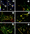
Confocal micrograph of double staining with NCAM (Texas Red) and Bcl-2 (FITC), showing coincident expression between these two markers on atypical ductules. In a the level of coincidence between NCAM (left side) and Bcl-2 (right side) is analyzed by dismerging and color banding in a biliary atresia tissue sample: the highest intensities are shown in red, and the lowest intensities are in blue. NCAM and Bcl-2 share the same distribution and concentration on biliary epithelium: the highest and the lowest levels of binding of NCAM and Bcl-2 are localized at the same places within the atypical ductular structures. All control reactions demonstrated the specificity of the double staining. In b–f, NCAM and Bcl-2 coexpression on ductular reaction is reported for different chronic hepatobiliary diseases: cryptogenic cirrhosis (b), PSC (c), biliary atresia (d), polycystic liver (e), and Caroli’s disease (f). Areas of coincident labeling appear yellow. Bcl-2 noncoincident staining (green) is observed in lymphocytes. In parenchymal liver cirrhosis (b) only a small subset of reactive ductules, characterized by a marginal distribution, coexpressed NCAM and Bcl-2. In primary cholangiopathies (c and d) and developmental liver diseases related to ductal plate malformation (e and f), coincident immunoreactivity for NCAM and Bcl-2 was extensively observed among atypical ductules. In c granular NCAM staining (red) was found in scattered periseptal hepatocytes.
Figure 4.
Figure 4.
Double staining of NCAM (Texas Red) with the hepatocellular marker LKM-1 (FITC) shows that NCAM-positive cells form periportal sprouting cords that are organized into irregularly shaped and poorly formed ductules, directly anastomosing with LKM-1-positive hepatocytes, as seen in PBC (a, original magnification ×40). In alcoholic liver cirrhosis double labeling of LKM-1 (FITC) with NCAM (Texas Red) (b: original magnification, ×40) and HEA-125 (Texas Red) (c: original magnification, ×40) showed that periportal hepatocytes, which were negative for NCAM, began to express HEA-125 immunoreactivity on the plasma membrane, as seen in serial consecutive sections. In a and b some periportal hepatocytes (green) contain small NCAM-positive cytoplasmic granules (red, arrow). In alcoholic liver disease HEA-125 (c) reveals many more ductular structures than NCAM (b), thus confirming the reduced number of atypical ductules in parenchymal liver cirrhosis.
Figure 5.
Figure 5.
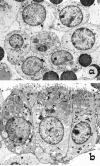
Ultrastructural analysis by TEM of cells freshly isolated from Percoll gradients by immunomagnetic separation, using anti-NCAM (a) and HEA-125 (b) on the NCAM-depleted population. Note the polar columnar morphology, with basal nuclei and well-formed apical microvilli of mature biliary epithelial cells (b), compared with the rounded shape, high nucleus-cytoplasmic ratio, short scattered microvilli, and fewer junctional complexes of NCAM-positive cells (a).

Similar articles

Cited by

References

    1. Popper H, Schaffner F, Stein R: Ductular cell reaction in the liver in hepatic injury. J Mt Sinai Hosp 1957, 24:551-556 - PubMed
    1. Masuko K, Rubin E, Popper H: Proliferation of bile ducts in cirrhosis. Arch Pathol 1964, 78:421-431 - PubMed
    1. Desmet VJ, Roskams T, Van Eyken P: Ductular reaction in the liver. Pathol Res Pract 1995, 191:513-524 - PubMed
    1. Roskams T, Rosenbau J, De Vos R, David G, Desmet VJ: Heparan sulfate proteoglycan expression in chronic cholestatic human liver diseases. Hepatology 1996, 24:524-532 - PubMed
    1. Matsumoto K, Fuji H, Michalopoulos G, Fung JJ, Demetris AJ: Human biliary epithelial cells secrete and respond to cytokines and hepatocyte growth factors in vitro: interleukin-6, hepatocyte growth factor and epidermal growth factor promote DNA synthesis in vitro. Hepatology 1994, 20:376-382 - PubMed

Publication types

MeSH terms

Grants and funding