. 2000 May;156(5):1663-72.
doi: 10.1016/S0002-9440(10)65037-7.

Up-regulation of AT(1) and AT(2) receptors in postinfarcted hypertrophied myocytes and stretch-mediated apoptotic cell death

Affiliations

Up-regulation of AT(1) and AT(2) receptors in postinfarcted hypertrophied myocytes and stretch-mediated apoptotic cell death

A Leri et al. Am J Pathol. 2000 May.

Abstract

To determine whether up-regulation of AT(1) and AT(2) receptors occurred in hypertrophied myocytes after infarction and whether AT(2) played a role in stretch-mediated apoptosis, left ventricular myocytes were dissociated from the surviving portion of the wall 8 days after coronary occlusion and cardiac failure in rats. Control cells were obtained from sham-operated animals. Myocytes were stretched in an equibiaxial stretch apparatus and angiotensin II (Ang II) formation and cell death were measured 3 and 12 hours later. AT(1) and AT(2) proteins were evaluated in freshly isolated myocytes and after stretch. The effects of AT(1) and AT(2) antagonists on stretch-induced Ang II synthesis and apoptosis were also established. Myocardial infarction increased AT(1) and AT(2) in myocytes and stretch further up-regulated these receptors. Ang II levels were higher in postinfarcted myocytes and this peptide increased with the duration of stretch in both groups of cells. Similarly, apoptosis increased with time in control and postinfarcted myocytes. Absolute values of Ang II and apoptosis were greater in myocytes from infarcted hearts at 3 and 12 hours after stretch. Addition of AT(1) blocker to cultures inhibited stretch-activated apoptosis in both myocyte populations as well as the generation of Ang II in postinfarcted myocytes. In contrast, AT(2) antagonists had no impact on these cellular events. In conclusion, Ang II stimulated cell death through AT(1) receptor activation, whereas ligand binding to AT(2) receptor did not alter Ang II concentration and apoptosis in normal and postinfarcted hypertrophied myocytes.


Figures

Figure 1.
Figure 1.
Culture of nonstretched (A, C) and stretched (B, D) left ventricular myocytes obtained from an infarcted heart at 8 days. Nuclei are stained by propridium iodide (yellow) and the cytoplasm by α-sarcomeric actin (red). Arrows in A and C indicate myocytes which are shown at higher magnification in B and D. Arrowheads in B and D define groups of 10 sarcomeres each. Confocal microscopy: A and B, ×100; C and D, ×800.
Figure 2.
Figure 2.
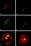
Stretched ventricular myocytes (A and B) obtained from two infarcted hearts at 8 days. Myocyte nuclei labeled by EMB (yellow; arrows) are apparent; myocyte cytoplasm is stained by α-sarcomeric actin (red). EMB-negative myocytes are also shown. Confocal microscopy, ×300.
Figure 3.
Figure 3.
Western blot of AT1 (A) and AT2 (B) receptors in freshly (F) isolated, nonstretched (NS), and stretched (S) myocytes for 3 (3) and 12 (12) hours, obtained from SO and MI hearts. Loading of proteins is illustrated by Coomassie blue staining. AT1R, human cloned AT1 receptor protein. Optical density values for AT1 and AT2 are shown in C; n = 6 in each determination. *, difference, P < 0.05, from myocytes obtained from SO animals; †, difference, P < 0.05, from NS myocytes.
Figure 4.
Figure 4.
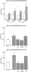
Effects of 3 (3h) and 12 (12h) hours of stretch (S) on the generation of Ang II from myocytes obtained from SO and MI hearts (A). B and C illustrate the impact of losartan (Los) and PD123319 (PD) on the formation of Ang II in myocytes from MI hearts stretched for 3 (B) and 12 (C) hours. NS, nonstretched myocytes. Results are means ± SD; n = 5 in each determination with two exceptions: n = 6 in nontreated S-myocytes from MI hearts in B and C. *, difference from NS-myocytes. In A, † indicates a difference from myocytes from SO hearts. In B and C, † indicates a difference from nontreated S-myocytes from MI hearts.
Figure 5.
Figure 5.
(Starts on facing page) Apoptosis in two binucleated (A–C; G–I) and one mononucleated (D–F) myocyte from post-MI cells stretched for 3 (A–C; G–I) and 12 (D–F) hours. Apoptosis, characterized by nuclear fragmentation, cellular shrinkage, and partial loss of contractile material, was apparent in the two cells shown in panels A–F, exhibiting advanced phases of cell death. Cellular morphology was much more preserved in the early stages of apoptosis depicted in panels G–I. Confocal microscopy: A–F, ×1,200; G–I, ×1,000.
Figure 5.
Figure 5.
(Starts on facing page) Apoptosis in two binucleated (A–C; G–I) and one mononucleated (D–F) myocyte from post-MI cells stretched for 3 (A–C; G–I) and 12 (D–F) hours. Apoptosis, characterized by nuclear fragmentation, cellular shrinkage, and partial loss of contractile material, was apparent in the two cells shown in panels A–F, exhibiting advanced phases of cell death. Cellular morphology was much more preserved in the early stages of apoptosis depicted in panels G–I. Confocal microscopy: A–F, ×1,200; G–I, ×1,000.
Figure 6.
Figure 6.
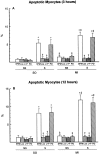
Effects of 3 (A) and 12 (B) hours of stretch (S) on Taq-labeled myocytes obtained from SO and MI hearts in the presence of losartan (Los), PD123319 (PD), and losartan and PD123319 (L+P). NS, nonstretched myocytes. Results are means ± SD; n = 8 in each group. *, difference from NS-myocytes; †, difference from S-myocytes kept in SFM; ‡, difference from S-myocytes from SO hearts.

References

    1. Pfeffer MA, Braunwald E: Ventricular remodeling after myocardial infarction: experimental observations and clinical implications. Circulation 1990, 81:1161-1172 - PubMed
    1. Pfeffer MA, Braunwald E, Moyé LA, Basta L, Jr, Brown EJ, Cuddy TE, Davis BR, Geltman EM, Goldman S, Flaker GC, Klein M, Lamas GA, Packer M, Rouleau J, Rouleau JL, Rutherford J, Wertheimer JH, Hawkins CM, : on behalf of the SAVE Investigators: Effect of captropril on mortality and morbidity in patients with left ventricular dysfunction after myocardial infarction. N Engl J Med 1992, 327:669-677 - PubMed
    1. Itoh G, Tamura J, Suzuki M, Suzuki Y, Ikeda H, Kloke M, Nomura M, Jie T, Ito K: DNA fragmentation of human infarcted myocardial cells demonstrated by the nick end labeling method and DNA agarose gel electrophoresis. Am J Pathol 1995, 146:1235-1331 - PMC - PubMed
    1. Olivetti G, Quaini F, Sala R, Lagrasta C, Corradi D, Bonacina E, Gambert SR, Cigola E, Anversa P: Acute myocardial infarction in humans is associated with activation of programmed myocyte cell death in the surviving portion of the heart. J Mol Cell Cardiol 1996, 28:2005-2016 - PubMed
    1. Narula J, Haider N, Virmani R, DiSalvo TG, Kolodgie FD, Hajjar RJ, Schmidt U, Semigran MJ, Dec GW, Khaw BA: Apoptosis in myocytes in end-stage heart failure. N Engl J Med 1996, 335:1182-1189 - PubMed

Publication types

MeSH terms