. 2000 May;156(5):1733-9.
doi: 10.1016/S0002-9440(10)65044-4.

Vascular endothelial growth factor (VEGF)-induced retinal vascular permeability is mediated by intercellular adhesion molecule-1 (ICAM-1)

Affiliations

Vascular endothelial growth factor (VEGF)-induced retinal vascular permeability is mediated by intercellular adhesion molecule-1 (ICAM-1)

K Miyamoto et al. Am J Pathol. 2000 May.

Abstract

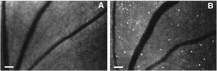
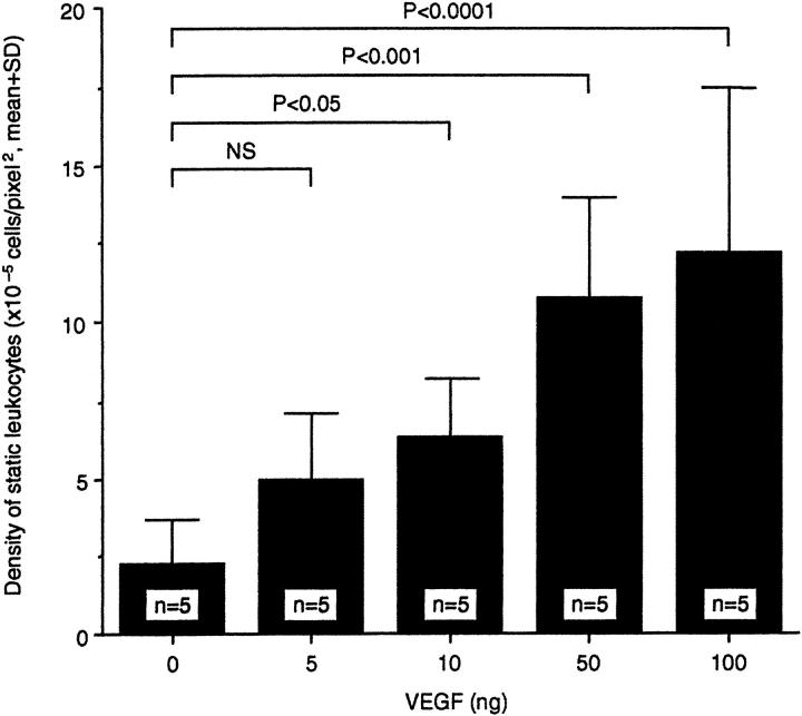
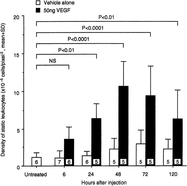
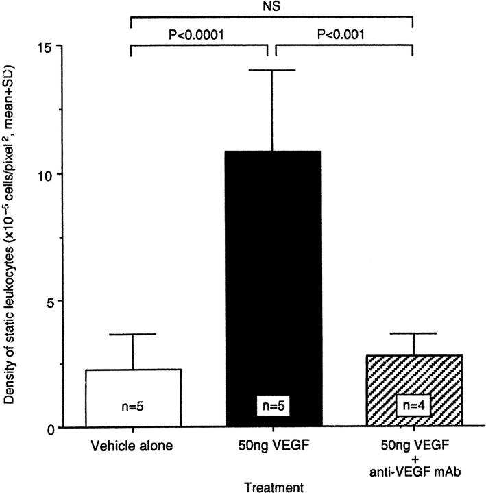
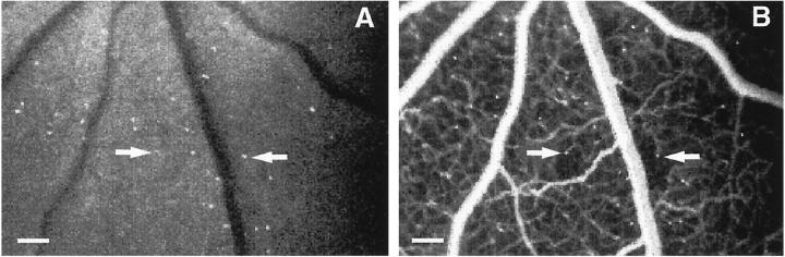
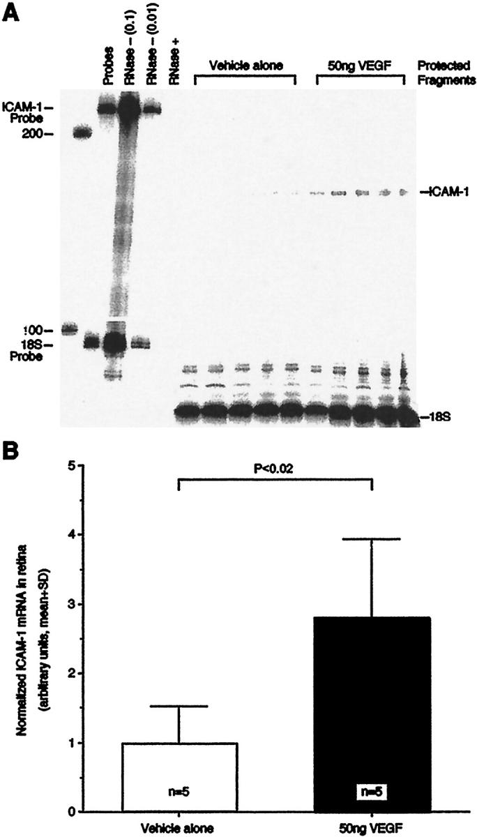
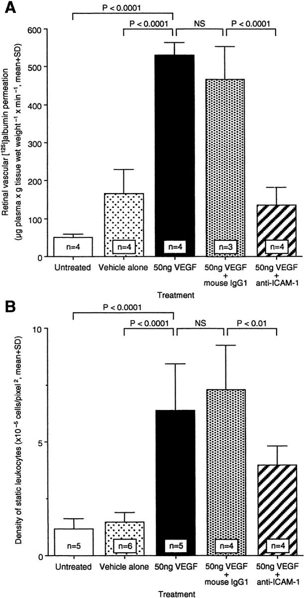
Two prominent vascular endothelial growth factor (VEGF)-induced retinal effects are vascular permeability and capillary nonperfusion. The mechanisms by which these effects occur are not completely known. Using a rat model, we show that intravitreous injections of VEGF precipitate an extensive retinal leukocyte stasis (leukostasis) that coincides with enhanced vascular permeability and capillary nonperfusion. The leukostasis is accompanied by the up-regulation of intercellular adhesion molecule-1 expression in the retina. The inhibition of intercellular adhesion molecule-1 bioactivity with a neutralizing antibody prevents the permeability and leukostasis increases by 79% and 54%, respectively. These data are the first to demonstrate that a nonendothelial cell type contributes to VEGF-induced vascular permeability. Additionally, they identify a potential mechanism for VEGF-induced retinal capillary nonperfusion.


Figures

Figure 1.
Figure 1.
VEGF-induced retinal leukostasis. A: AOLF appearance of a normal retinal before injection of 50 ng VEGF. B: AOLF appearance of the same retinal area 48 hours after intravitreous VEGF injection. Numerous static leukocytes are visible (white dots), as well as vessel dilation and tortuosity. Mobile leukocytes are not visible in this phase of the study. Scale bar, 100 μm (3.2 pixels = 1 μm).
Figure 2.
Figure 2.
Dose-response graph of VEGF-induced retinal leukostasis. The indicated amount of VEGF was delivered via intravitreous injection and leukostasis was quantitated with AOLF 48 hours later.
Figure 3.
Figure 3.
Time course of VEGF-induced retinal leukostasis. Fifty nanograms of VEGF or an equivalent volume of vehicle alone was delivered via intravitreous injection and leukostasis was quantitated with AOLF at the indicated time points.
Figure 4.
Figure 4.
Specificity of VEGF-induced leukostasis. Fifty nanograms of VEGF were delivered intravitreously either alone or combined a 50 molar excess of an anti-VEGF neutralizing mAb (clone A4.6.1). Leukostasis was quantitated with AOLF 48 hours later.
Figure 5.
Figure 5.
Leukocyte-induced capillary nonperfusion. A: Fifty nanograms of VEGF were delivered via intravitreous injection and AOLF was performed 48 hours later. B: AOLF was immediately followed by fluorescein angiography and showed areas of capillary nonperfusion downstream from static leukocytes (arrows). Scale bar, 100 μm (3.2 pixels = 1 μm).
Figure 6.
Figure 6.
VEGF-induced retinal ICAM-1 gene expression. A: The ribonuclease protection assay demonstrated that retinal ICAM-1 levels were significantly increased 20 hours after the intravitreous delivery of 50 ng VEGF. Control animals received 5 μl of PBS solvent alone. Each lane shows the signal from one retina of one animal. The lane labeled “Probes” shows a 100-fold dilution of the full-length ICAM-1 and 18S riboprobes. The lanes labeled “RNase− (0.1)” and “RNase− (0.01)” show the 10-fold and 100-fold dilutions, respectively, of the full-length riboprobes without sample or RNase. The lane labeled “RNase+” shows the full-length riboprobes with RNase, but without sample. B: ICAM-1 protection assay data normalized to 18S signal.
Figure 7.
Figure 7.
Effect of anti-ICAM-1 mAb on permeability and leukostasis after intravitreous VEGF injection. ICAM-1 bioactivity was inhibited via intravenous administration of ICAM-1 neutralizing antibody, and retinal permeability (A) or leukostasis (B) were evaluated, respectively. NS, not significant.

References

    1. Miyamoto K, Khosrof S, Bursell S-E, Rohan R, Murata T, Clermon A, Aiello LP, Ogura Y, Adamis AP: Prevention of leukostasis and vascular leakage in streptozotocin-induced diabetic retinopathy via intercellular adhesion molecule-1 inhibition. Proc Natl Acad Sci USA 1999, 96:10836-10841 - PMC - PubMed
    1. Tolentino MJ, Miller JW, Gragoudas ES, Jakobiec FA, Flynn E, Chatzistefanou K, Ferrara N, Adamis AP: Intravitreal injections of vascular endothelial growth factor produce retinal ischemia and microangiopathy in an adult primate. Ophthalmology 1996, 103:1820-1828 - PubMed
    1. Okamoto N, Tobe T, Hacket SF, Ozaki H, Vinores MA, LaRochelle W, Zack DJ, Campochiaro PA: Transgenic mice with increased expression of vascular endothelial growth factor in the retina: a new model of intraretinal and subretinal neovascularization. Am J Pathol 1997, 151:281-291 - PMC - PubMed
    1. Aiello LP, Bursell SE, Clermont A, Duh E, Ishii H, Takagi C, Mori F, Ciulla TA, Ways K, Jirousek M, Smith LE, King GL: Vascular endothelial growth factor-induced retinal permeability is mediated by protein kinase C in vivo and suppressed by an orally effective beta-isoform-selective inhibitor. Diabetes 1997, 46:1473-1480 - PubMed
    1. McLeod DS, Lefer DJ, Merges C, Lutty GA: Enhanced expression of intracellular adhesion molecule-1 and P-selectin in the diabetic human retina and choroid. Am J Pathol 1995, 147:642-653 - PMC - PubMed

Publication types

MeSH terms