

Eosinophilia of dystrophin-deficient muscle is promoted by perforin-mediated cytotoxicity by T cell effectors
- PMID: 10793090
- PMCID: PMC1876906
- DOI: 10.1016/S0002-9440(10)65050-X
Eosinophilia of dystrophin-deficient muscle is promoted by perforin-mediated cytotoxicity by T cell effectors
Abstract
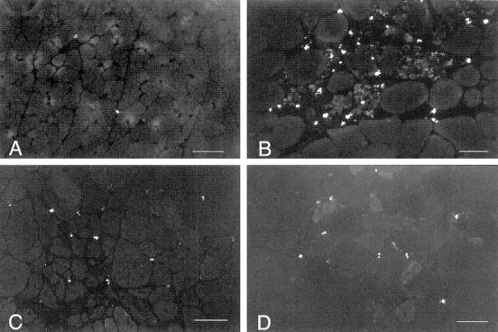
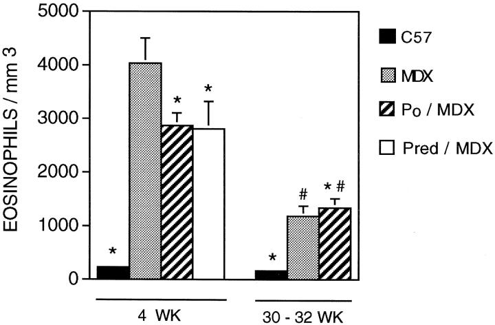
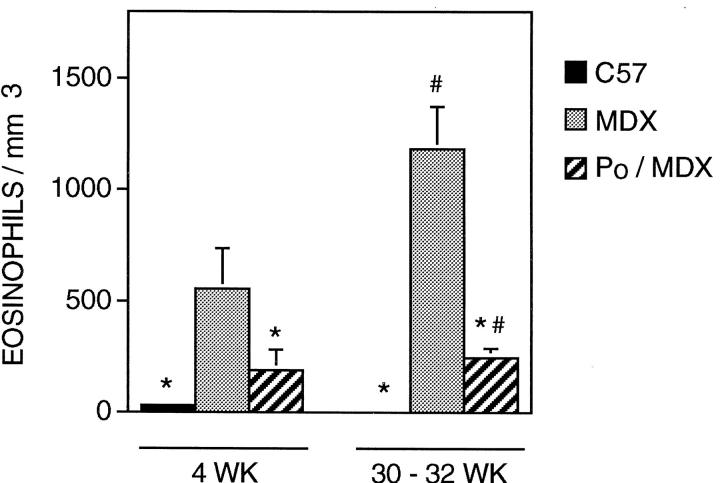
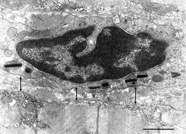
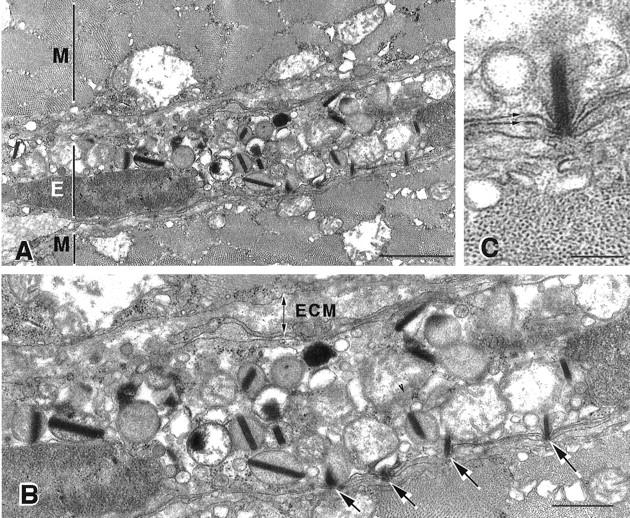
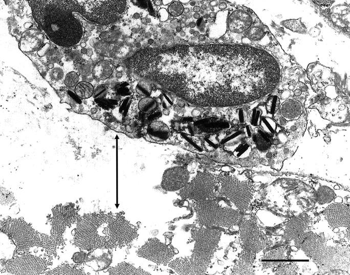
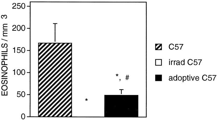
Previous investigations have shown that cytotoxic T lymphocytes (CTLs) contribute to muscle pathology in the dystrophin-null mutant mouse (mdx) model of Duchenne muscular dystrophy through perforin-dependent and perforin-independent mechanisms. We have assessed whether the CTL-mediated pathology includes the promotion of eosinophilia in dystrophic muscle, and thereby provides a secondary mechanism through which CTLs contribute to muscular dystrophy. Quantitative immunohistochemistry confirmed that eosinophilia is a component of the mdx dystrophy. In addition, electron microscopic observations show that eosinophils traverse the basement membrane of mdx muscle fibers and display sites of close apposition of eosinophil and muscle membranes. The close membrane apposition is characterized by impingement of eosinophilic rods of major basic protein into the muscle cell membrane. Transfer of mdx splenocytes and mdx muscle extracts to irradiated C57 mice by intraperitoneal injection resulted in muscle eosinophilia in the recipient mice. Double-mutant mice lacking dystrophin and perforin showed less eosinophilia than was displayed by mdx mice that expressed perforin. Finally, administration of prednisolone, which has been shown previously to reduce the concentration of CTLs in dystrophic muscle, produced a significant reduction in eosinophilia. These findings indicate that eosinophilia is a component of the mdx pathology that is promoted by perforin-dependent cytotoxicity of effector T cells. However, some eosinophilia of mdx muscle is independent of perforin-mediated processes.
Figures
Similar articles
-
Myonuclear apoptosis in dystrophic mdx muscle occurs by perforin-mediated cytotoxicity.J Clin Invest. 1997 Jun 1;99(11):2745-51. doi: 10.1172/JCI119464. J Clin Invest. 1997. PMID: 9169505 Free PMC article.
-
Helper (CD4(+)) and cytotoxic (CD8(+)) T cells promote the pathology of dystrophin-deficient muscle.Clin Immunol. 2001 Feb;98(2):235-43. doi: 10.1006/clim.2000.4966. Clin Immunol. 2001. PMID: 11161980
-
Coisogenic all-plus-one immunization: a model for identifying missing proteins in null-mutant conditions. Antibodies to dystrophin in mdx mouse after transplantation of muscle from normal coisogenic donor.Neuropediatrics. 1994 Aug;25(4):176-82. doi: 10.1055/s-2008-1073019. Neuropediatrics. 1994. PMID: 7824089
-
How does dystrophin deficiency lead to muscle degeneration?--evidence from the mdx mouse.Neuromuscul Disord. 1995 Nov;5(6):445-56. doi: 10.1016/0960-8966(95)00001-4. Neuromuscul Disord. 1995. PMID: 8580726 Review.
-
Cell-mediated cytotoxicity in perforin-less mice.Int Rev Immunol. 1995;13(1):1-14. doi: 10.3109/08830189509061734. Int Rev Immunol. 1995. PMID: 7494105 Review.
Cited by
-
Molecular feature of neutrophils in immune microenvironment of muscle atrophy.J Cell Mol Med. 2022 Sep;26(17):4658-4665. doi: 10.1111/jcmm.17495. Epub 2022 Jul 27. J Cell Mol Med. 2022. PMID: 35899367 Free PMC article. Review.
-
Major basic protein-1 promotes fibrosis of dystrophic muscle and attenuates the cellular immune response in muscular dystrophy.Hum Mol Genet. 2008 Aug 1;17(15):2280-92. doi: 10.1093/hmg/ddn129. Epub 2008 Apr 21. Hum Mol Genet. 2008. PMID: 18430716 Free PMC article.
-
Matrix Metalloproteinases and Tissue Inhibitor of Metalloproteinases in Inflammation and Fibrosis of Skeletal Muscles.J Neuromuscul Dis. 2016 Nov 29;3(4):455-473. doi: 10.3233/JND-160183. J Neuromuscul Dis. 2016. PMID: 27911334 Free PMC article. Review.
-
From innate to adaptive immune response in muscular dystrophies and skeletal muscle regeneration: the role of lymphocytes.Biomed Res Int. 2014;2014:438675. doi: 10.1155/2014/438675. Epub 2014 Jun 16. Biomed Res Int. 2014. PMID: 25028653 Free PMC article. Review.
-
The expanding role(s) of eosinophils in health and disease.Blood. 2012 Nov 8;120(19):3882-90. doi: 10.1182/blood-2012-06-330845. Epub 2012 Aug 30. Blood. 2012. PMID: 22936660 Free PMC article. Review.
References
-
- Silberstein D: Eosinophil function in health and disease. Crit Rev Oncol Hematol 1994, 19:47-77 - PubMed
-
- Laitinen L, Laitenen A, Neino M, Haahtela T: Eosinophilic airway inflammation during exacerbation of asthma and its treatment with inhaled corticosteroids. Am Rev Respir Dis 1991, 143:423-427 - PubMed
-
- Kornfield H, Berman J, Beer D, Center D: Induction of human T-lymphocyte motility by interleukin-2. J Immunol 1985, 134:3887-3890 - PubMed
MeSH terms
Substances
LinkOut - more resources
Full Text Sources
Medical
