Comparative Study
. 2000 May;11(5):1509-21.
doi: 10.1091/mbc.11.5.1509.

Identification of a novel member of the chloride intracellular channel gene family (CLIC5) that associates with the actin cytoskeleton of placental microvilli

Affiliations
Free PMC article
Comparative Study

Identification of a novel member of the chloride intracellular channel gene family (CLIC5) that associates with the actin cytoskeleton of placental microvilli

M Berryman et al. Mol Biol Cell. 2000 May.
Free PMC article

Abstract

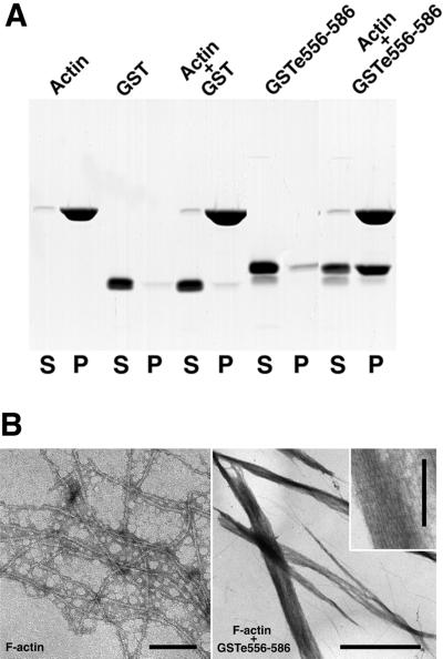
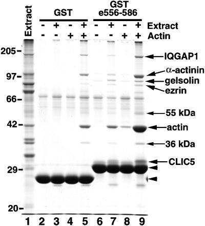
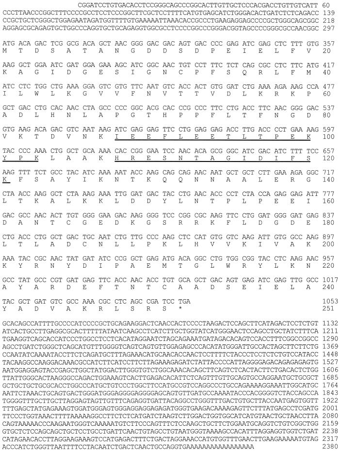
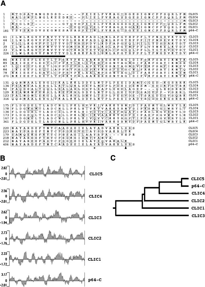
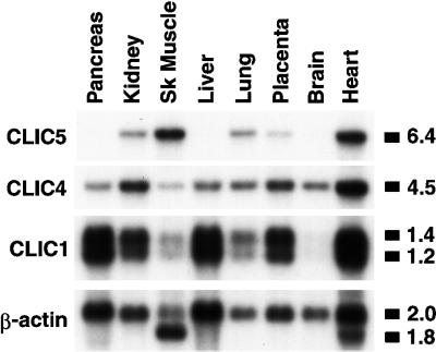
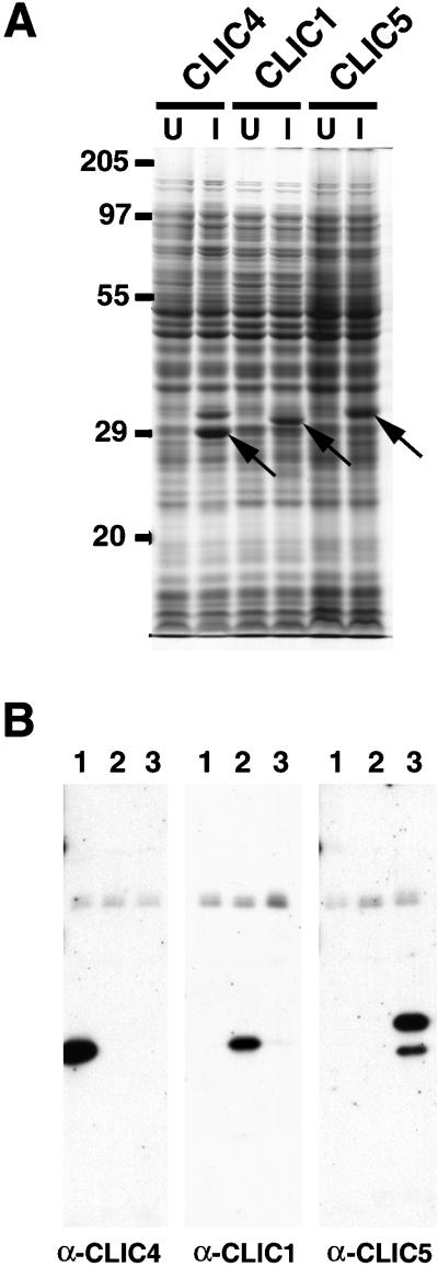
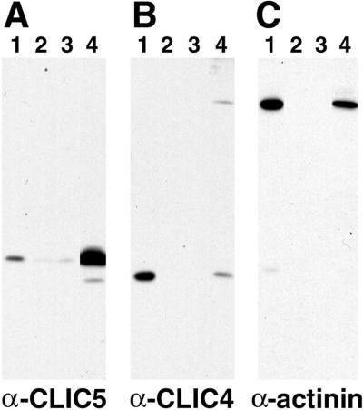
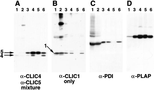
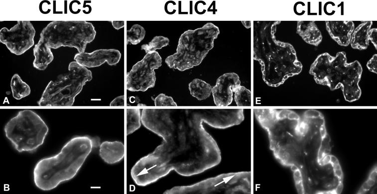
The chloride intracellular channel (CLIC) gene family has been implicated in chloride ion transport within various subcellular compartments. We report here the molecular, biochemical, and cellular characterization of a new member of this gene family termed CLIC5. CLIC5 was isolated from extracts of placental microvilli as a component of a multimeric complex consisting of several known cytoskeletal proteins, including actin, ezrin, alpha-actinin, gelsolin, and IQGAP1. We cloned human cDNAs and generated antibodies specific for CLIC5, CLIC1/NCC27, and CLIC4/huH1/p64H1. CLIC5 shares 52-76% overall identity with human CLIC1, CLIC2, CLIC3, and CLIC4. Northern blot analysis showed that CLIC5 has a distinct pattern of expression compared with CLIC1 and CLIC4. Immunoblot analysis of extracts from placental tissues demonstrated that CLIC4 and CLIC5 are enriched in isolated placental microvilli, whereas CLIC1 is not. Moreover, in contrast to CLIC1 and CLIC4, CLIC5 is associated with the detergent-insoluble cytoskeletal fraction of microvilli. Indirect immunofluorescence microscopy revealed that CLIC4 and CLIC5 are concentrated within the apical region of the trophoblast, whereas CLIC1 is distributed throughout the cytoplasm. These studies suggest that CLIC1, CLIC4, and CLIC5 play distinct roles in chloride transport and that CLIC5 interacts with the cortical actin cytoskeleton in polarized epithelial cells.


Figures

Figure 1
Figure 1
Direct binding of skeletal muscle actin to the C-terminal 30 residues of ezrin. (A) Purified actin or buffer alone was incubated with GST or GST-ezrin fusion protein (GSTe556–586), followed by ultracentrifugation. Fractions corresponding to the supernatant (S) and pellet (P) were resolved by SDS-PAGE and visualized with Coomassie blue staining. (B) F-actin incubated alone (bar, 0.1 μm) or in combination with GST-ezrin 556–586 (bar, 1.0 μm) was visualized by negative-stain electron microscopy as described in MATERIALS AND METHODS. The inset shows part of a bundle of parallel actin filaments at high magnification (bar, 0.1 μm).
Figure 2
Figure 2
Identification of placental microvillus proteins that associate with the C-terminal 30 residues of ezrin. Glutathione–agarose affinity beads containing GST or GST-ezrin 556–586 (GSTe556–586) were incubated with buffer alone, microvillus extracts, or actin. After washing, bound proteins were eluted, run on 12% SDS-PAGE, and stained with Coomassie blue. The mobilities of the fusion proteins are indicated by arrowheads. The mobilities of protein standards are indicated on the left in kilodaltons.
Figure 3
Figure 3
Nucleotide and deduced amino acid sequences of human CLIC5 cDNA. Residues corresponding to the two placental CLIC5 peptide sequences are underlined. This sequence is available from GenBank under accession number AF216941.
Figure 4
Figure 4
Human CLIC5 has homology to CLIC4, CLIC3, CLIC2, CLIC1, and the C-terminal half of bovine p64 (p64-C). (A) Sequence alignment. Amino acid residues are numbered on the left. Identities are boxed. The two conserved hydrophobic domains are underlined. Three highly conserved tyrosine residues are indicated by asterisks. The arrowhead indicates a potential phosphorylation site for cAMP-dependent protein kinase that is conserved in CLIC5, CLIC4, and p64-C. (B) Kyte-Doolittle hydrophobicity plots. (C) Dendrogram of CLIC proteins.
Figure 5
Figure 5
Comparison of the mRNA expression of CLIC5, CLIC4, and CLIC1 in human tissues. A commercially available Northern blot was probed sequentially with radiolabeled antisense DNA corresponding to the coding sequences of CLIC5, CLIC4, and CLIC1. A β-actin probe was used as a control. The sizes of the major transcripts are indicated on the right in kilobases.
Figure 6
Figure 6
Expression of recombinant CLIC proteins and characterization of monospecific antibodies. (A) Expression of untagged CLIC4, CLIC1, and CLIC5. Bacterial extracts from uninduced (U) and induced (I) cultures were run on 12% SDS-PAGE and stained with Coomassie blue. The major induced bands are indicated by arrows. The mobility of standard proteins is indicated on the left in kilodaltons. (B) Immunoblot analysis of induced bacterial lysates expressing untagged CLIC4 (lanes 1), CLIC1 (lanes 2), and CLIC5 (lanes 3). Lysates containing equal amounts (20 ng) of the induced CLIC proteins were resolved by 12% SDS-PAGE, transferred to membranes, and probed with specific antibodies, as indicated at the bottom of each panel. Note that there is an additional induced band that migrates slower than CLIC4 in the lysate shown in A that is not recognized in the corresponding immunoblot in B. In addition, the band that migrates faster than CLIC5 in the CLIC5 immunoblot in B presumably represents a bacterially derived degradation product of CLIC5.
Figure 7
Figure 7
Association of CLIC5 with cytoskeletal components in the affinity chromatography assay. A soluble extract of placental microvilli (lanes 1) was incubated with glutathione–agarose affinity beads containing GST (lanes 2), GST-ezrin 475–584 (lanes 3), and GST-ezrin 475–586 (lanes 4). The beads were washed and eluted, and the bound proteins were run on 12% SDS-PAGE and blotted. (A) Blot probed with antibody against CLIC5. (B) Blot probed with antibody against CLIC4. (C) Blot probed with antibody against α-actinin.
Figure 8
Figure 8
Immunoblot analysis of CLIC proteins in various fractions of human placenta. Approximately equal amounts (7 μg) of total protein from whole placenta (lanes 1), total soluble proteins (lanes 2), placental membranes (lanes 3), and isolated total microvilli (lanes 4) were separated on 12% SDS-PAGE and transferred to membranes. Equal volumes of total (lanes 4), detergent-insoluble (lanes 5), and detergent-soluble (lanes 6) microvillus fractions were also examined. (A) Blot probed with a mixture of antibodies specific for CLIC4 and CLIC5. (B) Blot probed with antibody to CLIC1. (C) Blot probed with a mAb to protein disulfide isomerase (PDI). (D) Blot probed with a mAb to placental alkaline phosphatase (PLAP). The mobilities of the CLIC proteins are indicated by arrows.
Figure 9
Figure 9
Indirect immunofluorescence localization of CLIC proteins in human placenta. Cryosections were fixed, permeabilized, treated with an antigen-unmasking solution, and stained with primary antibodies specific for CLIC5 (A and B), CLIC4 (C and D), and CLIC1 (E and F). Fluorescein-conjugated anti-rabbit immunoglobulin G was used to detect the primary antibodies. The arrows indicate staining in the apical cytoplasm. The images in the top row were taken with a 16× objective lens (bar in A, 40 μm), and the images in the bottom row were taken with a 40× objective lens (bar in B, 16 μm).

References

    1. Al-Awqati Q. Chloride channels of intracellular organelles. Curr Opin Cell Biol. 1995;7:504–508. - PubMed
    1. Altschul SF, Gish W, Miller W, Myers EW, Lipman DJ. Basic local alignment search tool. J Mol Biol. 1990;215:403–410. - PubMed
    1. Begenisich T, Melvin JE. Regulation of chloride channels in secretory epithelia. J Membr Biol. 1998;163:77–85. - PubMed
    1. Berryman M, Gary R, Bretscher A. Ezrin oligomers are major cytoskeletal components of placental microvilli: a proposal for their involvement in cortical morphogenesis. J Cell Biol. 1995;131:1231–1242. - PMC - PubMed
    1. Bretscher A. Purification of an 80,000-dalton protein that is a component of the isolated microvillus cytoskeleton, and its localization in nonmuscle cells. J Cell Biol. 1983;97:425–432. - PMC - PubMed

Publication types

MeSH terms

Associated data