. 2000 May;11(5):1905-17.
doi: 10.1091/mbc.11.5.1905.

A novel selection regime for differentiation defects demonstrates an essential role for the stumpy form in the life cycle of the African trypanosome

Affiliations
Free PMC article

A novel selection regime for differentiation defects demonstrates an essential role for the stumpy form in the life cycle of the African trypanosome

M Tasker et al. Mol Biol Cell. 2000 May.
Free PMC article

Abstract

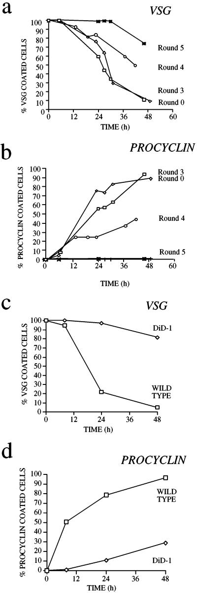
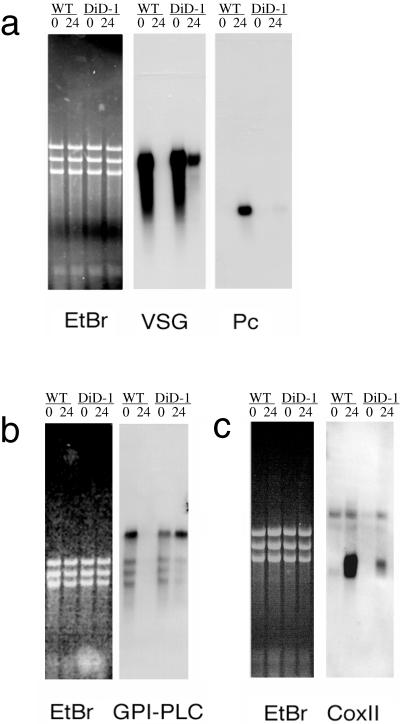
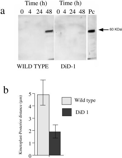
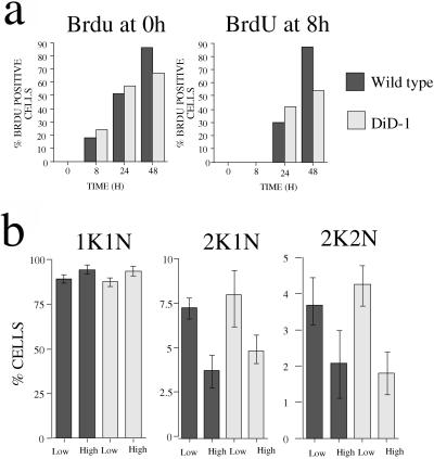
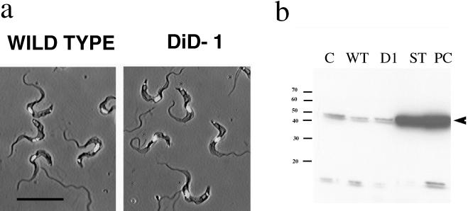
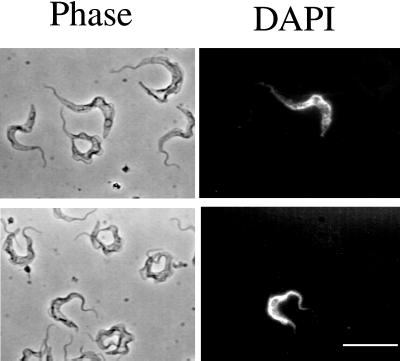
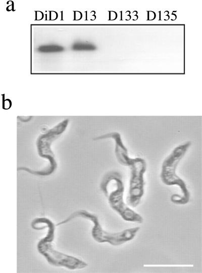
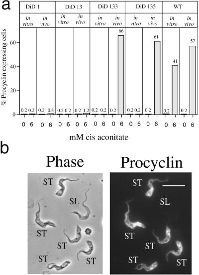
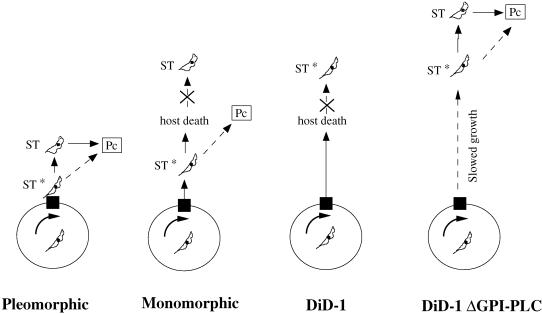
A novel selection scheme has been developed to isolate bloodstream forms of Trypanosoma brucei, which are defective in their ability to differentiate to the procyclic stage. Detailed characterization of one selected cell line (defective in differentiation clone 1 [DiD-1]) has demonstrated that these cells are indistinguishable from the wild-type population in terms of their morphology, cell cycle progression, and biochemical characteristics but are defective in their ability to initiate differentiation to the procyclic form. Although a small proportion of DiD-1 cells remain able to transform, deletion of the genes for glycophosphatidyl inositol-phospholipase C demonstrated that this enzyme was not responsible for this inefficient differentiation. However, the attenuated growth of the Delta-glycophosphatidyl inositol-phospholipase C DiD-1 cells in mice permitted the expression of stumpy characteristics in this previously monomorphic cell line, and concomitantly their ability to differentiate efficiently was restored. Our results indicate that monomorphic cells retain expression of a characteristic of the stumpy form essential for differentiation, and that this is reduced in the defective cells. This approach provides a new route to dissection of the cytological and molecular basis of life cycle progression in the African trypanosome.


Figures

Figure 1
Figure 1
Selection for differentiation-defective trypanosome lines. (a) VSG coat loss for trypanosome lines subjected to zero (Round 0; T. b. rhodesiense EATRO 2340 GUP2965.3; the starting wild-type population), three, four, and five rounds of in vitro differentiation and in vivo selection. (b) Expression of the procyclic stage-specific coat procyclin during the selection regimen. (c) VSG coat loss for DiD-1, a clonal line derived from Round 4 of the selection regimen, shown in comparison with the wild-type population. (d) Differentiation of the DiD-1 population assessed by gain of procyclin.
Figure 2
Figure 2
Stage-regulated RNA expression in the DiD-1 and wild-type populations 24 h after incubation in culture at 27°C containing 6 mM cis-aconitate. To demonstrate relative loading, an ethidium-stained image of each gel is shown, together with its corresponding Northern blot. (a) VSG and procyclin gene expression; (b) GPI-PLC gene expression; (c) cytochrome oxidase subunit II gene expression. The respective sizes for each transcript are VSG, ∼1.6 kb; procyclin, ∼0.8 kb; GPI-PLC, ∼4 kb, and coxII, 0.7 kb.
Figure 3
Figure 3
Glycosomal development and morphological restructuring of DiD-1. (a) Glycosomal development of DiD-1 cells 4, 24, and 48 h after culture at 27°C in 6 mM cis-aconitate. The left panel shows equal protein loadings of the wild-type population undergoing differentiation, and the right panel shows DiD-1 samples under the same conditions. Each blot was probed with a rat anti-PEPCK antibody (a kind gift from Prof. T. Seebeck, University of Bern, Bern, Switzerland). A procyclic protein sample is shown at the extreme right. (b). Quantitative analysis of the relative distance between the kinetoplast and cell posterior in wild-type or DiD-1 populations 24 h after incubation in differentiation conditions. n = 100 cells for each cell type.
Figure 4
Figure 4
DiD-1 does not show a cell cycle defect. (a) BrdU incorporation of DiD-1 and wild-type cells in culture at 27°C, with 6 mM cis-aconitate. In the left graph, cells were incubated in the presence of 50 mM BrdU and 50 mM 2′-deoxycytidine from the outset. In the right graph, BrdU and 2′-deoxycytidine were not added until 8 h after the cells had been placed into differentiation conditions. (b) Cell cycle progression of wild-type and DiD-1 cells during the course of a bloodstream parasitemia. The proportions of cells with a configuration of one kinetoplast and one nucleus (1K1N), 2K1N, and 2K2N were scored for each population when the parasite density was 5 × 10⁷ cells/ml (low) or 5 × 10⁸ cells/ml (high). The data represent four individual parasitemias for each cell type.
Figure 5
Figure 5
Characteristics of DiD-1 in the bloodstream. (a) Phase-contrast images of either wild-type (left panel) or DiD-1(right panel) cells from a bloodstream parasitemia of 5 × 10⁸ cells/ml. In each case cells have been counterstained with DAPI to reveal the nucleus and kinetoplast. Bar, 20 μm. (b) Expression of DHLADH in cultured monomorphic cells (C), wild-type cells isolated from a bloodstream parasitemia (WT), DiD-1 cells isolated from a bloodstream parasitemia (D1), bloodstream stumpy forms (ST), and cells induced to differentiate from the stumpy form to the procyclic form and isolated after 24 h (Pc). Proteins from the wild-type and DiD-1 populations were prepared from a parasitemia at a cell density of 7 × 10⁸ cells/ml. Note that stumpy and differentiated procyclic form cells express abundant DHLADH, whereas a uniformly low level is detected in each of the other populations. A small cross-reacting band is also detected with this antibody of ∼15 kDa.
Figure 6
Figure 6
A small proportion of DiD-1 cells are able to differentiate. The left panels shows the morphology of DiD-1 cells 4 h after incubation in culture at 27°C with 6 mM cis-aconitate. In each case cells were sought that demonstrated the expression of procyclin (right panel), and their morphology was examined for the expression of stumpy cell characteristics. Bar, 30 μm.
Figure 7
Figure 7
GPI-PLC gene deletion in DiD-1. (a) Genomic Southern blots of DiD-1 cells (DI), DiD-1 cells deleted for one allele of GPI-PLC (D13), or DiD-1 cells deleted for both alleles of GPI-PLC (D133 and D135). Each was digested with BamHI and probed with the gene for GPI-PLC. (b) Development of stumpy cell characteristics after prolonged growth of the DiD-1–3.5 GPI-PLC null mutant in the bloodstream. Bar, 10 μm.
Figure 8
Figure 8
Differentiation of wild-type, DiD-1, and DiD-1 ΔGPI-PLC mutants grown in vitro and in vivo. (a) Percentage of procyclin expressers in the respective cell lines either 72 h (in vitro) or 24 h (in vivo) after incubation in differentiation conditions; the concentration of cis-aconitate used (0 or 6 mM) is shown in each case. Note that all parasites isolated in vivo were grown in mice immunocompromised with cyclophosphamide. (b) Representative image of the GPI-PLC null mutant DiD-1–3.5 cells 4 h after incubation in differentiation conditions, when bloodstream morphology is preserved. Note that those cells with a stumpy or stumpy-like morphology (ST) have initiated the expression of procyclin, whereas the morphologically slender cell (SL) does not express this marker.
Figure 9
Figure 9
Model for the ability of bloodstream cells to differentiate based on their progression toward a differentiation-competent ST* state. For each cell type (pleomorphic, monomorphic, and differentiation-defective DiD-1; Did-1ΔGPI-PLC) the circle represents the cell cycle of the trypanosome, with the black box indicating the receptive window of the cell cycle at which parasites must accumulate to be able to differentiate. Cells must then progress toward an ST* state before transformation to procyclic cells either directly or via the morphologically stumpy form.

References

    1. Ashcroft MT. A comparison between a syringe passaged and a tsetse fly transmitted line of a strain of Trypanosoma rhodesiense. Ann Trop Med Parasitol. 1960;54:44–70. - PubMed
    1. Bangs JD, Ransom DM, McDowell MA, Brouch EM. Expression of bloodstream variant surface glycoproteins in procyclic stage Trypanosoma brucei: role of GPI anchors in secretion. EMBO J. 1997;16:4285–4294. - PMC - PubMed
    1. Bass KE, Wang CC. The in vitro differentiation of pleomorphic Trypanosoma brucei from bloodstream into procyclic form requires neither intermediary nor short-stumpy stage. Mol Biochem Parasitol. 1991;44:261–270. - PubMed
    1. Bass KE, Wang CC. Transient inhibition of protein synthesis accompanies differentiation of Trypanosoma brucei from bloodstream to procyclic forms. Mol Biochem Parasitol. 1992;56:129–140. - PubMed
    1. Brown RC, Evans DA, Vickerman K. Changes in oxidative metabolism and ultrastructure accompanying differentiation of the mitochondrion in Trypanosoma brucei. Int JParasitol. 1973;3:691–704. - PubMed

Publication types

LinkOut - more resources