. 2000 Jun;74(11):4988-98.
doi: 10.1128/jvi.74.11.4988-4998.2000.

Synergistic action of GA-binding protein and glucocorticoid receptor in transcription from the mouse mammary tumor virus promoter

Affiliations

Synergistic action of GA-binding protein and glucocorticoid receptor in transcription from the mouse mammary tumor virus promoter

K Aurrekoetxea-Hernández et al. J Virol. 2000 Jun.

Abstract

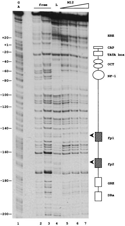
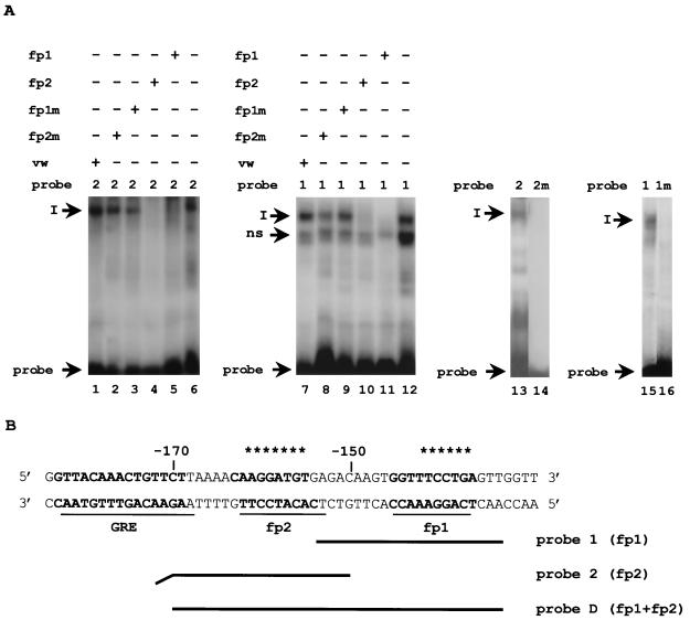
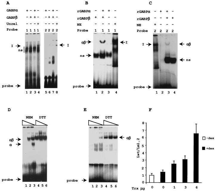
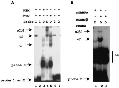
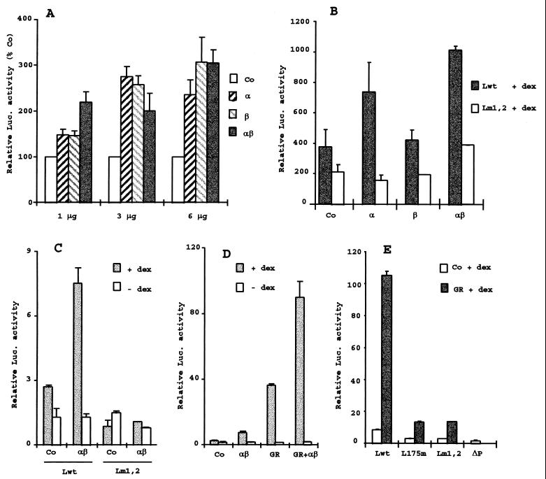
B lymphocytes are among the first cells to be infected by mouse mammary tumor virus (MMTV), and they play a crucial role in its life cycle. To study transcriptional regulation of MMTV in B cells, we have analyzed two areas of the long terminal repeat (LTR) next to the glucocorticoid receptor binding site, fp1 (at position -139 to -146 from the cap site) and fp2 (at -157 to -164). Both showed B-cell-specific protection in DNase I in vitro footprinting assays and contain binding sites for Ets transcription factors, a large family of proteins involved in cell proliferation and differentiation and oncogenic transformation. In gel retardation assays, fp1 and fp2 bound the heterodimeric Ets factor GA-binding protein (GABP) present in B-cell nuclear extracts, which was identified by various criteria: formation of dimers and tetramers, sensitivity to pro-oxidant conditions, inhibition of binding by specific antisera, and comigration of complexes with those formed by recombinant GABP. Mutations which prevented complex formation in vitro abolished glucocorticoid-stimulated transcription from an MMTV LTR linked to a reporter gene in transiently transfected B-cell lines, whereas they did not affect the basal level. Exogenously expressed GABP resulted in an increased level of hormone response of the LTR reporter plasmid and produced a synergistic effect with the coexpressed glucocorticoid receptor, indicating cooperation between the two. This is the first example of GABP cooperation with a steroid receptor, providing the opportunity for studying the integration of their intracellular signaling pathways.


Figures

FIG. 1
FIG. 1
B-cell factors binding in vitro to the HRE of MMTV. DNase I footprinting analysis with nuclear extracts of the M12 B-cell line shows two protected sites, fp1 and fp2 (arrowheads), not seen with nuclear extracts of the fibroblastic Ltk cell line. A DNA fragment comprising the sequences from the StyI restriction site at positions −303 to +133, where a synthetic BamHI linker was inserted, was 5′ end labeled at the StyI site. The fragment was incubated without added proteins (lanes 2 and 3), with 60 μg of nuclear proteins from L cells (lane 4), or with 48 (lane 5), 72 (lane 6), or 104 (lane 7) μg of nuclear proteins from M12 cells. The complexes were subjected to DNase I digestion and separated on a sequencing gel. Lane 1 contains a purine sequencing reaction of the probe, and the numbers on the left indicate the number of nucleotides from the transcription start site (+1). Also represented in the scheme on the right are known factor-binding sites in the HRE of the MMTV LTR that are protected by L-cell extracts: RNA initiation site (CAP), TATA box, binding sites for octamer factors (OCT), for CTF/NF-1, for the glucocorticoid receptor (the distal GRE), and for a tissue-specific factor (DRa [14, 47]).
FIG. 2
FIG. 2
B-cell nuclear extracts form similar complexes on probes with the fp1 or fp2 sequences. Gel retardation assays (A) with M12 nuclear extracts and end-labeled oligonucleotide probes (B) are shown. Mutated oligonucleotides (1m and 2m) contained transversions at the positions denoted by asterisks in the sequence in panel B, where the GRE and the B-cell footprints fp1 and fp2 are underlined and in bold. (A) Protein-DNA complexes were resolved on nondenaturing polyacrylamide gels, followed by autoradiography. Where indicated (+; above the probes), a 300-fold excess of competitor DNA, either homologous, heterologous, mutated (m), or nonspecific (vw, an oligonucleotide with the sequence of the adenovirus-2 promoter), was included in the preincubation mixture. Specific complexes with similar mobilities (I) on fp1 (1) or fp2 (2) showed cross competition (lanes 4, 5, 10, and 11). Mutations in the fp1 (1m) or fp2 (2m) sites abolished the formation of complex I (lanes 14 and 16), which was not affected by adding fp1m (lanes 3 and 9) or fp2m (lanes 2 and 8) in the competition experiment. ns, nonspecific complex.
FIG. 3
FIG. 3
Factors binding in fp1 and fp2 cooperate in the transcriptional response to glucocorticoids in B cells. pGL-3 luciferase reporter plasmids were transiently transfected into A20 cells (A, C, and D) or M12 cells (B) and stimulated (+ dex) or not (− dex) with the glucocorticoid hormone dexamethasone (50 nM) for 5 h. The relative luciferase (Luc.) activity was calculated as the ratio to the activity of the cotransfected SV40-Renilla luciferase internal standard. Individual experiments are shown, which have been repeated with the same results at least twice. The error bars show the standard deviations between duplicate samples. (A and B) Luciferase activities of plasmids containing a wild-type LTR (Lwt) or LTRs with mutations in fp1 and fp2 (Lm1,2), in fp1 (Lm1), or in fp2 (Lm2). Plasmid ΔP contained an LTR lacking the sequences downstream of position −105, including the promoter, and serves as a negative control. (C) Luciferase activity (+ dex) of plasmids containing LTRs truncated at position −303 (Twt and Tm1,2). (D) LTRs with mutations in the GRE are unresponsive to dexamethasone in A20 B cells. In the mutated LTRs an octameric HindIII linker replaces the sequences between −193 and −162 (in L193m) or between −175 and −166 (in L175m).
FIG. 4
FIG. 4
GABP binds to fp1 and fp2 and is present in complex I of B-cell extracts in a redox-sensitive form. (A to E) Gel retardation assays with end-labeled oligonucleotide probes. (A) Specific antibodies against GABPα and/or GABPβ (lanes 1 to 3 and 5 to 7) abolished the formation of complex I-GABPαβ (lanes 4 and 8) on 32P-labeled oligonucleotides containing either fp1 (1) or fp2 (2). A20 nuclear extracts were preincubated with the polyclonal antibodies (purified IgGs) prior to addition of the probes. A similarly purified unrelated (Unrel) antibody was used as a negative control (lanes 4 and 8). (B and C) Recombinant GABPα (rGABPα) and/or GABPβ was incubated with the fp1 probe (B, lanes 1 to 3) or the fp2 probe (C, lanes 2 to 4) in parallel with A20 nuclear extracts (NE) (B, lane 4, and C, lane 1). ns, nonspecific complex (also present in the non-DNA-binding GABPβ sample). (D and E) Redox sensitivity of complex formation in vitro. Treatment with NEM (0.25, 0.5, or 5 mM) or DTT (2, 3, or 4 mM) for 20 min at 20°C was carried out in the binding reaction with A20 nuclear extracts and the fp1 (D) or the fp2 (E) probe. (F) Redox sensitivity of reporter activity in vivo. M12 cells were cotransfected with a plasmid expressing thioredoxin (Trx; 0, 1, 3, or 6 μg) and a vector plasmid (to a total of 6 μg) plus 0.75 pmol of a luciferase reporter under the control of either the wild-type LTR (Lwt) or an LTR with mutations in fp1 and fp2 (Lm1,2). The cells were cultured in the presence of 1 mM H2O2 for 24 h, and dexamethasone (50 nM) was added to the +dex samples during the last 4 h before harvesting. The firefly luciferase activity of the extracts was measured and normalized to the SV40-Renilla luciferase activity of a cotransfected internal standard. The ratio of the activity of Lwt to that of Lm1,2 was plotted as a function of the amount of cotransfected thioredoxin plasmid. The error bars indicate standard deviations. +, present; −, absent.
FIG. 5
FIG. 5
Redox-sensitive GABP tetramers are formed by B-cell nuclear extracts on a probe containing both fp1 and fp2. (A) Gel retardation assay with A20 nuclear extracts and oligonucleotide probe fp1 (1), fp2 (2), or fp1-fp2 (D; see Fig. 2B). With probe D, a slower-migrating band is detected (lane 4). By analogy to the complex formed by recombinant GABP, and to those observed with similarly arranged Ets sites of known genes, it is tentatively identified as the tetrameric from of GABP and is labeled α2β2. Treatment with alkylating agents eliminated the tetrameric complex, in vitro by adding NEM (20 min; 20°C) to the binding reaction (lane 3) or in vivo by using a nuclear extract of DEM-treated cells (lane 5). On the single-site probes as well, DEM treatment of the cells used for the nuclear extract eliminated the αβ complex (lanes 2 versus 1 and 7 versus 6). (B) For comparison, tetramer formation on probe D by a mixture of recombinant GABPα (rGABPα) and GABPβ (lane 2). +, present; −, absent. ns, nonspecific complex.
FIG. 6
FIG. 6
Exogenous GABP increases glucocorticoid-stimulated reporter activity in M12 cells (A and C) and L cells (B) in an fp1-fp2-dependent manner (B and C); exogenous glucocorticoid receptor synergizes with GABP (D) and requires fp1 and fp2 (E) in M12 cells. (A) The expression plasmids sRSV-GABPα and/or sRSV-GABPβ1 (0, 1, 3, or 6 μg) plus the control plasmid pRSV-TK (Co) to a total amount of 12 μg were transiently cotransfected with 0.75 pmol of wild-type LTR-(firefly) luciferase plasmid into M12 cells, which were treated with dexamethasone (50 nM). (B) 6 μg of pRSV-TK (Co) or 3 μg of sRSV-GABPα and/or sGABPβ1 plus pRSV-TK to a total of 6 μg were cotransfected with 0.75 pmol of wild-type (Lwt) or fp1-fp2-mutated (Lm1,2) LTR-luciferase reporter constructs into Ltk cells, which were treated with 100 nM dexamethasone (+ dex). (C) Three micrograms each of sRSV-GABPα and sRSV-GABPβ1 expression plasmids (αβ) or 6 μg of pRSV-TK (Co) was cotransfected with 0.75 pmol of wild-type (Lwt) or fp1-fp2-mutated (Lm1,2) LTR-luciferase constructs into M12 cells, which were treated (+ dex) or not (− dex) with dexamethasone (50 nM). (D) Synergistic effect of GABP and the glucocorticoid receptor (GR). M12 cells were transfected with 0.75 pmol of Lwt plus 9 μg of pRSV-TK (Co) or 3 μg each of sRSV-GABPα and sRSV-GABPβ1 (αβ) and/or 3 μg of pRSVhGRα (GR). pRSV-TK was added to a total amount of 9 μg per transfection. The cells were treated (+ dex) or not (− dex) with 50 nM dexamethasone. (E) Overexpressed glucocorticoid receptor required fp1 and fp2 as well as the GRE for glucocorticoid stimulation in M12 cells. Three micrograms of pSG5mGR (GR) or of pSV2-neo control plasmid (Co) was cotransfected with 0.75 pmol of LTR-luciferase constructs containing either the wild-type LTR (Lwt), an LTR with the mutation LS 175/−166 in the GRE (L175m), the fp1-fp2-mutated LTR (Lm1,2), or the promoterless ΔP plasmid as a negative control. The cells were treated with 50 nM dexamethasone. In panel E, the firefly luciferase (Luc.) activities of the extracts were measured and normalized to the SV40-Renilla luciferase activity of a cotransfected internal standard. In panels A to D, the luciferase activity is expressed in arbitrary units with respect to the protein content. Individual experiments are shown, which have been repeated with the same results at least twice. The error bars show the standard deviations between duplicate samples.

References

    1. Acha-Orbea H, MacDonald H R. Superantigens of mouse mammary tumor virus. Annu Rev Immunol. 1995;13:459–486. - PubMed
    1. Arroyo J, Winchester E, McLellan B S, Huber B T. Shared promoter elements between a viral superantigen and the major histocompatibility complex class II-associated invariant chain. J Virol. 1997;71:1237–1245. - PMC - PubMed
    1. Avots A, Hoffmeyer A, Flory E, Cimanis A, Rapp U R, Serfling E. GABP factors bind to a distal interleukin 2 (IL-2) enhancer and contribute to c-Raf-mediated increase in IL-2 induction. Mol Cell Biol. 1997;17:4381–4389. - PMC - PubMed
    1. Bannert N, Avots A, Baier M, Serfling E, Kurth R. GA-binding protein factors, in concert with the coactivator CREB binding protein/p300, control the induction of the interleukin 16 promoter in T lymphocytes. Proc Natl Acad Sci USA. 1999;96:1541–1546. - PMC - PubMed
    1. Basuyaux J P, Ferreira E, Stehelin D, Butticè G. The Ets transcription factors interact with each other and with the c-Fos/c-Jun complex via distinct protein domains in a DNA-dependent and -independent manner. J Biol Chem. 1997;272:26188–26195. - PubMed

Publication types

MeSH terms

LinkOut - more resources