. 2000 May 23;97(11):6108-13.
doi: 10.1073/pnas.100133697.

A DNA transfection system for generation of influenza A virus from eight plasmids

Affiliations

A DNA transfection system for generation of influenza A virus from eight plasmids

E Hoffmann et al. Proc Natl Acad Sci U S A. .

Abstract

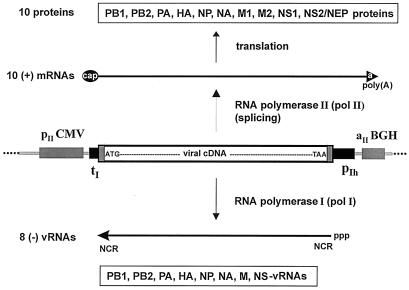
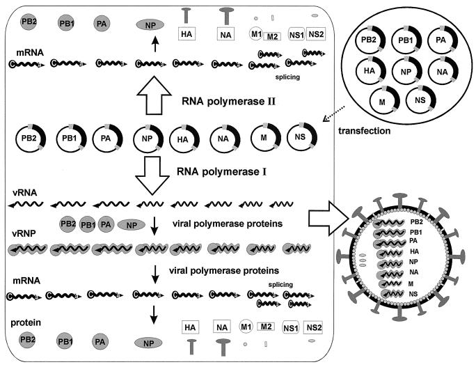
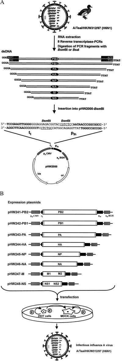
We have developed an eight-plasmid DNA transfection system for the rescue of infectious influenza A virus from cloned cDNA. In this plasmid-based expression system, viral cDNA is inserted between the RNA polymerase I (pol I) promoter and terminator sequences. This entire pol I transcription unit is flanked by an RNA polymerase II (pol II) promoter and a polyadenylation site. The orientation of the two transcription units allows the synthesis of negative-sense viral RNA and positive-sense mRNA from one viral cDNA template. This pol I-pol II system starts with the initiation of transcription of the two cellular RNA polymerase enzymes from their own promoters, presumably in different compartments of the nucleus. The interaction of all molecules derived from the cellular and viral transcription and translation machinery results in the generation of infectious influenza A virus. The utility of this system is proved by the recovery of the two influenza A viruses: A/WSN/33 (H1N1) and A/Teal/HK/W312/97 (H6N1). Seventy-two hours after the transfection of eight expression plasmids into cocultured 293T and MDCK cells, the virus yield in the supernatant of the transfected cells was between 2 x 10(5) and 2 x 10(7) infectious viruses per milliliter. We also used this eight-plasmid system for the generation of single and quadruple reassortant viruses between A/Teal/HK/W312/97 (H6N1) and A/WSN/33 (H1N1). Because the pol I-pol II system facilitates the design and recovery of both recombinant and reassortant influenza A viruses, it may also be applicable to the recovery of other RNA viruses entirely from cloned cDNA.


Figures

Figure 1
Figure 1
Schematic representation of the pol I–pol II transcription system for synthesis of vRNA and mRNA. The cDNA of each of the eight influenza virus segments is inserted between the pol I promoter (pIh) and the pol I terminator (tI). This pol I transcription unit is flanked by the pol II promoter (pIICMV) of the human cytomegalovirus and the polyadenylation signal (aIIBGH) of the gene encoding bovine growth hormone. After transfection of the eight expression plasmids, two types of molecules are synthesized. From the human pol I promoter, negative-sense vRNA is synthesized by cellular pol I. The synthesized vRNA contains the noncoding regions (NCR) at the 5′ and 3′ ends. Transcription by pol II yields mRNAs with 5′ cap structures and 3′ poly(A) tails; these mRNAs are translated into viral proteins. The ATG of the viral cDNA is the first ATG downstream of the pol II transcription start site.
Figure 2
Figure 2
The eight-plasmid pol I–pol II system for the generation of influenza A virus. Eight expression plasmids containing the eight viral cDNAs inserted between the human pol I promoter and the pol II promoter (see Fig. 1) are transfected into eukaryotic cells. Because each plasmid contains two different promoters, both cellular pol I and pol II will transcribe the plasmid template, presumably in different nuclear compartments, which will result in the synthesis of viral mRNAs and vRNAs. After synthesis of the viral polymerase complex proteins (PB1, PB2, PA, nucleoproteins), the viral replication cycle is initiated. Ultimately, the assembly of all viral molecules directly (pol II transcription) or indirectly (pol I transcription and viral replication) derived from the cellular transcription and translation machinery results in the interaction of all synthesized molecules (vRNPs and the structural proteins HA, NA, M1, M2, NS2/NEP) to generate infectious influenza A virus.
Figure 3
Figure 3
Schematic representation of the method developed for the construction and transfection of the eight expression plasmids to recover A/Teal/HK/W312/97 (H6N1). (A) Viral RNA was extracted from virus particles. RT-PCR was performed with primers containing segment-specific nucleotides and sequences for the type IIs restriction endonucleases BsmBI or BsaI. The eight viral PCR fragments were digested with BsmBI or BsaI and inserted into pHW2000 (linearized with BsmBI). This insertion resulted in eight expression constructs where the viral cDNAs are precisely fused to the pol I promoter and terminator (the viral terminal sequences AGC… ACT are shown for the PB2 segment in the black rectangles). (B) The eight expression plasmids with a pol I promoter and a pol II promoter contain one copy of each of the viral cDNAs of the eight segments. The open reading frames for the 10 viral proteins are flanked by the segment-specific noncoding regions (gray boxes). Because the used human pol I promoter shows high activity only in cell lines derived from humans or related species, we cocultured human 293T cells together with the standard cell line used for influenza A (MDCK cells). Viruses produced in the 293T cells after transfection can then infect MDCK cells and replicate.
Figure 4
Figure 4
Characterization of the recovered viruses by RT-PCR. (A) RNA was extracted from virus particles after two passages of the supernatant of transfected cells (see Tables 1 and 2) on MDCK cells. RT-PCR was performed with primers specific for the NS gene segment and with vRNA extracted from virions. The NS primers we used were not strain specific, thus allowing the amplification of any influenza A NS segment. The reaction products were subjected to electrophoresis on a 2% agarose gel. To ensure that the amplified DNA fragments were derived from vRNA and not from plasmid DNA carried over from transfected cells, one reaction was performed without the addition of reverse transcriptase (RT−). Lanes 1 and 2, recombinant A/Teal/HK/W312/97 (Table 1); lanes 3 and 4, M reassortant (Table 2); lanes 5 and 6, NS reassortant (Table 2); lanes 7 and 8, recombinant A/WSN/33 virus (Table 1); lanes 9 and 10, quadruple reassortant (Table 2). (B) NcoI digestion of the fragments shown in A (only the WSN-NS-cDNA has an NcoI site). The identity of the NS fragments was also verified by sequence analysis of the amplified product (not shown).

References

    1. Webster R G, Bean W J, Gorman O T, Chambers T M, Kawaoka Y. Microbiol Rev. 1992;56:152–179. - PMC - PubMed
    1. Scholtissek C, Bürger H, Kistner O, Shortridge K F. Virology. 1985;147:287–294. - PubMed
    1. Claas E C, Osterhaus A D, van Beek R, De Jong J C, Rimmelzwaan G F, Senne D A, Krauss S, Shortridge K F, Webster R G. Lancet. 1998;351:472–477. - PubMed
    1. Suarez D L, Perdue M L, Cox N, Rowe T, Bender C, Huang J, Swayne D E. J Virol. 1998;72:6678–6688. - PMC - PubMed
    1. Subbarao K, Klimov A, Katz J, Regnery H, Lim W, Hall H, Perdue M, Swayne D, Bender C, Huang J, et al. Science. 1998;279:393–396. - PubMed

Publication types

MeSH terms

LinkOut - more resources