. 2000 May 23;97(11):5972-7.
doi: 10.1073/pnas.100448997.

Increased metalloproteinase activity, oxidant production, and emphysema in surfactant protein D gene-inactivated mice

Affiliations

Increased metalloproteinase activity, oxidant production, and emphysema in surfactant protein D gene-inactivated mice

S E Wert et al. Proc Natl Acad Sci U S A. .

Abstract

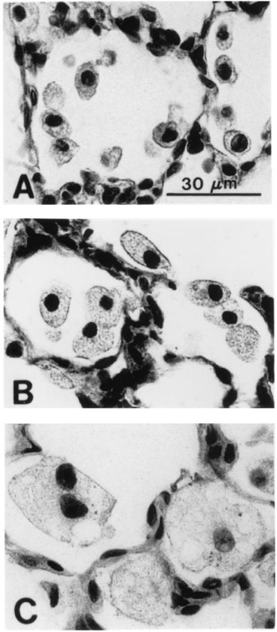
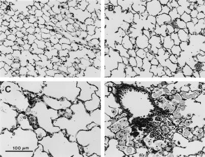
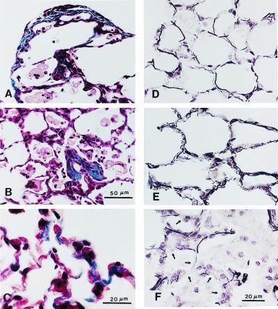
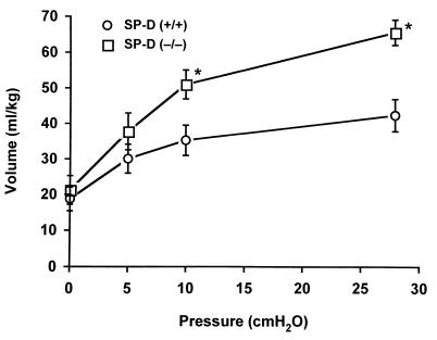
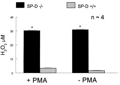
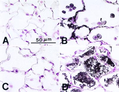
Targeted ablation of the surfactant protein D (SP-D) gene caused chronic inflammation, emphysema, and fibrosis in the lungs of SP-D (-/-) mice. Although lung morphology was unperturbed during the first 2 weeks of life, airspace enlargement was observed by 3 weeks and progressed with advancing age. Inflammation consisted of hypertrophic alveolar macrophages and peribronchiolar-perivascular monocytic infiltrates. These abnormalities were associated with increased activity of the matrix metalloproteinases, MMP2 and MMP9, and immunostaining for MMP9 and MMP12 in alveolar macrophages. Hydrogen peroxide production by isolated alveolar macrophages also was increased significantly (10-fold). SP-D plays a critical role in the suppression of alveolar macrophage activation, which may contribute to the pathogenesis of chronic inflammation and emphysema.


Figures

Figure 1
Figure 1
Progressive enlargement of alveolar macrophages from SP-D (−/−) mice. (A) Increased numbers of normal-appearing macrophages were observed by 2 weeks of age in SP-D (−/−) mice. (B) Enlarged, foamy, alveolar macrophages were readily observed by 3 weeks of age in SP-D (−/−) mice. (C) By 6 weeks of age, multiple foci of large, foamy, alveolar macrophages were observed throughout the lungs of SP-D (−/−) mice. (Scale bar = 30 μm.)
Figure 2
Figure 2
Lung tissue from SP-D (+/+) (A) and SP-D (−/−) (BD) mice killed at 7 months of age. Moderate (B) to severe (C) emphysematous changes were observed in SP-D (−/−) mice compared with age-matched SP-D (+/+) controls (A). Focal areas of macrophage accumulation and perivascular/peribronchiolar monocytic infiltrates also were observed in the lungs of 7-month-old SP-D (−/−) mice (D). (Scale bar = 100 μm.)
Figure 3
Figure 3
Collagen (AC) and elastin (DF) staining of lung tissue from 7-month-old SP-D (−/−) mice. Increased collagen deposition was observed at the pleural surface (A), in focal areas of macrophage accumulation associated with localized fibrosis (B), and in alveolar septa (C) of SP-D (−/−) mice. Elastin staining was increased in alveolar septa of SP-D (−/−) mice (E) compared with age-matched SP-D (+/+) controls (D), the elastic fibers appearing shorter, thicker, and more highly coiled in the SP-D (−/−) mice. Elastin staining was decreased, however, in alveolar septa (arrows) adjacent to foci of macrophage accumulation and fibrosis (F). [Scale bars = 50 μm (A, B, D, and E) and 20 μm (C and F).]
Figure 4
Figure 4
Comparison of changes in fractional areas (% fx area) of airspace (a) and respiratory parenchyma (b) with age in SP-D (−/−) mice and age-matched SP-D (+/+) controls. No significant differences were observed in the fractional area of airspace and parenchyma found in the lungs of SP-D (−/−) mice when compared with wild-type controls at 5 days (0.5 wk), 14 days (2 wk), or 17 days (2.5 wk) of age. The fractional area devoted to both airspace and parenchyma diverged significantly between the two different genotypes at 3 weeks (*, P = 0.013), 6 weeks (*, P = 0.0007), and 28 weeks (*, P = 0.004) of age. Data are expressed as % fractional area and represent the mean ± SE.
Figure 5
Figure 5
Deflation limbs of pressure-volume curves from SP-D (+/+) and SP-D (−/−) mice. Pressure-volume curves were generated in 5–6 mice at 12 weeks of age. Lung volumes associated with the deflation limbs of pressure-volume curves were significantly greater for 12-week-old SP-D (−/−) mice compared with age-matched SP-D (+/+) mice at 10 cm H2O and at the maximum pressure of 28 cm H2O (*, P < 0.05). Data are expressed as ml/kg and represent the mean ± SE.
Figure 6
Figure 6
Increased hydrogen peroxide production in alveolar macrophages from SP-D (−/−) mice. Hydrogen peroxide production was assessed from 1 × 10⁶ macrophages isolated from BALF as described in Experimental Procedures. Macrophages from SP-D (−/−) mice (solid bar) generated significantly more hydrogen peroxide than macrophages from SP-D (+/+) mice (hatched bar) with and without PMA stimulation. Data are expressed as μM of H2O2 and represent the mean ± SE with n = 4 determinations per group. A total of 8–10 mice was used for each determination, *, P < 0.05, compared with SP-D (+/+) mice.
Figure 7
Figure 7
Production of MMP by alveolar macrophages in SP-D (−/−) mice. MMP activity was assessed in macrophage-conditioned media. Pooled alveolar macrophages (1 × 10⁵ cells/well) were incubated in 100 μl of serum-free media for 24 h. The supernatant was collected, concentrated, and analyzed by gelatin zymography. Production of 72-kDa (MMP2) and 105-kDa (MMP9) gelatinases was significantly increased in SP-D (−/−) mice, whereas there was no detectable enzyme activity in SP-D (+/+) mice. Alveolar macrophages were pooled from four SP-D (−/−) mice and eight SP-D (+/+) mice. The assay was performed in three different sets of animals. Data shown are for one assay and are presented as the reverse image of the original zymogram.
Figure 8
Figure 8
Immunohistochemical detection of MMP12 and MMP9 in 6- to 7-month-old SP-D (−/−) mice. Immunostaining for both MMP12 (A and B) and MMP9 (C and D) was enhanced in the lungs of SP-D (−/−) mice (B and D) compared with age-matched SP-D (+/+) mice (A and C). MMP12 immunopositive material was detected in regions of parenchyma composed of thickened alveolar septa and in adjacent macrophages (C). MMP9 immunopositive material was detected in alveolar spaces containing large, foamy and/or disintegrating alveolar macrophages (D). (Scale bar = 50 μm.)

References

    1. Crouch E C. Biochim Biophys Acta. 1998;1408:278–289. - PubMed
    1. Reid K B M. Biochem Biophys Acta. 1998;1408:290–295. - PubMed
    1. Mason R J, Greene K, Voelker D R. Am J Physiol. 1998;275:L1–L13. - PubMed
    1. Crouch E, Rust K, Marienchek W, Parghi D, Chang D, Persson A. Am J Respir Cell Mol Biol. 1991;5:13–18. - PubMed
    1. Fisher J G, Mason R. Am J Respir Cell Mol Biol. 1995;12:13–18. - PubMed

Publication types

MeSH terms