Case Reports
. 2000 May 23;97(11):6167-72.
doi: 10.1073/pnas.100510697.

Beyond re-membering: phantom sensations of congenitally absent limbs

Affiliations
Case Reports

Beyond re-membering: phantom sensations of congenitally absent limbs

P Brugger et al. Proc Natl Acad Sci U S A. .

Abstract

Phantom limbs are traditionally conceptualized as the phenomenal persistence of a body part after deafferentation. Previous clinical observations of subjects with phantoms of congenitally absent limbs are not compatible with this view, but, in the absence of experimental work, the neural basis of such "aplasic phantoms" has remained enigmatic. In this paper, we report a series of behavioral, imaging, and neurophysiological experiments with a university-educated woman born without forearms and legs, who experiences vivid phantom sensations of all four limbs. Visuokinesthetic integration of tachistoscopically presented drawings of hands and feet indicated an intact somatic representation of these body parts. Functional magnetic resonance imaging of phantom hand movements showed no activation of primary sensorimotor areas, but of premotor and parietal cortex bilaterally. Movements of the existing upper arms produced activation expanding into the hand territories deprived of afferences and efferences. Transcranial magnetic stimulation of the sensorimotor cortex consistently elicited phantom sensations in the contralateral fingers and hand. In addition, premotor and parietal stimulation evoked similar phantom sensations, albeit in the absence of motor evoked potentials in the stump. These data indicate that body parts that have never been physically developed can be represented in sensory and motor cortical areas. Both genetic and epigenetic factors, such as the habitual observation of other people moving their limbs, may contribute to the conscious experience of aplasic phantoms.


Figures
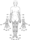
Figure 1
Figure 1
Phantoms (shaded areas) in a subject with limb amelia. The numbers are vividness ratings (means of three measurements) for the felt presence of different phantom body parts on a 7-point scale from 0 (no awareness) to 6 (most vivid impression).
Figure 2
Figure 2
Implicit reaching task. Displayed are task demands and conditions of stimulus presentation along with two sample stimuli for each condition. Mean latencies (SD in brackets) of correct decisions for subject A.Z. and three control subjects also are listed. In all subjects, mental rotation of a limb during implicit reaching was reflected by significantly longer decision times.
Figure 3
Figure 3
Cortical activation areas during self-paced movements of phantom fingers. Two representative sections through the sensorimotor cortical hand areas during fingers-to-thumb opposition with the left (Top) and right (Bottom) phantom fingers. Arrows indicate the anatomical region corresponding to the hand representation in normal subjects (21). The color scale on the left of each section represents Student's t test values [yellow, t = 3.6 (P ≤ 0.001); red, t = 6.9). The anatomical locations of activated areas are listed next to the corresponding sections both as Talairach and Tournoux coordinates and probable Brodmann areas (BA).
Figure 4
Figure 4
Cortical activation areas during movements of existing body parts. Three contiguous sections covering the sensorimotor cortex from the hand representation up to the vertex during movements of the left (Top) and right (Bottom) upper arm. BA, Brodmann areas. (Color scale and coordinates as in Fig. 3.)
Figure 5
Figure 5
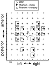
Overlay of MEP map and the subject's reports of phantom hand sensations in response to TMS. Each rectangle represents a stimulation site from which positive responses were obtained. Crosses indicate stimulation sites with neither MEPs nor phantom sensations. The curved line depicts the approximate location of the central sulci.

Similar articles

Cited by

References

    1. Ramachandran V S, Hirstein W. Brain. 1998;121:1603–1630. - PubMed
    1. Valentin G. Repertorium Anat Physiol. 1836;1:328–337.
    1. Simmel M L. Am J Psychol. 1961;74:467–470.
    1. Weinstein S, Sersen E A. Neurology. 1961;11:905–911. - PubMed
    1. Poeck K. Cortex. 1964;1:269–275.

Publication types