Review
. 2000 May;181 Suppl 2(Suppl 2):S295-302.
doi: 10.1086/315584.

The discovery of the 27-nm Norwalk virus: an historic perspective

Affiliations
Review

The discovery of the 27-nm Norwalk virus: an historic perspective

A Z Kapikian. J Infect Dis. 2000 May.

Abstract

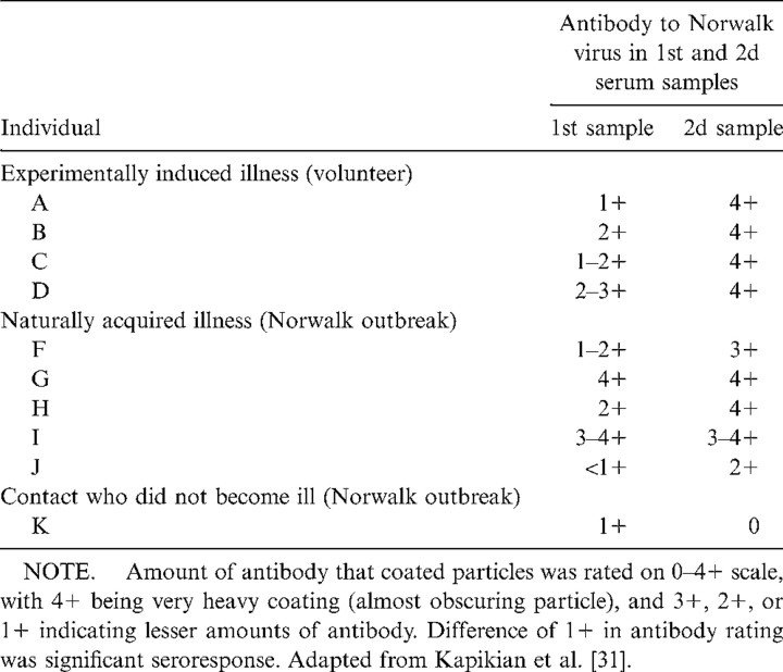
In 1972, a 27-nm virus-like particle was discovered by use of immune electron microscopy (IEM) in an infectious stool filtrate derived from an outbreak of gastroenteritis in an elementary school in Norwalk, Ohio. IEM enabled the direct visualization of antigen-antibody interaction, as the particles were aggregated and coated by specific antibodies. This allowed the recognition and identification of a 27-nm virus-like particle that did not have a distinctive morphology, was low-titered, and was among the smallest viruses known. Serum antibody responses to the 27-nm particle were demonstrated in key individuals infected under natural or experimental conditions; this and other evidence suggested that this virus-like particle was the etiologic agent of the Norwalk gastroenteritis outbreak. The fastidious 27-nm Norwalk virus is now considered to be the prototype strain of a group of noncultivatable viruses that are important etiologic agents of epidemic gastroenteritis in adults and older children.


Figures

Figure 1
Figure 1
Single particle of rhinovirus 1A from control preparation in which rhinovirus 1A suspension was incubated with PBS prior to further preparation for electron microscopy. Particles were randomly distributed, and it was difficult to determine whether certain objects were virus particles. Bar = 100 nm. (From Kapikian et al. [27], bar added.)
Figure 2
Figure 2
Micrograph illustrating large, distinctive aggregate of rhinovirus particles in rhinovirus 1A suspension, which was incubated with 1 : 180 dilution of rhinovirus 1A goat antiserum prior to further preparation for electron microscopy. Bar = 100 nm. (From Kapikian et al. [27], bar added.)
Figure 3
Figure 3
A, An aggregate observed after incubation of 0.8 mL of Norwalk (8FIIa) stool filtrate with 0.2 mL of 1 : 5 dilution of prechallenge serum of volunteer A and further preparation for electron microscopy. Quantity of antibody on these glistening particles was rated as 1+. B, Aggregate observed after incubation of 0.8 mL of Norwalk (8FIIa) stool filtrate with 1 : 5 dilution of postchallenge serum from volunteer B and further preparation for electron microscopy. Particles were very heavily coated with antibody. Heavily coated particles were usually found in small aggregates, whereas those with less antibody were usually in larger aggregates. Quantity of antibody on these particles was rated as 4+. Bar = 100 nm and applies to both A and B. (From Kapikian et al. [31], bar added.)
Figure 4
Figure 4
Aggregate observed after incubation of 0.8 mL of Norwalk stool filtrate with 0.2 mL of 1 : 5 dilution of volunteer's prechallenge serum and further preparation for electron microscopy. Volunteer developed gastroenteritis after challenge with 2d-passage Norwalk filtrate, which had been heated for 30 min at 60°C [22]. Quantity of antibody on particles in this aggregate was rated 1-2-2+, and prechallenge serum was given overall rating of 1–2+. Bar = 100 nm and applies to entire figure. B, Single particle. C, 3 single particles observed after incubating 0.8 mL of Norwalk stool filtrate with 0.2 mL of 1 : 5 dilution of same volunteer's convalescent serum and further preparation for electron microscopy. These particles are heavily coated with antibody. Quantity of antibody on these particles was rated 4+, and serum was given overall rating of 4+ also. Difference in quantity of antibody coating particles in prechallenge and postchallenge sera is clearly evident. (From Kapikian et al. [30], bar added.)
Table 1
Table 1
Antibody responses to the 27-nm Norwalk virus, as determined by immune electron microscopy.

References

    1. Bell JA, Huebner RJ, Rosen L, et al. Illness and microbial experiences of nursery children at Junior Village. Am J Hyg. 1961;74:267–92.
    1. Connor JD, Barrett-Connor E. Infectious diarrheas. Pediatr Clin North Am. 1967;14:197–221. - PubMed
    1. Yow MD, Melnick JL, Blattner RJ, Stephenson NB, Robinson NM, Burkhardt MA. The association of viruses and bacteria with infantile diarrhea. Am J Epidemiol. 1970;92:33–9. - PubMed
    1. Reimann HA, Prince AH, Hodges JH. The cause of epidemic diarrhea, nausea and vomiting (viral dysentery?) Proc Soc Exp Biol Med. 1945;59:8–9.
    1. Gordon I, Ingraham HS, Korns RF. Transmission of epidemic gastroenteritis to human volunteers by oral administration of fecal filtrates. J Exp Med. 1947;86:409–22. - PMC - PubMed

MeSH terms