. 2000 May 15;20(10):3537-43.
doi: 10.1523/JNEUROSCI.20-10-03537.2000.

Protein phosphatase-1 regulation in the induction of long-term potentiation: heterogeneous molecular mechanisms

Affiliations

Protein phosphatase-1 regulation in the induction of long-term potentiation: heterogeneous molecular mechanisms

P B Allen et al. J Neurosci. .

Abstract

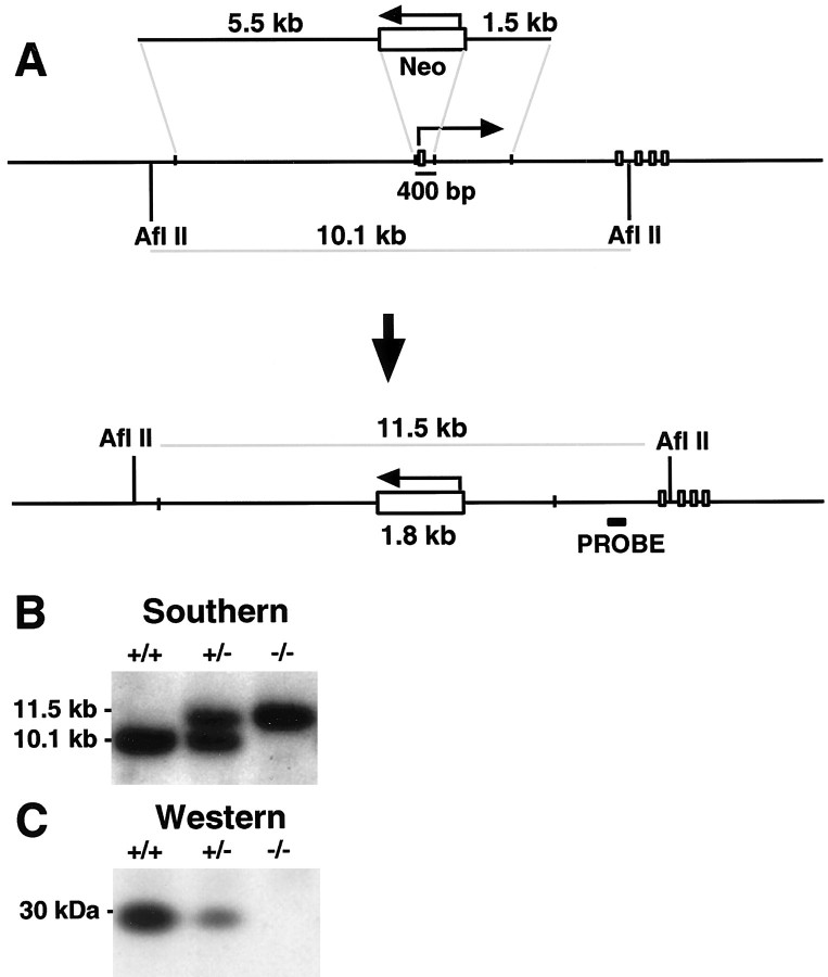
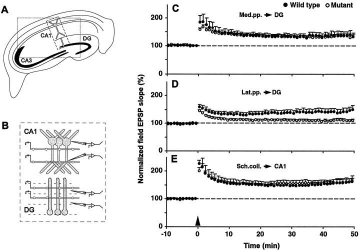
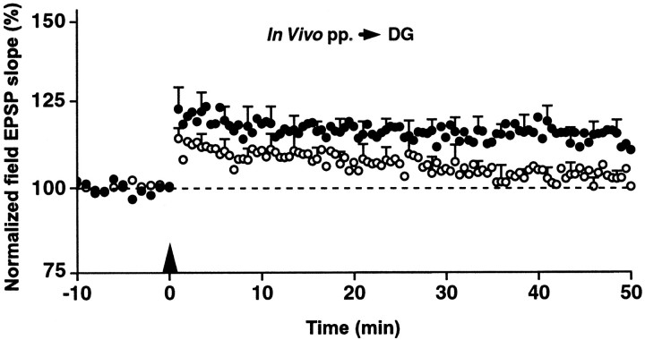
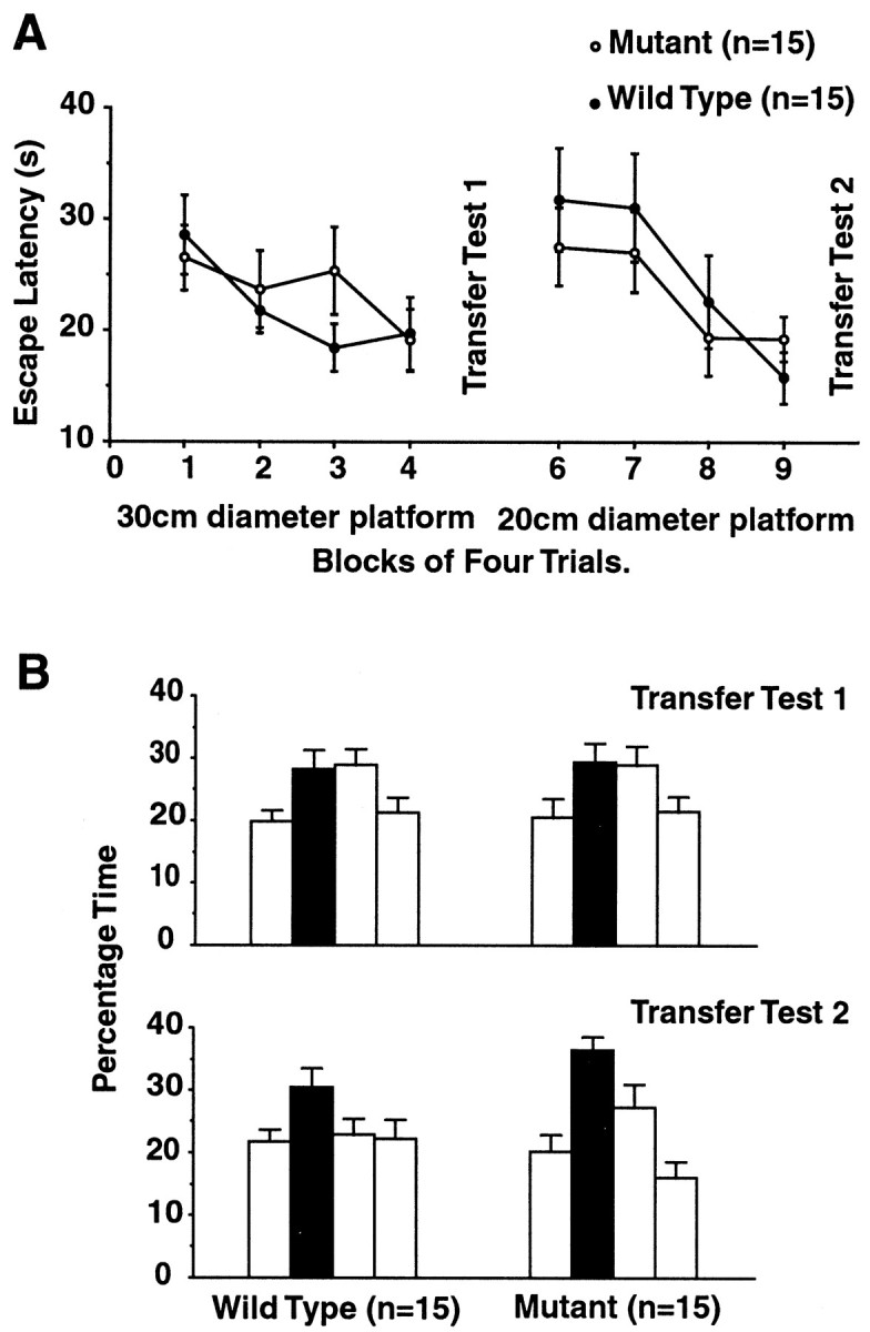
Protein phosphatase inhibitor-1 (I-1) has been proposed as a regulatory element in the signal transduction cascade that couples postsynaptic calcium influx to long-term changes in synaptic strength. We have evaluated this model using mice lacking I-1. Recordings made in slices prepared from mutant animals and also in anesthetized mutant animals indicated that long-term potentiation (LTP) is deficient at perforant path-dentate granule cell synapses. In vitro, this deficit was restricted to synapses of the lateral perforant path. LTP at Schaffer collateral-CA1 pyramidal cell synapses remained normal. Thus, protein phosphatase-1-mediated regulation of NMDA receptor-dependent synaptic plasticity involves heterogeneous molecular mechanisms, in both different dendritic subregions and different neuronal subtypes. Examination of the performance of I-1 mutants in spatial learning tests indicated that intact LTP at lateral perforant path-granule cell synapses is either redundant or is not involved in this form of learning.


Figures

Fig. 1.
Fig. 1.
A, Depiction of the mouseI-1 genomic locus and the targeting strategy used for gene disruption; a replacement vector was designed to delete a ∼400 bp fragment containing the first coding exon of the I-1 gene and to replace it with a ∼1.8 kb fragment containing the neomycin resistance gene. Homologous recombination was detected by a reduction in the mobility of a genomic AflII restriction fragment because of an increase in size of ∼1.4 kb. B, Southern blot analysis of genomic tail DNA isolated from an I-1 mutant pedigree and digested with AflII. The probe is depicted inA. Wild type, +/+; heterozygote, +/−; homozygote, −/−. C, Immunoblot analysis using anti-I-1 antibody. SDS homogenates (1%) were prepared from hippocampi taken from animals of the indicated genotype.
Fig. 2.
Fig. 2.
Immunoperoxidase labeling of I-1 in the hippocampal formation (see large box in Fig.3A) of I-1 homozygous mutant and wild-type mice.A, In the mutant, there is no immunoreactivity. The darkness in the alveus (A) and the stratum lacunosum-moleculare (LM) is attributable to light scattering by the myelinated fibers abundant in these zones; to be visible, the photomicrograph of A was printed at twice the exposure time for wild type. B, In the wild type, the staining in the dentate gyrus, mossy fiber layer in CA3 (MF) terminal zones and LM is much stronger than in other regions. The cellular localization of I-1 in the dentate gyrus dorsal blade is shown by differential interference contrast (C, D) and electron microscopy (E). Granule cell perikarya (g), dendrites (arrows), and mossy fiber boutons (arrowheads) are strongly immunoreactive.E, A labeled spine (s) protruding from an immunopositive dendrite (arrow) receives an asymmetric synapse from an immunonegative nerve ending.A, Alveus; H, hilus; O, stratum oriens; P, stratum pyramidale; R, stratum radiatum; LM, stratum lacunosum-moleculare;MF, mossy fiber layer in CA3; G, stratum granulare; Mi, Mm, Mo, inner, middle, and outer thirds of stratum moleculare of the dentate gyrus. R contains the Schaffer collateral terminals, andMo and Mm contain the terminals of the lateral and medial perforant path, respectively.Asterisks mark the obliterated hippocampal fissure. Scale bars: A, B, 200 μm;C, 50 μm; D, 10 μm; E, 0.5 μm.
Fig. 3.
Fig. 3.
I-1 regulates synaptic plasticity at a subset of synapses. A, Schematic of a hippocampal slice. The large box shows the region from which the immunohistochemical section in Figure 2 is taken. The small box indicates the part of CA1 and dentate gyrus in which LTP was studied. B, Diagram of stimulating and recording electrode arrangement in the CA1 (top panel) and in the upper blade of the dentate gyrus (bottom panel). C–E, Pooled data of the extracellular fEPSP slopes evoked in the wild-type (filled circles) and mutant (open circles) mice in the medial perforant path (C), in the lateral perforant path (D), and in the CA1 region (E). For the sake of clarity, nontetanized control pathway responses are not shown. Arrow indicates the time of tetanic stimulation. Vertical bars indicate SEM.
Fig. 4.
Fig. 4.
In vivo LTP of the fEPSP in dentate gyrus. After tetanization of the perforant path, LTP in the wild-type group shows significant potentiation, whereas LTP in the mutants declines to baseline over the course of the experiment.Arrow indicates the time of tetanic stimulation. Vertical bars indicate SEM.
Fig. 5.
Fig. 5.
Performance in a water maze test.A, Mean escape latencies for mutant and wild-type mice. Initial training was performed using a 30 cm platform (left). ANOVA revealed an effect of blocks of four trials (F(3,84) = 2.76;p < 0.05), indicating that learning occurred over the 4 d of training, whereas no difference was seen between mutant and wild type (F < 1; p > 0.5). After transfer test 1, subsequent trial blocks were performed using a 20 cm platform (right), and this produced a similar pattern of results: a more pronounced trial effect (F(3,84) = 7.61; p< 0.001) and again no difference between mutant and wild-type groups (F < 1; p > 0.5).B, Percentage time spent in each of the pool quadrants during transfer tests. After the first training set (top panel), an effect of quadrant was seen (F(3,84) = 4.63; p< 0.05), but a t test revealed that this was between the adjacent left–training and the adjacent right–training quadrants (p < 0.05). No difference was found between the training quadrant and the adjacent right quadrant. This suggested that the spatial bias was not yet directed toward the training quadrant alone. No genotype group difference between any of the quadrants was seen (F < 1; p > 0.3). Transfer tests performed after the second set of trials with the 20 cm platform (bottom panel) indicated that performance had substantially improved. Again, an effect of quadrant was seen (F(3,84) = 8.53;p < 0.001). The percentage time spent in the training quadrant was significantly greater relative to both adjacent quadrants (t test: 0.001 < p< 0.05). However, no genotype group difference was found (F = 1; p > 0.3).

Similar articles

Cited by

References

    1. Allen PB, Ouimet CC, Greengard P. Spinophilin, a novel protein phosphatase 1 binding protein localized to dendritic spines. Proc Natl Acad Sci USA. 1997;94:9956–9961. - PMC - PubMed
    1. Barbas H, Gustafson EL, Greengard P. Comparison of the immunocytochemical localization of DARPP-32 and I-1 in the amygdala and hippocampus of the rhesus monkey. J Comp Neurol. 1993;334:1–18. - PubMed
    1. Bito H, Deisseroth K, Tsien RW. CREB phosphorylation and dephosphorylation: a Ca2+- and stimulus duration-dependent switch for hippocampal gene expression. Cell. 1996;87:1203–1214. - PubMed
    1. Blank T, Nijholt I, Teichert U, Kugler H, Behrsing H, Fienberg A, Greengard P, Spiess J. The phosphoprotein DARPP-32 mediates cAMP-dependent potentiation of striatal N-methyl-d-aspartate responses. Proc Natl Acad Sci USA. 1997;94:14859–14864. - PMC - PubMed
    1. Bliss TVP, Collingridge GL. A synaptic model of memory: long-term potentiation in the hippocampus. Nature. 1993;361:31–39. - PubMed

Publication types

MeSH terms

LinkOut - more resources