. 2000 May 15;20(10):3552-62.
doi: 10.1523/JNEUROSCI.20-10-03552.2000.

The mechanisms of hsp27 antibody-mediated apoptosis in retinal neuronal cells

Affiliations

The mechanisms of hsp27 antibody-mediated apoptosis in retinal neuronal cells

G Tezel et al. J Neurosci. .

Abstract

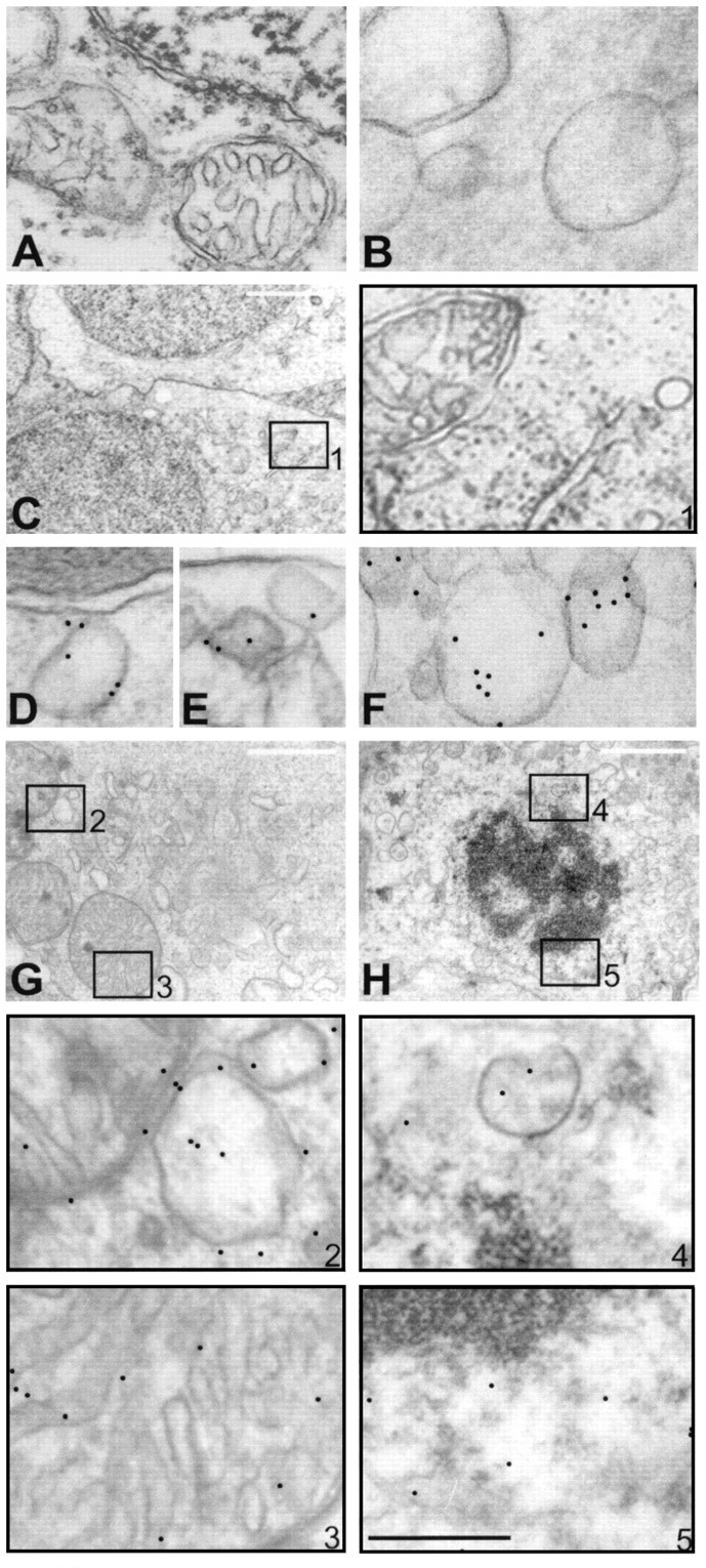
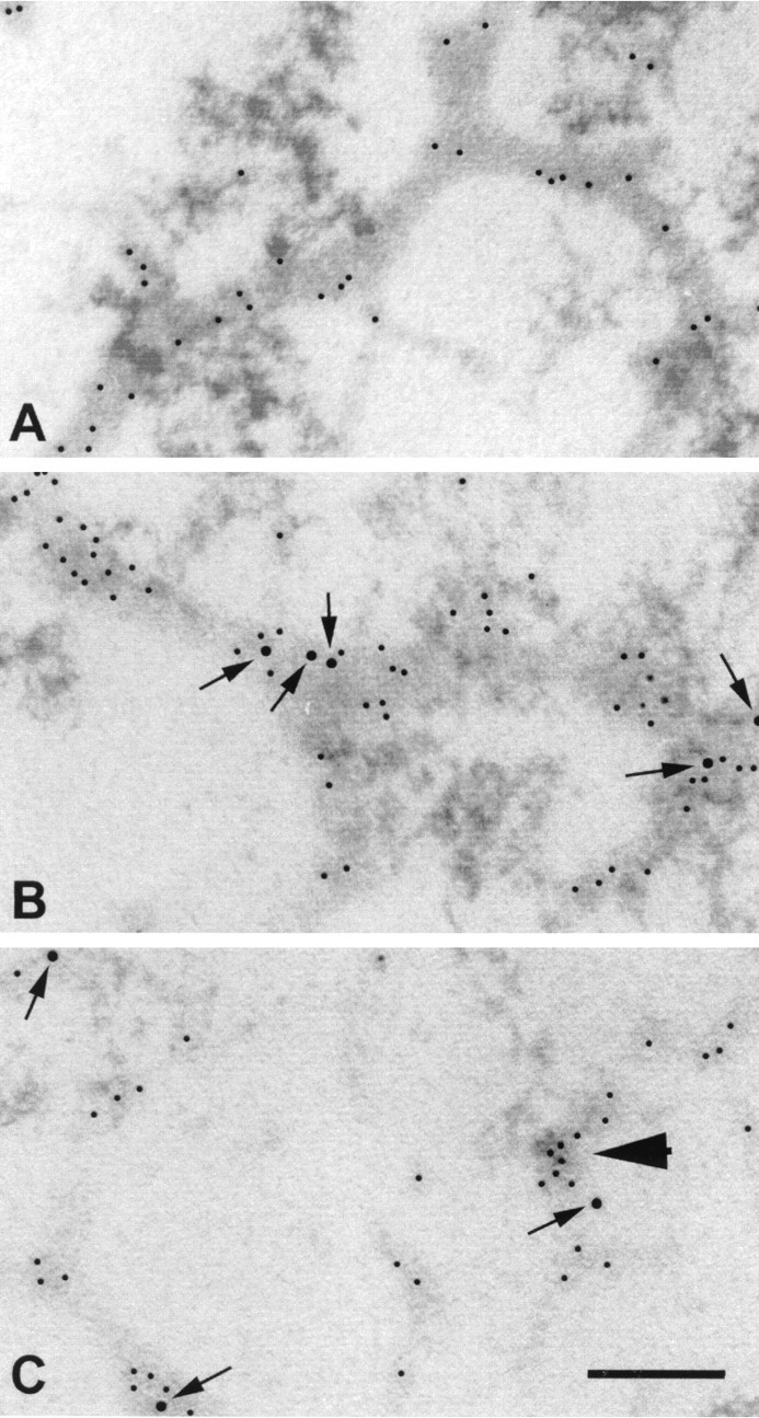
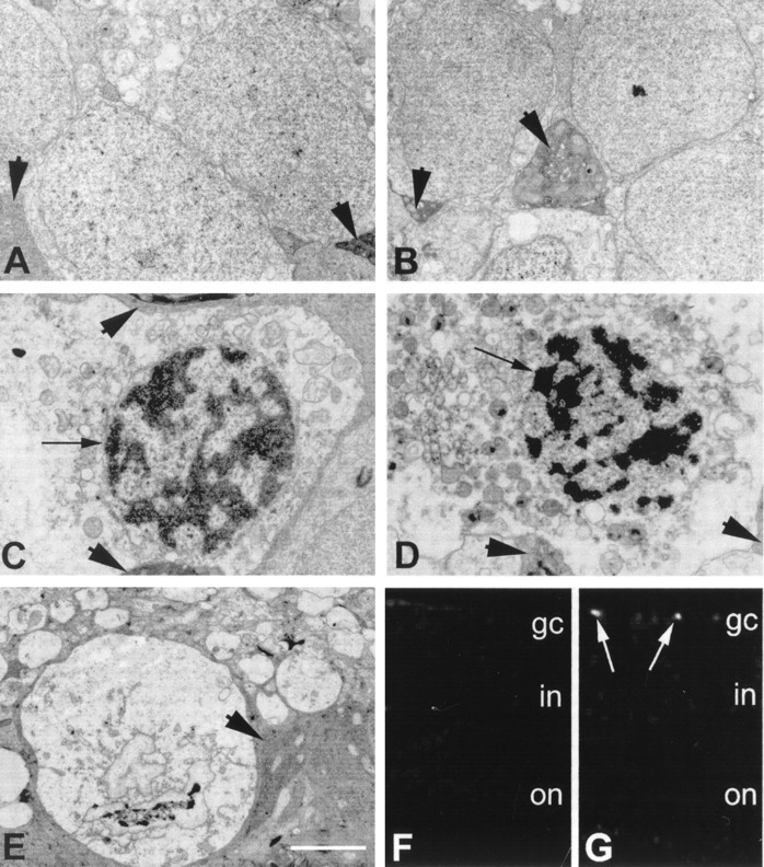
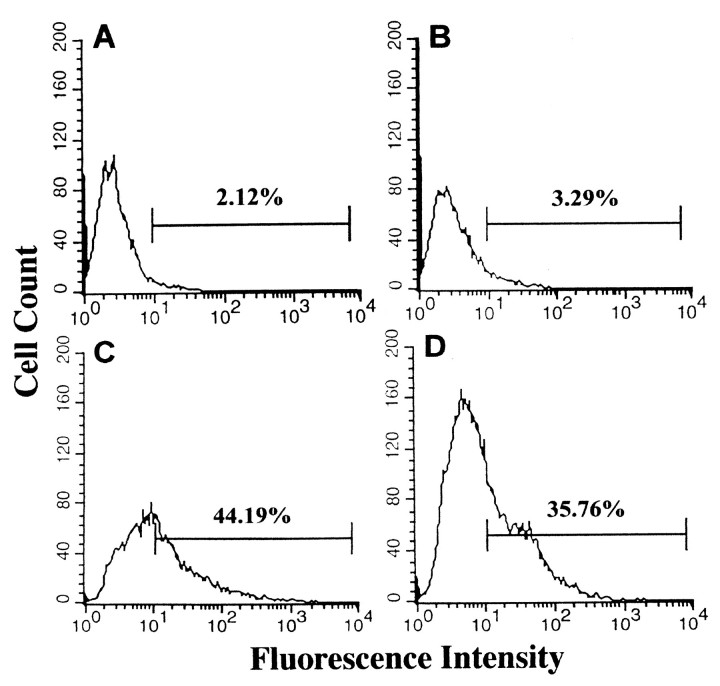
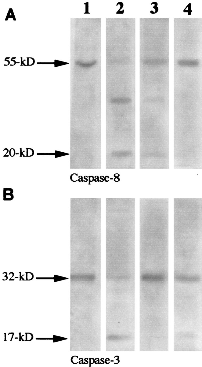
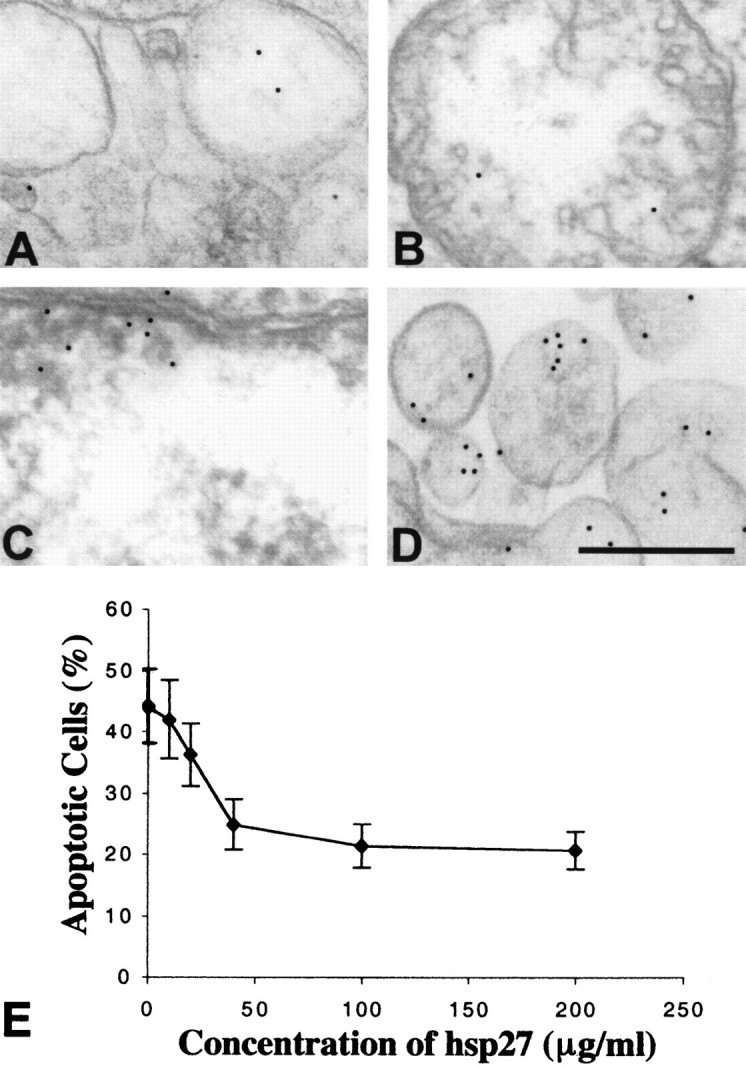
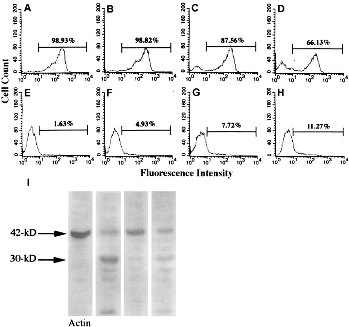
Although elevated titers of serum antibodies to hsp27 accompany human diseases such as cancer and glaucoma, evidence of their pathogenic effects is lacking. Here we present novel evidence that exogenously applied hsp27 antibody enters neuronal cells in human retina by an endocytic mechanism. Subsequent to internalization, hsp27 antibody facilitates apoptotic cell death as characterized by morphological assessment, DNA fragmentation, and the activation of cysteine aspartic acid proteases. In addition, we demonstrate that after internalization, hsp27 antibody is detected in discrete cytoplasmic and nuclear structures and colocalizes to actin cytoskeleton. Hsp27 antibody binding to actin results in depolymerization and proteolytic cleavage of actin in a dose-dependent manner. These results suggest that exogenous hsp27 antibody may induce neuronal apoptosis by inactivating or attenuating the ability of native hsp27 to stabilize actin cytoskeleton, thereby providing a novel mechanism by which autoantibodies to hsp27 may impair cell survival in selective human diseases.


Figures

Fig. 1.
Fig. 1.
Internalization of hsp27 antibody by human retina observed by immunogold labeling and electron microscopy. No gold particles were observed within neuronal cells in control retina incubated without antibody (A) or incubated with control antibodies, anti-IgG (B), or anti-calbindin-D (C). C, Micrograph of retinal ganglion cells after incubation with calbindin-D antibody for 12 hr. Higher magnification micrograph of selected area inA is shown in box 1, which indicates negative gold labeling. However, after incubation with hsp27 antibody, intracellular gold particles were observed in a time-dependent pattern. Immunolabeling was positive in endosomes (D,E) and vesicles (F) of the retinal ganglion cells incubated with hsp27 antibody for 30 min. Micrographs of two retinal cells incubated with hsp27 antibody for 6 and 12 hr, respectively, are shown in G and H. Higher magnification micrographs of selected areas in G andH are shown in boxes with corresponding numbers.Boxes B2 and 3 show mitochondrion and vesicules, and boxes 4 and 5 show perinuclear area. After incubation with hsp27 antibody for 6 or 12 hr, gold particles were mostly observed in vesicular structures of various size and mitochondria (G, H). In addition, perinuclear areas and nuclei of the retinal cells incubated with hsp27 antibody exhibited immunogold labeling (H). Notice the double membrane and internal cristae of mitochondria in G and condensed nuclear chromatin in H. Black scale bar:A, B, DF, 0.5 μm; white scale bar: C, G,H, 2 μm.
Fig. 2.
Fig. 2.
Double immunogold labeling of internalized hsp27 antibody and actin in human retina. A, Immunoelectron microscopy of control retina incubated in the absence of hsp27 antibody exhibited actin antibody-labeled microfilaments in retinal ganglion cells (5 nm gold particles). B, After incubation of retinas with hsp27 antibody for 6 hr, immunoelectron microscopy revealed colocalization of hsp27 antibody (10 nm gold particles) with actin (5 nm gold particles). Notice the close proximity of hsp27 antibody (arrows) with actin-labeled areas in the human retina. C, In retinas incubated with hsp27 antibody for 12 hr, actin microfilaments disappeared in some of the ganglion cells, leaving irregular actin aggregations (arrowhead). Scale bar, 0.25 μm.
Fig. 3.
Fig. 3.
Evidence of apoptosis in human retina incubated with hsp27 antibody. A, Electron micrograph of normal-appearing ganglion cells in human retina incubated without antibody. B, Electron micrograph of normal-appearing ganglion cells in human retina incubated with control antibody. Ganglion cells are sheathed by electron-dense glial processes (arrowheads) that originate from Müller cells characterized by dark cytoplasm containing different organelles and parallel filaments. Ganglion cells in human retina incubated with hsp27 antibody for 6 hr (C) or 12 hr (D) illustrated abnormal nuclear morphology.Black arrows show nuclei with clumped dense chromatin in cells with condensed cytoplasm and intact cytoplasmic organelles.E, An apoptotic body with a dense clump of presumed chromatin. Cytoplasmic organelles and surrounding membranes, however, are still intact. Scale bar: AE, 2 μm. F, G, Fluorescence microscopy images (original magnification 40×) using TUNEL technique in control retina incubated without antibody and retina incubated with hsp27 antibody, respectively. The TUNEL technique showed brightly fluorescein-stained nuclei representing fragmented DNA in human retina incubated with hsp27 antibody (white arrows).
Fig. 4.
Fig. 4.
Flow cytometric analysis of apoptotic cell death in E1A.NR3 rat retinal cells. Histograms obtained after fluorescein staining of DNA breaks are shown in control cells incubated without hsp27 antibody (A), in cells incubated with anti-IgG control antibody (B), in cells incubated with hsp27 antibody (C), or in cells incubated with hsp27 antibody in the presence of heat-inactivated serum (D). There was no difference between apoptosis rates of the control cells incubated without hsp27 antibody and cells incubated with control antibody (Fisher's exact test,p = 0.65). However, although 2–3% of control cells demonstrated a fluorescence intensity above 101, the same fluorescence intensity occurred in as much as 44% of the cell population incubated with hsp27 antibody (Fisher's exact test, p < 0.0001). The use of heat-inactivated serum did not markedly change the rate of apoptosis induced by hsp27 antibody (Fisher's exact test, p= 0.25). Data are representative of three independent experiments that were collected using logarithmic amplification of at least 10,000 cells.
Fig. 5.
Fig. 5.
Western blot findings demonstrating caspase cleavage: column 1, control retinal cells (E1A.NR3);column 2, retinal cells incubated with hsp27 antibody (100 μg/ml) for 24 hr; column 3, retinal cells incubated with hsp27 antibody in the presence of a nonselective caspase inhibitor, B-D-FMK (50 μm); column 4, retinal cells incubated with hsp27 antibody in the presence of the caspase-8 inhibitor Z-IETD-FMK (20 μm). Although no cleavage of caspase-8 (A, column 1) or caspase-3 (B, column 1) was detected using the lysates of the control retinal cells, cleavage of caspase-8 and caspase-3 was observed using retinal cells incubated with hsp27 antibody. A 55 kDa immunoreactive band corresponding to caspase-8 cleaved to 30 and 20 kDa products is shown in A,column B. A 32 kDa pro-enzyme caspase-3 cleaved to a 17 kDa subunit is shown in B, column 2. Incubation of the retinal cells with hsp27 antibody in the presence of caspase inhibitors prevented specific caspase cleavage. B-D-FMK effectively inhibited caspase-3 and partially prevented caspase-8 cleavage (column 3); Z-IETD-FMK inhibited the cleavage of caspase-8 and only partially inhibited the cleavage of caspase-3 (column 4).
Fig. 6.
Fig. 6.
Regulation of hsp27 antibody internalization in retinal cells. Decreased internalization of hsp27 antibody in human retinal ganglion cells preincubated with purified hsp27 is shown. Approximately 50% decreased numbers of gold particles in vesicular structures (A), in a mitochondrion (B), and in the nucleus (C) are shown. Fc receptor blockade with Fc fragments of IgG did not change the internalization of hsp27 antibody by retinal neuronal cells (D). Scale bar, 0.5 μm. Flow cytometric analysis of cultured retinal cells preincubated with purified hsp27 revealed that the percentage of cells having fluorescein-labeled DNA fragments after incubation with hsp27 antibody decreased in a dose-dependent manner (E). The percentage of cells exhibiting a fluorescence intensity above 101was obtained from fluorescence histograms and used to generate the graphic. Data are representative of three independent experiments that were collected using logarithmic amplification of at least 10,000 cells.
Fig. 7.
Fig. 7.
Flow cytometric analysis of F- and G-actin in retinal cells incubated with hsp27 antibody. F- and G-actin in retinal cells were examined by double staining using phalloidin and Dnase I.A, B, C, andD represent fluorescein isothiocyanate fluorescence intensity of phalloidin staining, and E,F, G, and H represent Texas Red fluorescence intensity of Dnase I staining. A,E, Control cells; B,F, retinal cells incubated with 50 μg/ml; C,G, 100 μg/ml of hsp27 antibody; D,H, 200 μg/ml of hsp27 antibody. A shift in the population distribution of the phalloidin-stained cells, which corresponds to depolymerization of actin, was dependent on the concentration of hsp27 antibody. Although 99% of control cells demonstrated a fluorescence intensity above 101, the same fluorescence intensity occurred in only 66% of the cell population incubated with 200 μg/ml of hsp27 antibody and exhibited a second peak below 101 (Fisher's exact test,p < 0.0001). There was a concurrent increase in G-actin in these cells as assessed by Dnase I staining (Fisher's exact test, p = 0.01). Data are representative of three separate experiments. I, In addition, Western blot analysis revealed a proteolytic cleavage of actin in retinal cells incubated with hsp27 antibody. I, Column 1, control retinal cells (E1A.NR3); column 2, retinal cells incubated with hsp27 antibody (100 μg/ml) for 24 hr;column 3, retinal cells incubated with hsp27 antibody in the presence of a nonselective caspase inhibitor, B-D-FMK (50 μm); column 4, retinal cells incubated with hsp27 antibody in the presence of the caspase-8 inhibitor Z-IETD-FMK (20 μm). In retinal cells incubated with hsp27 antibody, 42 kDa actin (G-actin) (column 1) cleaved to ∼30 and 10 kDa fragments (column 2). Simultaneous incubation with B-D-FMK inhibited the cleavage of actin in retinal cells after incubation with hsp27 antibody (column 3).

Similar articles

Cited by

References

    1. Adamus G, Machnicki M, Seigel GM. Apoptotic retinal cell death induced by autoantibodies of cancer-associated retinopathy. Invest Ophthalmol Vis Sci. 1997;38:283–291. - PubMed
    1. Alarcon-Segovia D, Lorente L. Antibody penetration into living cells IV. Different effects of anti-native DNA and anti-ribonucleoprotein IgG on the cell cycle of activated T cells. Clin Exp Immunol. 1983;52:365–371. - PMC - PubMed
    1. Alarcon-Segovia D, Ruiz-Argüelles A, Fishbein E. Antibody to nuclear ribonucleoprotein penetrates live human mononuclear cells through Fc receptors. Nature. 1978;271:67–69. - PubMed
    1. Alarcon-Segovia D, Ruiz-Argüelles A, Lorente L. Broken dogma: penetration of autoantibodies into living cells. Immunol Today. 1996;17:163–164. - PubMed
    1. Aquino DA, Capello E, Weisstein J, Sanders V, Lopez C, Tourtellotte WW, Brosnan CF, Raine CS, Norton WT. Multiple sclerosis: altered expression of 70- and 27-kDa heat shock proteins in lesions and myelin. J Neuropathol Exp Neurol. 1997;56:664–672. - PubMed

Publication types

MeSH terms