

A novel nervous system beta subunit that downregulates human large conductance calcium-dependent potassium channels
- PMID: 10804197
- PMCID: PMC6772688
- DOI: 10.1523/JNEUROSCI.20-10-03563.2000
A novel nervous system beta subunit that downregulates human large conductance calcium-dependent potassium channels
Abstract
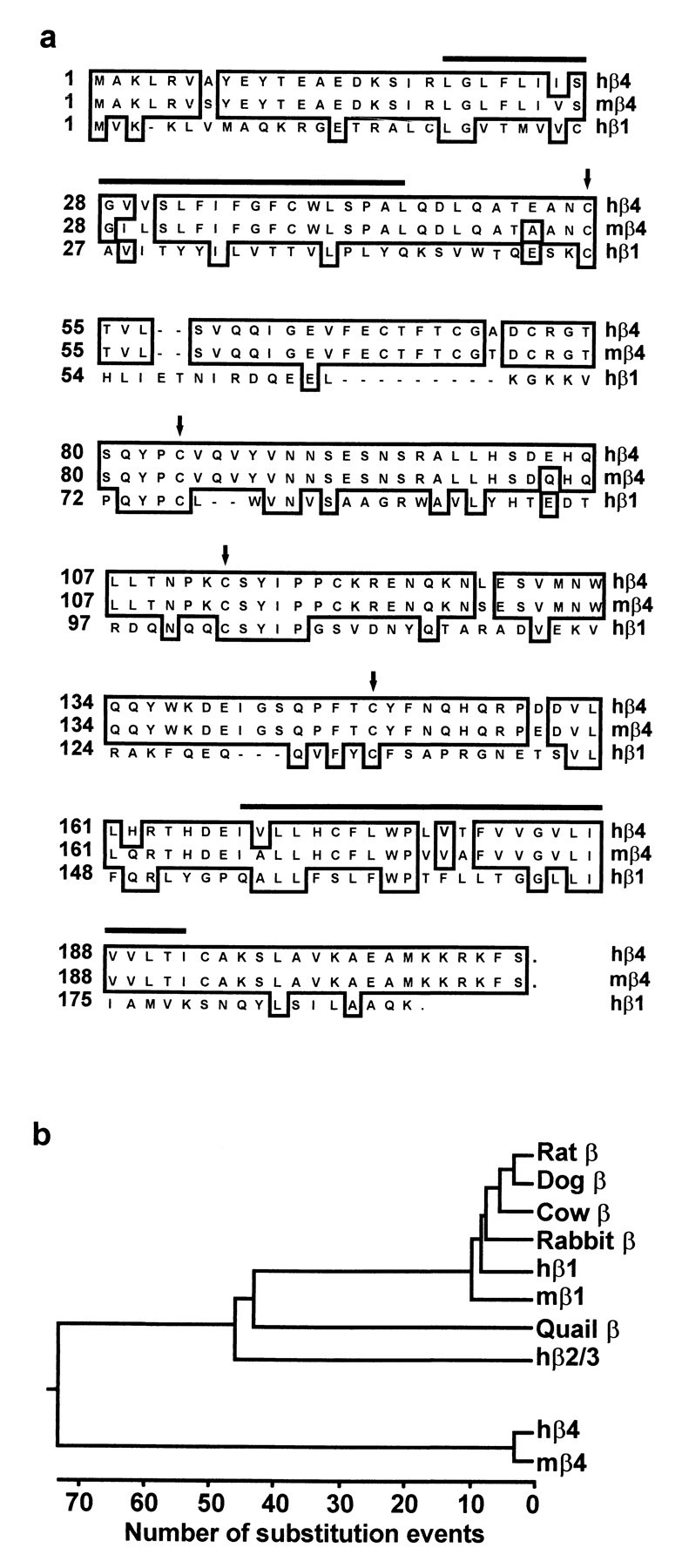
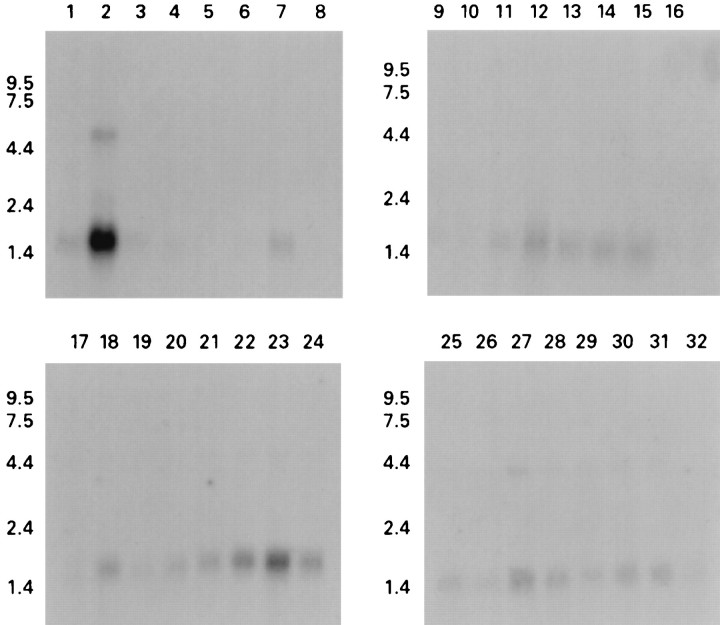
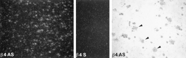
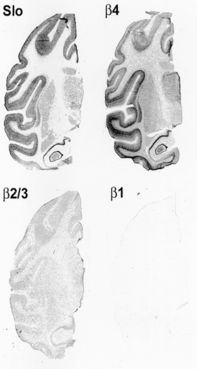
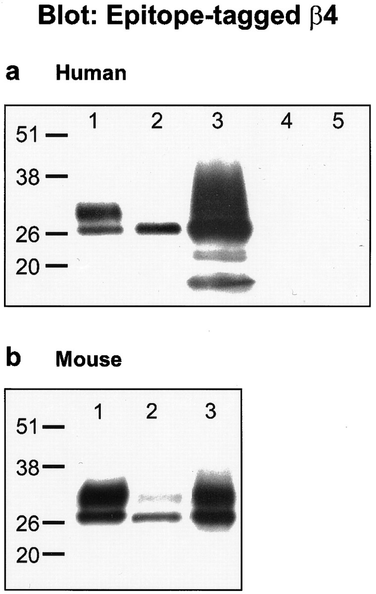
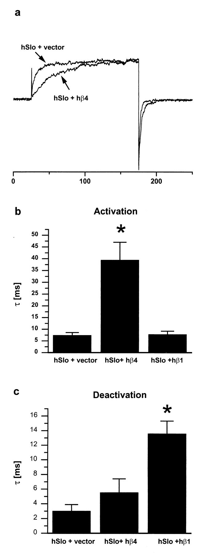
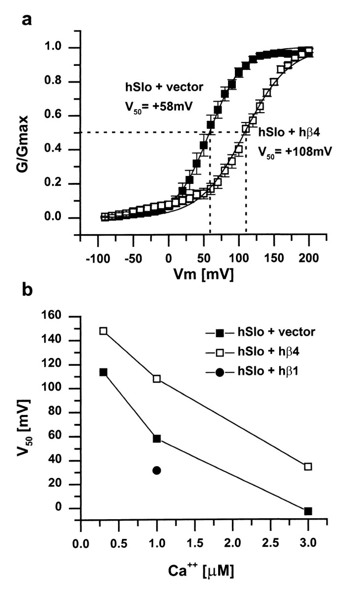
The pore-forming alpha subunits of many ion channels are associated with auxiliary subunits that influence channel expression, targeting, and function. Several different auxiliary (beta) subunits for large conductance calcium-dependent potassium channels of the Slowpoke family have been reported, but none of these beta subunits is expressed extensively in the nervous system. We describe here the cloning and functional characterization of a novel Slowpoke beta4 auxiliary subunit in human and mouse, which exhibits only limited sequence homology with other beta subunits. This beta4 subunit coimmunoprecipitates with human and mouse Slowpoke. beta4 is expressed highly in human and monkey brain in a pattern that overlaps strikingly with Slowpoke alpha subunit, but in contrast to other Slowpoke beta subunits, it is expressed little (if at all) outside the nervous system. Also in contrast to other beta subunits, beta4 downregulates Slowpoke channel activity by shifting its activation range to more depolarized voltages and slowing its activation kinetics. beta4 may be important for the critical roles played by Slowpoke channels in the regulation of neuronal excitability and neurotransmitter release.
Figures
Similar articles
-
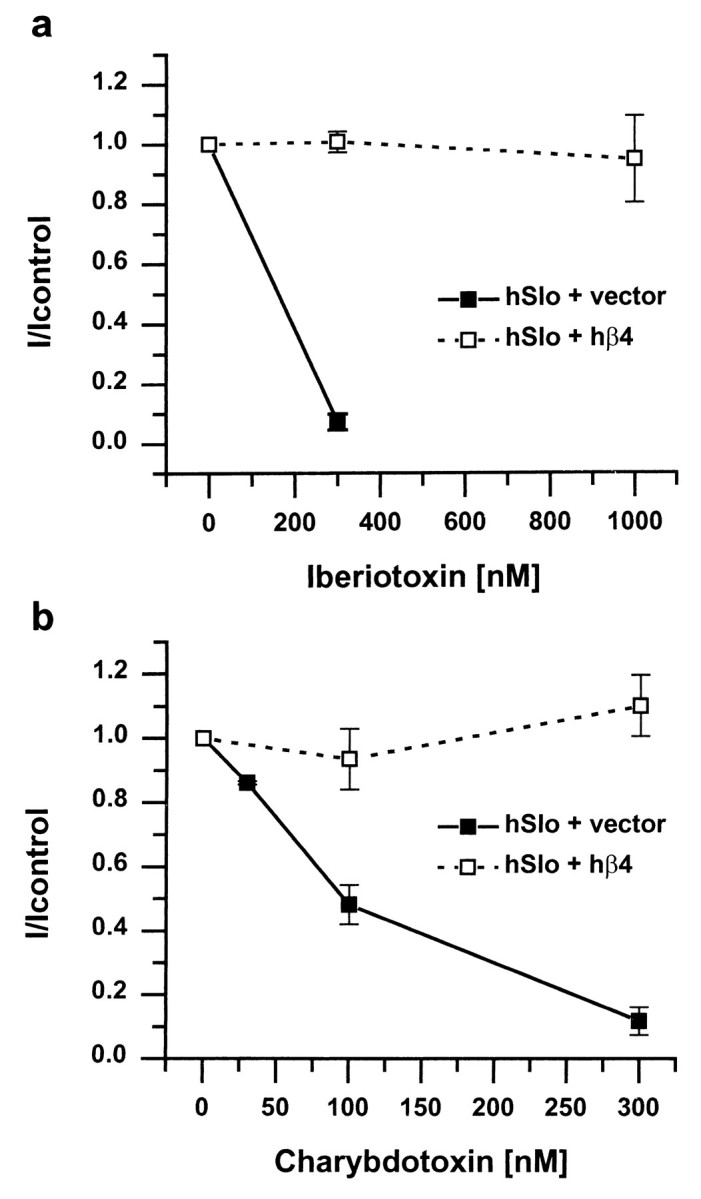
A neuronal beta subunit (KCNMB4) makes the large conductance, voltage- and Ca2+-activated K+ channel resistant to charybdotoxin and iberiotoxin.Proc Natl Acad Sci U S A. 2000 May 9;97(10):5562-7. doi: 10.1073/pnas.100118597. Proc Natl Acad Sci U S A. 2000. PMID: 10792058 Free PMC article.
-
Functional effects of auxiliary beta4-subunit on rat large-conductance Ca(2+)-activated K(+) channel.Biophys J. 2004 May;86(5):2871-82. doi: 10.1016/S0006-3495(04)74339-8. Biophys J. 2004. PMID: 15111404 Free PMC article.
-
Molecular basis for the inactivation of Ca2+- and voltage-dependent BK channels in adrenal chromaffin cells and rat insulinoma tumor cells.J Neurosci. 1999 Jul 1;19(13):5255-64. doi: 10.1523/JNEUROSCI.19-13-05255.1999. J Neurosci. 1999. PMID: 10377337 Free PMC article.
-
High-conductance calcium-activated potassium channels; structure, pharmacology, and function.J Bioenerg Biomembr. 1996 Jun;28(3):255-67. doi: 10.1007/BF02110699. J Bioenerg Biomembr. 1996. PMID: 8807400 Review.
-
Purification and characterization of epithelial Ca(2+)-activated K+ channels.Kidney Int. 1995 Oct;48(4):1047-56. doi: 10.1038/ki.1995.388. Kidney Int. 1995. PMID: 8569066 Review.
Cited by
-
BK in Double-Membrane Organelles: A Biophysical, Pharmacological, and Functional Survey.Front Physiol. 2021 Oct 26;12:761474. doi: 10.3389/fphys.2021.761474. eCollection 2021. Front Physiol. 2021. PMID: 34764886 Free PMC article. Review.
-
Molecular investigations of BK(Ca) channels and the modulatory beta-subunits in porcine basilar and middle cerebral arteries.J Mol Histol. 2009 Apr;40(2):87-97. doi: 10.1007/s10735-009-9216-3. Epub 2009 Apr 1. J Mol Histol. 2009. PMID: 19337844
-
Big conductance calcium-activated potassium channel openers control spasticity without sedation.Br J Pharmacol. 2017 Aug;174(16):2662-2681. doi: 10.1111/bph.13889. Epub 2017 Jul 7. Br J Pharmacol. 2017. PMID: 28677901 Free PMC article. Clinical Trial.
-
Gating and ionic currents reveal how the BKCa channel's Ca2+ sensitivity is enhanced by its beta1 subunit.J Gen Physiol. 2005 Oct;126(4):393-412. doi: 10.1085/jgp.200509346. J Gen Physiol. 2005. PMID: 16186565 Free PMC article.
-
Revealing stage-specific expression patterns of long noncoding RNAs along mouse spermatogenesis.RNA Biol. 2020 Mar;17(3):350-365. doi: 10.1080/15476286.2019.1700332. Epub 2019 Dec 23. RNA Biol. 2020. PMID: 31869276 Free PMC article.
References
-
- Adelman JP, Shen K-Z, Kavanaugh MP, Warren RA, Wu Y-N, Lagrutta A, Bond CT, North RA. Calcium- activated potassium channels expressed from cloned complementary DNAs. Neuron. 1992;9:209–216. - PubMed
-
- Butler A, Tsunoda S, McCobb DP, Wei A, Salkoff L. mSlo, a complex mouse gene encoding “maxi” calcium-activated potassium channels. Science. 1993;261:221–224. - PubMed
-
- Dunlap K, Luebke JI, Turner TJ. Exocytotic Ca2+ channels in mammalian central neurons. Trends Neurosci. 1995;18:89–98. - PubMed
-
- Dworetzky SI, Trojnacki JT, Gribkoff VK. Cloning and expression of a human large-conductance calcium-activated potassium channel. Mol Brain Res. 1994;27:189–193. - PubMed
Publication types
MeSH terms
Substances
Associated data
- Actions
LinkOut - more resources
Full Text Sources
Molecular Biology Databases