. 2000 May 15;20(10):3631-40.
doi: 10.1523/JNEUROSCI.20-10-03631.2000.

N-CAM binding inhibits the proliferation of hippocampal progenitor cells and promotes their differentiation to a neuronal phenotype

Affiliations

N-CAM binding inhibits the proliferation of hippocampal progenitor cells and promotes their differentiation to a neuronal phenotype

M C Amoureux et al. J Neurosci. .

Abstract

Cell adhesion molecules (CAMs) play important roles during the development of the nervous system. On the basis of our previous observations that binding of the neural CAM (N-CAM) inhibits astrocyte proliferation and alters gene expression, we hypothesized that N-CAM may influence the balance between the proliferation and the differentiation of neural progenitor cells. Rat and mouse hippocampal progenitor cells were cultured and showed dependence on basic FGF for proliferation, immunoreactivity for nestin, the presence of limited numbers of differentiated cells, and the ability to generate glial cells and neurons under different culture conditions. Addition of soluble N-CAM reduced cell proliferation in a dose-dependent manner with no evidence of apoptosis. The inhibition of proliferation by N-CAM was accompanied by an induction of differentiation to the neuronal lineage, as indicated by a twofold increase in the percentage of microtubule-associated protein 2-positive cells even in the presence of mitogenic growth factors. Experiments using hippocampal cells from N-CAM knock-out mice indicated that N-CAM on the cell surface is not required for these effects, suggesting the existence of heterophilic signaling. These results support a role for N-CAM and N-CAM ligands in the inhibition of proliferation and the induction of neural differentiation of hippocampal neural progenitor cells.


Figures

Fig. 1.
Fig. 1.
Characterization of primary E17–E18 rat hippocampal progenitor cells. A, Bright-field view of proliferating rat hippocampal cells cultured on poly-l-lysine- and laminin-coated substrates in NB/B27 medium in the presence of 20 ng/ml bFGF after 4 d in culture.B, C, BrdU incorporation (B) and immunolabeling (C) of the intermediate filament protein nestin. D, Phenotype of cells cultured for 4 d in NB/B27 + 20 ng/ml bFGF followed by 2 d of culture in the same medium without bFGF to stimulate differentiation. Cells were double-labeled for GFAP (FITC; green) and MAP2 (Texas Red; red) and counterstained with the nuclear stain DAPI (blue). Magnification: A, 10×;B—D, 40×.
Fig. 2.
Fig. 2.
Inhibition of rat hippocampal progenitor cell proliferation by N-CAM. A, Dose response of the effect of N-CAM on rat hippocampal progenitor cell proliferation measured by BrdU incorporation stimulated by 20 ng/ml bFGF. Thevalues indicated on the graph represent the relative BrdU incorporation compared with that of untreated cultures. Values represent the average ± SD of a minimum of two experiments. B, BrdU incorporation stimulated by 20 ng/ml bFGF measured after treatment with N-CAM, the extracellular domain of N-CAM, the recombinant Ig III domain of N-CAM, or N-CAM preabsorbed on an N-CAM monoclonal antibody as described in Materials and Methods. The values are normalized to the BrdU incorporation in untreated cultures. Each valuerepresents the average ± SEM of a minimum of three experiments (***p < 0.001; Student's ttest).
Fig. 3.
Fig. 3.
Absence of apoptotic cell death after N-CAM treatment. Hippocampal cells were plated in NB/B27 with 20 ng/ml bFGF for 48 hr and treated for 2 additional days with 10 μg/ml N-CAM. Apoptosis was assessed using the TUNEL method as described in Materials and Methods. Green fluorescent nuclei correspond to apoptotic cells in which the terminal transferase has incorporated fluorescent dUTP (Bodipy-dUTP) into fragmented DNA. All the cells were counterstained with the nuclear stain DAPI shown in blue. A minimum of 10 independent fields was used to assess the percentage of apoptotic cells (see Results).
Fig. 4.
Fig. 4.
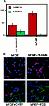
Stimulation of differentiation of rat and mouse hippocampal neural progenitor cells toward a neuronal phenotype by N-CAM. A, Quantitation of differentiation of rat hippocampal progenitor cells. Cells were grown for 4 d on poly-l-lysine- and laminin-coated glass multichamber slides in NB/B27 media in the presence of bFGF alone (20 ng/ml) and then treated for 3 d with N-CAM (10 μg/ml) or left untreated. In each experiment, the number of MAP2+ cells and GFAP+ cells is shown as a percentage of the total number of cells. A minimum of 200 cells in five different fields per condition were counted. The values are expressed as the average ± SD from a representative experiment.B, Immunocytochemistry for MAP2 (Texas Red) and GFAP (FITC) showing N-CAM induction of MAP2+ cells and CNTF or FCS induction of GFAP+ cells in mouse progenitor cell culture after treatment for 3 d with N-CAM (10 μg/ml), CNTF (100 ng/ml), or FCS (10%). Cells were counterstained with DAPI.
Fig. 5.
Fig. 5.
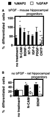
Comparison of the effect of N-CAM, BDNF, NT-3, IGF-I, PDGF, and CNTF on the differentiation of hippocampal progenitor cells. A, The protocol was exactly the same as that used in Figure 4. BDNF, NT-3, IGF-I, PDGF, and CNTF were used at 100 ng/ml, N-CAM was used at 10 μg/ml, and factors were added in the presence of bFGF (20 ng/ml) after 4 d of culture of mouse hippocampal progenitor cells (***p < 0.001; *p < 0.05; Student's t test).B, Cells were cultured for 4 d in the presence of bFGF, after which BDNF (100 ng/ml) or N-CAM (10 μg/ml) was added without bFGF and the cultures were grown for an additional 3 d. The counting was also done as described in Figure 4, and thevalues represent the average ± SD from a representative experiment (**p < 0.01; *p < 0.05; Student's ttest).
Fig. 6.
Fig. 6.
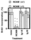
Inhibition of proliferation of hippocampal progenitor cells from N-CAM heterozygous (+/−) and knock-out (−/−) mice. Neural progenitor cells were prepared from the hippocampi of N-CAM heterozygous (+/−) or N-CAM knock-out (−/−) mice and incubated with 10 μg/ml N-CAM, Ig I–II, or Ig III, after 2 d in culture as described in Materials and Methods. BrdU incorporation was measured over the last 24 hr of treatment. Each valuerepresents the average ± SEM of a minimum of three experiments (***p < 0.001; Student's ttest).
Fig. 7.
Fig. 7.
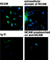
Binding of N-CAM and extracellular N-CAM to live hippocampal progenitor cells from N-CAM knock-out mice. Cells were treated with N-CAM, the extracellular domain of N-CAM, and the recombinant Ig III domain (10 μg/ml) for 2 hr at 37°C. The cells were washed with PBS, fixed with 4% PFA, and processed for immunocytochemistry using antibodies for N-CAM and N-CAM domains and FITC-labeled secondary antibodies as described in Materials and Methods. Nuclei were revealed by counterstaining with DAPI.
Fig. 8.
Fig. 8.
Saccharides and saccharide-modifying enzymes do not affect N-CAM binding to knock-out progenitor cells. Cells were pretreated (1 hr) with heparin (0.01 mg/ml), chondroitin sulfate (0.01 mg/ml), heparinase (0.01 U/ml), or glycosidases (4 U/ml; N-glycosidase endo F and O-glycosidase) and then treated with N-CAM (10 μg/ml) for 2 hr at 37°C. The cells were washed with PBS, fixed with 4% PFA, and processed for immunocytochemistry for N-CAM, as described in Materials and Methods.
Fig. 9.
Fig. 9.
N-CAM inhibitory activity on proliferation is not affected by known N-CAM heterophiles. The protocol was the same as that used in Figure 2, with N-CAM applied at 2 μg/ml. Cells were pretreated (1 hr) with heparin (0.01 mg/ml), chondroitin sulfate (0.01 mg/ml), heparinase (0.01 U/ml), anti-FGF receptor (1:200), or glycosidases (4 U/ml; N-glycosidase endo F and O-glycosidase). Thevalues represent the BrdU incorporation in a representative experiment. Values are expressed as the average ± SD of a minimum of three measurements.

References

    1. Acheson A, Sunshine JL, Rutishauser U. N-CAM polysialic acid can regulate both cell-cell and cell-substrate interactions. J Cell Biol. 1991;114:143–153. - PMC - PubMed
    1. Ahmed S, Reynolds BA, Weiss S. BDNF enhances the differentiation but not the survival of CNS stem cell-derived neuronal precursors. J Neurosci. 1995;15:5765–5778. - PMC - PubMed
    1. Arsenijevic Y, Weiss S. Insulin-like growth factor-I is a differentiation factor for postmitotic CNS stem cell-derived neuronal precursors: distinct actions from those of brain-derived neurotrophic factor. J Neurosci. 1998;18:2118–2128. - PMC - PubMed
    1. Azizeh BY, Cribbs DH, Kreng VM, Cotman CW. Cross-linking of N-CAM receptors on neurons induces programmed cell death. Brain Res. 1998;796:20–26. - PubMed
    1. Barnea A, Nottebohm F. Recruitment and replacement of hippocampal neurons in young and adult chickadees: an addition to the theory of hippocampal learning. Proc Natl Acad Sci USA. 1996;93:714–718. - PMC - PubMed

Publication types

MeSH terms