. 2000 May 15;20(10):3745-60.
doi: 10.1523/JNEUROSCI.20-10-03745.2000.

Olivocerebellar climbing fibers in the granuloprival cerebellum: morphological study of individual axonal projections in the X-irradiated rat

Affiliations

Olivocerebellar climbing fibers in the granuloprival cerebellum: morphological study of individual axonal projections in the X-irradiated rat

I Sugihara et al. J Neurosci. .

Abstract

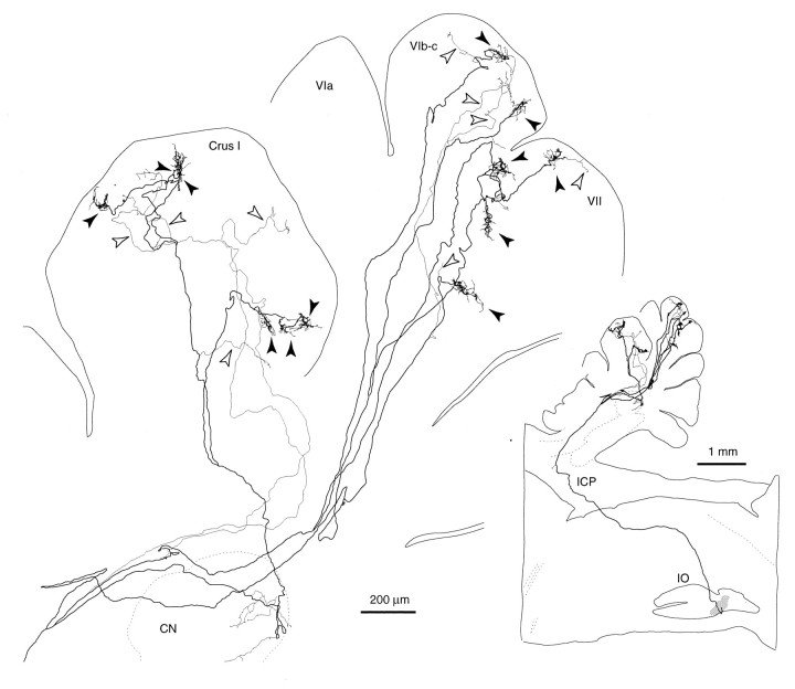
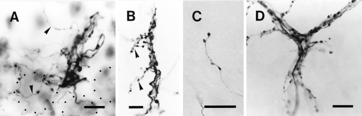
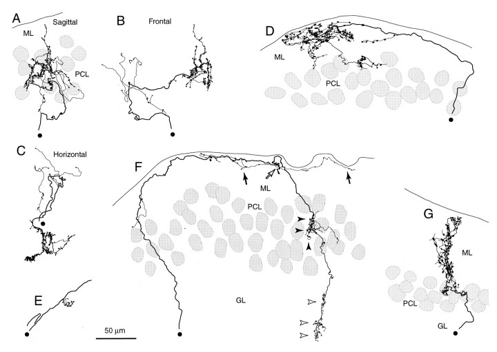
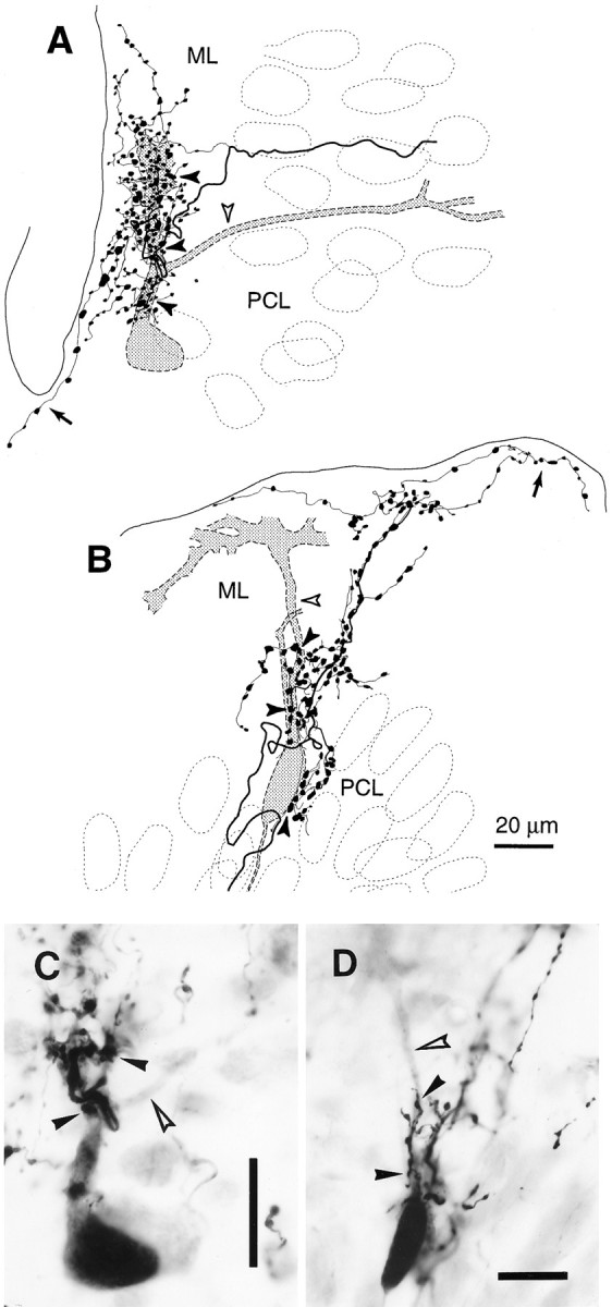
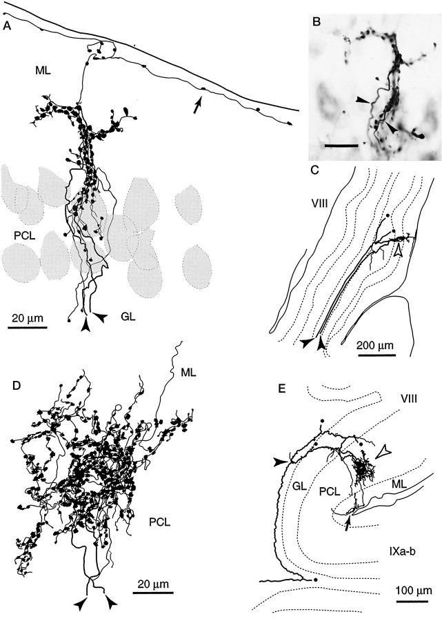
Elimination of cerebellar granule cells early during postnatal development produces abnormal neural organization that retains immature characteristics in the adult, including innervation of each Purkinje cell by multiple climbing fibers from the inferior olive. To elucidate mechanisms underlying development of the olivocerebellar projection, we studied light-microscopic morphology of single olivocerebellar axons labeled with biotinylated dextran amine in adult rats rendered agranular by a single postnatal X-irradiation. Each reconstructed olivocerebellar axon gave off approximately 12 climbing fibers, approximately twice as many as in normal rats. Terminal arborizations of climbing fibers made irregular tufts in most areas, whereas they were arranged vertically in a few mildly affected areas. Each climbing fiber terminal arborization innervated only part of the dendritic arbor of a Purkinje cell, and multiple climbing fibers innervated a single Purkinje cell. These climbing fibers originated either from the same olivocerebellar axon (pseudomultiple innervation) or from distinct axons (true multiple innervation). Abundant non-climbing fiber thin collaterals projected to all cortical layers. Although the longitudinal pattern of the zonal olivocerebellar projection was generally observed, lateral branching, including bilateral projections, was relatively frequent. These results suggest that the granule cell-parallel fiber system induces several important features of olivocerebellar projection: (1) organization of the climbing fiber terminal arborization tightly surrounding Purkinje cell dendrites, (2) elimination of pseudo- and true multiple innervations establishing one-to-one innervation, (3) retraction of non-climbing fiber thin collaterals from the molecular layer, and (4) probable refinement of the longitudinal projection domains by removing aberrant transverse branches.


Figures

Fig. 1.
Fig. 1.
Photomicrographs of counterstained sagittal sections of the cerebellar cortex of an X-irradiated rat (A) and the cerebellar cortex of a control rat (B). Tilted black bars indicate the molecular layer, and white bars indicate the Purkinje cell layer. Dots indicate the surface of the cerebellar folium of vermal lobule V. Scale bar, 200 μm.
Fig. 2.
Fig. 2.
Photomicrographs of abnormal Purkinje cells in an irradiated rat labeled by BDA injections into the fastigial and interposed cerebellar nuclei. A, A Purkinje cell located in the middle of the Purkinje cell layer whose dendrites extended into the molecular and granular layers. B, A Purkinje cell located in the superficial Purkinje cell layer. The dendrites of this Purkinje cell spread mainly in the molecular layer. C, A Purkinje cell in a control rat. All panels show sections in vermal lobule VI. The surface of the cerebellar cortex is toward thetop in each panel. Scale bar, 50 μm.
Fig. 3.
Fig. 3.
Photomicrographs of irregular, superficial, and vertical configurations of the abnormal mass olivocerebellar projection in an irradiated rat. A, Irregular projection in lobule VII. B, Predominantly superficial projection with some irregular projection in lobule V. C, Predominantly vertical projection in lobule IXc. D, Mixed irregular and superficial projections in lobule VII. E, Normal olivocerebellar projection in lobule VII in a control rat.F, BDA injection centered into the caudal inferior olive in an irradiated rat from which sections for A–D were obtained. All sections shown in this figure were counterstained. Each panel (A–E) shows a sagittal section of the cerebellar cortex (the surface is toward the top) in which many olivocerebellar axons were labeled. Dotsin F indicate the contour of the rostral and central inferior olive. Scale bars: E, 50 μm (applies toA–E); F, 500 μm.
Fig. 4.
Fig. 4.
Sagittal view of the trajectory of a single olivocerebellar axon innervating vermal lobule VI and VII and hemispheric crus I in an irradiated rat, reconstructed from 72 serial sagittal sections. Inset shows the nearly complete path of this axon from the ventral medulla near the injection site. This axon was labeled presumably by the uptake of BDA at the portion passing through the injection site (the right inferior olive). Because the number of labeled axons was small in the right cerebellar cortex, tracing all thin collaterals was possible. Black arrowheads indicate climbing fiber terminal arborizations, andopen arrowheads indicate non-climbing fiber thin collaterals. Abbreviations in this and subsequent figures:I-X, lobules I-X; a-d, sublobules a-d;C, caudal; CN, cerebellar nucleus;CP, copula pyramidis; Crus I, crus I ansiform lobule; Crus II, crus II ansiform lobule;D, dorsal; DPFL, dorsal paraflocculus;FL, flocculus; GL, granular layer;ICP, inferior cerebellar peduncle; IO, inferior olive; ML, molecular layer;Param, paramedian lobule; PCL, Purkinje cell layer; R, rostral; Sim, simple lobule; V, ventral; VPFL, ventral paraflocculus; WM, white matter.
Fig. 5.
Fig. 5.
Photomicrographs of significantly deformed single climbing fiber terminal arborizations and of a termination of a non-climbing fiber thin collateral in an irradiated rat.A, A terminal arborization in the Purkinje cell layer and molecular layer of an irregular projection area. The section inA only was counterstained in this figure. Some parts of labeled terminal arborizations were not in focus. Dotted lines indicate the contour of the three Purkinje cells closest to this terminal arborization. Arrowheads indicate thin collaterals given off from this terminal arborization.B, A terminal arborization in the molecular layer in a vertical type projection area. Arrowheads indicate horizontal branches in the terminal arborization that were presumably associated with secondary dendrites of the target Purkinje cell.C, En passant and terminal swellings of the non-climbing fiber collateral of an olivocerebellar axon in the molecular layer. D, Proximal portion of a terminal arborization in the control rat. Proximal side of the climbing fiber is to the bottom in each panel. Scale bars, 10 μm.
Fig. 6.
Fig. 6.
Single climbing fiber terminal arborizations entirely reconstructed in irradiated rats. A, Sagittal view of a terminal arborization mainly in the Purkinje cell layer in an irregular projection area (lobule IXa-b). Reconstructed from four serial sections. B, Frontal view of the same terminal arborization as in A. C, Horizontal view of the same terminal arborization as in A.D, A terminal arborization located mainly in the molecular layer in an area with some superficial mixed with predominantly irregular projections. E, A smallen passant terminal arborization in the middle of a thick branch of an olivocerebellar axon in the granular layer of an irregular projection area. F, A terminal arborization in the granular layer (open arrowheads) and an en passant terminal arborization in the Purkinje cell layer (filled arrowheads) on the same thick branch of an olivocerebellar axon in a presumed superficial plus irregular projection area. Several non-climbing fiber thin collaterals (filled arrows) and an en passantsmall terminal arborization (open arrow) were given off in the superficial molecular layer. G, A terminal arborization in a vertical projection area. Filled circles indicate the proximal side of the axon. See Figure 4, legend, for abbreviations.
Fig. 7.
Fig. 7.
Climbing fiber terminal arborization covering part of a Purkinje cell dendritic arbor. A, An entire climbing fiber terminal arborization and a Purkinje cell in an irregular projection area (lobule VIb-c) reconstructed from three sections. B, An entire climbing fiber terminal arborization and a Purkinje cell in a vertical projection area (lobule IXc) reconstructed from three sections. C, D, Photomicrographs of the same cases as in panels A andB, respectively. Open arrowheads inA–D indicate the dendrites of the labeled Purkinje cells that were not in contact with the labeled climbing fiber terminal arborizations. Filled arrowheads in A–Dindicate portions of the terminal arborization that were in contact with the thick dendrites and the somata of the Purkinje cells.Arrows in A and B indicate thin collaterals given off from the terminal arborization and running in the superficial molecular layer. Both the Purkinje cells and the climbing fibers in A–D were labeled by BDA injected into the cerebellar nuclei. The sections were counterstained. Scale bars: C, D, 20 μm.
Fig. 8.
Fig. 8.
Putative true multiple and pseudodouble innervation of a Purkinje cell by two climbing fibers.A, Entire combined climbing fiber terminal arborizations in an irregular projection area reconstructed from two sections.B, Photomicrograph of the same case as inA. Somata of neurons were visualized by counterstaining.Arrowheads in A and Bindicate two climbing fibers forming these terminal arborizations. The continuous arrangement of the combined terminal arborizations indicated that they together innervate a primary dendrite of a single Purkinje cell. C, Two axons (filled arrowheads) forming the combined terminal arborizations shown in A and B (open arrowhead) traced toward the proximal side, reconstructed from six sections. Further tracing was difficult in this case.Circles indicate thick branches given off from one of the axons, which were not reconstructed completely. D, Entire combined terminal arborizations of two climbing fibers (filled arrowheads) in an irregular projection area recon- structed from three sections. They are very closely combined, indicating that they can innervate one or a few Purkinje cells together. E, Trajectory of the climbing fibers inD traced proximally. Reconstructed from 28 sections. Two climbing fibers which form the combined terminal arborizations (open arrowheads) are branches of a single olivocerebellar axon (filled arrowhead). Some thick branches (circles), which ended as climbing fibers, were not drawn completely. The stem axon was traced down to the medulla (data not shown). Arrows inA and E indicate thin collaterals given off from terminal arborizations and running in the superficial molecular layer. Panels A, B, andD were rotated so that the surface of the cerebellum is toward the top. See Figure 4, legend, for abbreviations. Scale bar: B, 20 μm.
Fig. 9.
Fig. 9.
Reconstructions of well developed non-climbing fiber thin collaterals of olivocerebellar axons. A, Entire trajectory of two non-climbing fiber collaterals (filled arrowheads), reconstructed from 13 sections. They originated from a thick branch of an olivocerebellar axon that terminated as a climbing fiber (open arrowhead). B, A small en passantterminal arborization formed on a non-climbing fiber thin collateral in the granular layer. The distal side is toward the top. See Figure 4, legend, for abbreviations.
Fig. 10.
Fig. 10.
Zonal distribution of labeled climbing fiber terminal arborizations in the cerebellar cortex of irradiated and control rats. A, B, Irradiated rats. Two (A) and three (B) injections of BDA were made into the right inferior olive. Eachdot represents a climbing fiber terminal arborization.C, D, Control rats. Insets inA–D, parasagittal sections of the right inferior olive showing BDA injection sites. Distance from the midline for injection sites, 0.25 mm (A), 0.25 mm (B), 0.07 mm (C, top), 0.24 mm (C, bottom), and 0.2 mm (D). Arrows in A–D indicate the distributions of terminal arborizations in the intermediate area of the cerebellum (see Results). In each diagram of the cerebellum, the mediolateral distances for each terminal arborization and the cerebellar outline were to scale (measured by the number of the parasagittal sections). The rostrocaudal dimension of each cerebellar lobule was determined by measuring the length of the Purkinje cell layer in each lobule in parasagittal sections at the midline and at 1.5 and 3 mm lateral to the midline. Therefore, the diagrams represent the entire Purkinje cell layer approximately. The primary fissure was made straight arbitrarily. The rostrocaudal distances in the paraflocculus and flocculus were not to scale (enlarged). Broken outlines in the paraflocculus and flocculus indicate the continuation of the Purkinje cell layer. The position in the rostrocaudal axis for each terminal arborization was determined by measuring its relative distance from the borders of the lobule in parasagittal sections. See Figure 4, legend, for abbreviations.
Fig. 11.
Fig. 11.
Increased midline crossing by olivocerebellar axons in the cerebellum. A, Axons in a sagittal section of an irradiated rat (the case in Fig. 10B).B, Axons in a sagittal section of an control rat (the case in Fig. 10D). Only axons running transversely in the white matter in the sagittal section were drawn.
Fig. 12.
Fig. 12.
Distribution in the cerebellar cortex of all climbing fiber terminal arborizations originating from single olivocerebellar axons, indicating some lateral branching.A, Three axons terminating in crus I and vermal lobule VIa (a), in vermal lobules VIb-c and VII and in crus I (b), and in vermal lobule VIII and IX (c) in an irradiated rat. Nine climbing fiber terminal arborizations are located within a small area in lobule VIa, some of which made pseudomultiple innervations, in the case ofa. The case in b is the same axon as shown in Figure 4. B, An axon terminating in lobule IXb and X in a control rat. Dots surrounded by each broken contour represent individual climbing fiber terminal arborizations originating from a single axon. Single olivocerebellar axons for each (a–d) were completely reconstructed except for some thin collaterals. The diagrams of the unfolded cerebellar cortex are similar to those in Figure 10. See Figure 4, legend, for abbreviations.

Similar articles

Cited by

References

    1. Altman J, Anderson WJ. Experimental reorganization of the cerebellar cortex. I. Morphological effects of elimination of all microneurons with prolonged X-irradiation started at birth. J Comp Neurol. 1972;146:355–406. - PubMed
    1. Altman J, Bayer SA. Development of the cerebellar system in relation to its evolution, structure, and functions. CRC; Boca Raton, FL: 1997.
    1. Bailly Y, Schoen SW, Delhaye-Bouchaud N, Kreutzberg GW, Mariani J. Synaptic localization of 5′-nucleotidase activity in the cerebellar cortex of the adult rat after postnatal X-irradiation. C R Acad Sci III. 1990;311:487–493. - PubMed
    1. Bailly Y, Schoen SW, Delhaye-Bouchaud N, Kreutzberg GW, Mariani J. 5′-nucleotidase activity as a synaptic marker of parasagittal compartmentation in the mouse cerebellum. J Neurocytol. 1995;24:879–890. - PubMed
    1. Bailly Y, Kyriakopoulou K, Delhaye-Bouchaud N, Mariani J, Karagogeos D. Cerebellar granule cell differentiation in mutant and X-irradiated rodents revealed by the neural adhesion molecule TAG-1. J Comp Neurol. 1996;369:150–161. - PubMed

Publication types

LinkOut - more resources