. 2000 May 15;20(10):3785-97.
doi: 10.1523/JNEUROSCI.20-10-03785.2000.

Distribution, targeting, and internalization of the sst4 somatostatin receptor in rat brain

Affiliations

Distribution, targeting, and internalization of the sst4 somatostatin receptor in rat brain

M Schreff et al. J Neurosci. .

Abstract

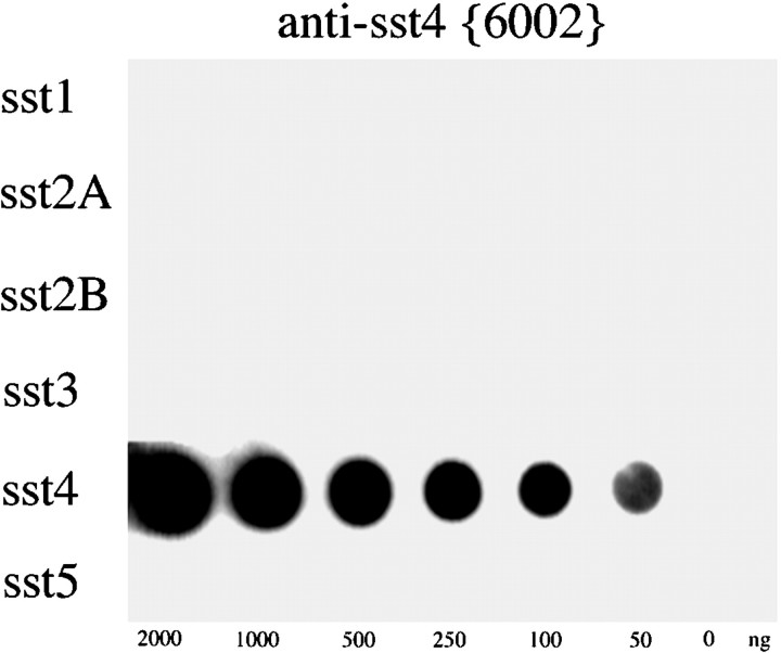
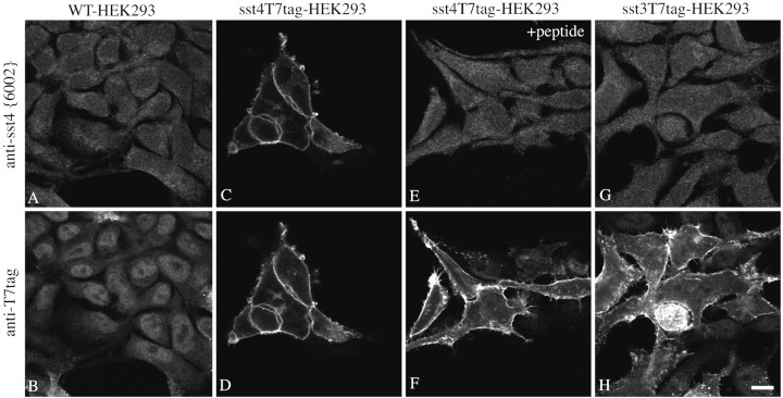
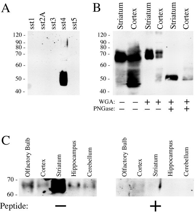
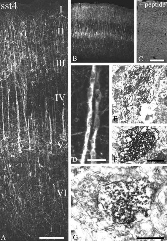
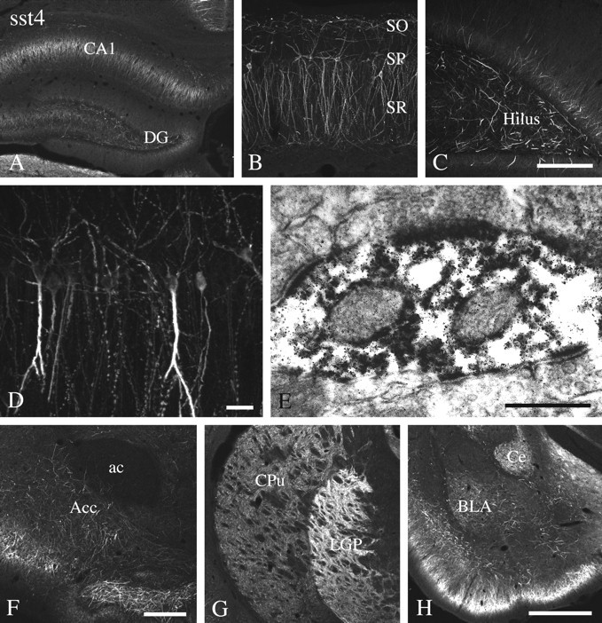
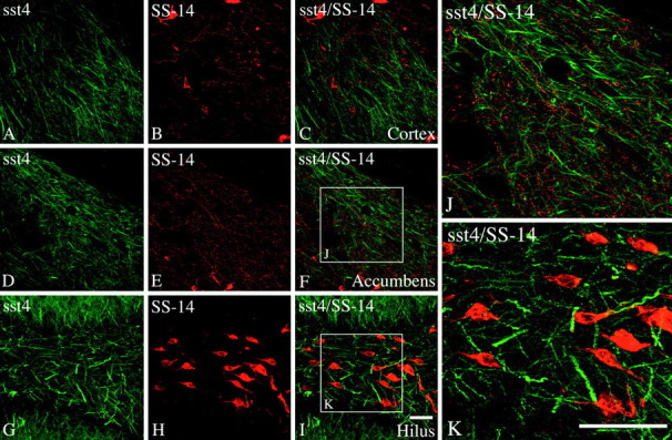
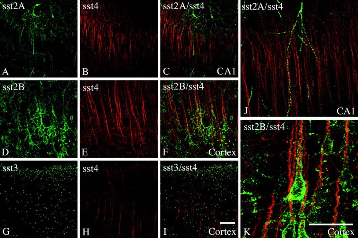
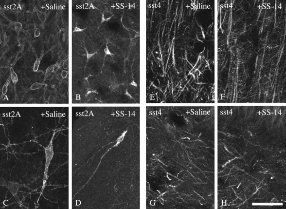
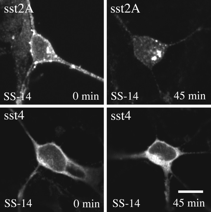
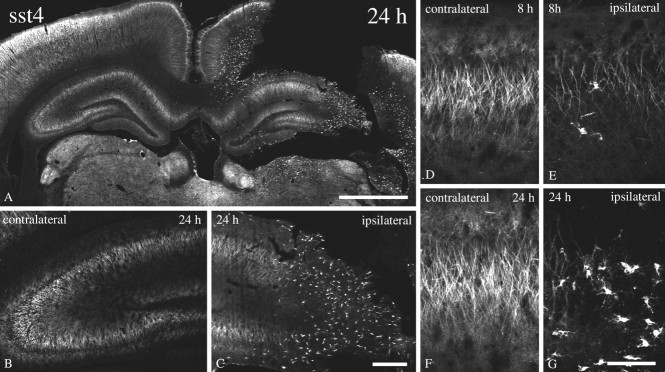
Somatostatin mediates its diverse physiological effects through a family of five G-protein-coupled receptors (sst(1)-sst(5)); however, knowledge about the distribution of individual somatostatin receptor proteins in mammalian brain is incomplete. In the present study, we have examined the regional and subcellular distribution of the somatostatin receptor sst(4) in the rat CNS by raising anti-peptide antisera to the C-terminal tail of sst(4). The specificity of affinity-purified antibodies was demonstrated using immunofluorescent staining of HEK 293 cells stably transfected with an epitope-tagged sst(4) receptor. In Western blotting, the antiserum reacted specifically with a broad band in rat brain, which migrated at approximately 70 kDa before and approximately 50 kDa after enzymatic deglycosylation. sst(4)-Like immunoreactivity was most prominent in many forebrain regions, including the cerebral cortex, hippocampus, striatum, amygdala, and hypothalamus. Analysis at the electron microscopic level revealed that sst(4)-expressing neurons target this receptor preferentially to their somatodendritic domain. Like the sst(2A) receptor, sst(4)-immunoreactive dendrites were often closely apposed by somatostatin-14-containing fibers and terminals. However, unlike the sst(2A) receptor, sst(4) was not internalized in response to intracerebroventricular administration of somatostatin-14. After percussion trauma of the cortex, neuronal sst(4) receptors progressively declined at the sites of damage. This decline coincided with an induction of sst(4) expression in cells with a glial-like morphology. Together, this study provides the first description of the distribution of immunoreactive sst(4) receptor proteins in rat brain. We show that sst(4) is strictly somatodendritic and most likely functions in a postsynaptic manner. In addition, the sst(4) receptor may have a previously unappreciated function during the neuronal degeneration-regeneration process.


Figures

Fig. 1.
Fig. 1.
Immunodot-blot analysis of the specificity of the anti-sst4 antiserum. Serial dilutions (0–2000 ng) of the peptides corresponding to the C-terminal regions of sst1, sst2A, sst2B, sst3, sst4, and sst5 were blotted onto a nitrocellulose membrane and incubated with affinity-purified anti-sst4 antibody (6002) at a concentration of 1 μg/ml. The blot was subsequently incubated with peroxidase-conjugated secondary antibodies and developed using enhanced chemiluminescence.
Fig. 2.
Fig. 2.
Characterization of the anti-sst4antiserum using stably transfected HEK 293 cells. Double immunofluorescent labeling and confocal imaging of wild-type HEK 293 cells (A, B) and HEK 293 cells transfected with a construct coding for sst4T7tag (C–F) or sst3T7tag (G, H) using a mixture of the anti-sst4 antiserum (6002) and the mouse monoclonal anti-T7 antibody. For absorption controls, this mixture was preincubated with 10 μg/ml of the homologous sst4 peptide (E, F). Note that the anti-sst4 antiserum yielded prominent immunofluorescence localized at the level of the plasma membrane only in sst4T7tag-expressing HEK 293 cells but not in wild-type or sst3T7tag-expressing HEK 293 cells. This staining is completely abolished by preincubation with homologous peptide.WT, Wild-type. Scale bar, 15 μm.
Fig. 3.
Fig. 3.
Western blot analysis of sst4-immunoreactivity in transfected HEK 293 cells and rat brain. A, Membrane preparations from HEK 293 cells transfected with sst1, sst2A, sst3, sst4, or sst5were separated on an 8% SDS polyacrylamide gel and blotted onto nitrocellulose. Membranes were then incubated with affinity-purified anti-sst4 antibodies (6002) at a concentration of 1 μg/ml. B, Crude membrane preparations, WGA-purified preparations, or PNGase F-treated WGA extracts prepared from striatum and cortex were separated on an 8% SDS polyacrylamide gel and blotted onto nitrocellulose. Membranes were then incubated with affinity-purified anti-sst4 antibodies (6002) at a concentration of 1 μg/ml. C, WGA extracts prepared from the olfactory bulb, cortex, striatum, hippocampus, and cerebellum were separated on an 8% SDS polyacrylamide gel and blotted onto nitrocellulose. Membranes were then incubated with affinity-purified anti-sst4 antibodies (6002) at a concentration of 1 μg/ml in either the absence (left panel) or presence (right panel) of the peptide antigen (10 μg/ml). Blots were developed using enhanced chemiluminescence. Ordinate, Migration of protein molecular weight markers (Mr × 10−3).
Fig. 4.
Fig. 4.
Immunofluorescent and electron micrographs showing the regional and subcellular localization of sst4-Li in rat neocortex. A,B, D, Coronal rat brain section immunofluorescently stained with affinity-purified anti-sst4 antibodies (6002). C, Corresponding adsorption control. The anti-sst4 antibody was preincubated with 10 μg/ml of its cognate peptide.E, F, Rat brain sections from cortical layer IV were processed for immunoperoxidase detection of the anti-sst4 antibody. G, Rat brain sections from cortical layer I were processed for immunoperoxidase detection of the anti-sst4 antibody. Note that sst4-Li is enriched throughout the layers of the cerebral cortex with prominent labeling of pyramidal cells in layers II/II and V, as well as their primary dendrites. This staining pattern is completely neutralized by preincubation with the immunizing peptide. At the electron microscopic level, immunoperoxidase product was mostly intracellular in large apical pyramidal cell dendrites in layer IV. In layer I in which sst4-immunopositve dendrites project into and ramify, immunolabeling was more densely distributed along neuronal plasma membranes. The neuronal profiles containing sst4-Li were dendrites and symmetrical synapses. Scale bars: A, 50 μm; B, C, 200 μm; D, 10 μm; E, F, 1.5 μm;G, 0.4 μm.
Fig. 5.
Fig. 5.
Immunofluorescent and electron micrographs showing the regional and subcellular localization of sst4-Li in rat forebrain. A–D, F–H, Coronal rat brain section immunofluorescently stained with affinity-purified anti-sst4 antibodies (6002). E, Rat brain sections from the hippocampal CA1 region were processed for immunoperoxidase detection of the anti-sst4 antibody. Note that sst4-Li is enriched in the hippocampal formation with high levels found in the Ammon's horn and the hilar region of the dentate gyrus. sst4-Li was also distributed along neuronal processes in the nucleus accumbens, striatum, and amygdala. At the electron microscopic level, immunoperoxidase product was always postsynaptic, and some instances of immunolabeling at asymmetrical synapses were found in the hippocampal CA1 region (E). ac, Anterior commissure;Acc, nucleus accumbens, BLA, basolateral amygdaloid nucleus; Ce, central amygdaloid nucleus,CPu, caudate-putamen; DG, dentate gyrus;LGP, lateral globus pallidus; SO, stratum oriens; SP, stratum pyramidale, SR, stratum radiatum. Scale bars: A, G,H, 250 μm; B, C, 50 μm; D, 10 μm; E, 0.2 μm;F, 100 μm.
Fig. 6.
Fig. 6.
Immunofluorescent confocal images of rat brain sections showing the spatial relationship of the sst4receptor and SS-14. A–K, Coronal rat brain sections double stained for sst4-Li (green) and SS-14-LI (red). Note that, in the cerebral cortex and nucleus accumbens, sst4-immunoreactive dendrites were often closely apposed by SS-14-containing fibers and terminals (C, F, J). In contrast, in the hilar region of the dentate gyrus, immunoreactive sst4 receptors appeared to decorate distal processes of SS-14-positive interneurons (I,K). Scale bars: A–I, 50 μm;J, K, 25 μm.
Fig. 7.
Fig. 7.
Immunofluorescent confocal images of rat brain sections showing the spatial relationships between the sst4receptor and other somatostatin receptor subtypes. A–C,J, Coronal rat brain sections double stained for sst2A-Li (green) and sst4-Li (red). D–F,K, Coronal rat brain sections double stained for sst2B-Li (green) and sst4-Li (red). G–I, Coronal rat brain sections double stained for sst3-Li (green) and sst4-Li (red). Note that a high degree of colocalization was seen between sst4-Li and sst2B-Li in layer V cortical pyramidal cells (K). Whereas sst2B-Li was distributed in a patch-like manner along the soma and proximal dendrites, sst4-Li was most dense at the distal portion of the apical dendrites of these neurons (K). No such colocalization was observed between either sst4 and sst2A or sst4 and sst3. Scale bars: A–I, 50 μm;J, K, 25 μm.
Fig. 8.
Fig. 8.
Differential internalization of sst2Aand sst4 in rat brain. Confocal micrographs showing the subcellular distribution of sst2A-Li (A–D) and sst4-Li (E–H) in rat brain after intracerebroventricular administration of either saline (A, C,E, G) or 1 μg of SS-14 (B, D, F,H). Coronal rat brain section immunofluorescently stained with affinity-purified anti-sst2A (6291) (A–D) or anti-sst4 (6002) (E–H) antibodies. Micrographs shown inA and B were taken from the lateral septum, C and D were taken from the central gray, E and F were taken from the cortex, and G and H were taken from the hilar region of the dentate gyrus. Note that sst2A rapidly redistributes from the plasma membrane into vesicle-like structures within the cytosol. In contrast, sst4 appears to be resistant against short-term downregulation via receptor internalization. Scale bar: A–H, 25 μm.
Fig. 9.
Fig. 9.
Differential internalization of sst2Aand sst4 in primary neuronal culture. Primary dissociated cultures were prepared from embryonic day 19 fetuses and grown on coverslips for 1–2 weeks. Cells were then exposed to 100 nm SS-14 for 0 or 45 min. Cells were subsequently fixed, fluorescently labeled with antibodies specific for either sst2A or sst4, and examined by confocal microscopy. Note that nearly all immunoreactive sst2Areceptors rapidly redistribute from the plasma membrane into the cytosol in the presence of somatostatin. Such a redistribution was not seen for the sst4 receptor. Very similar results were obtained in the presence of monensin. Representative results from three independent experiments performed in duplicate. Scale bar,A–D, 25 μm.
Fig. 10.
Fig. 10.
Dynamic changes of sst4-Li after percussion trauma of the cortex. Immunofluorescent confocal micrographs of coronal rat brain section of animals that had been subjected to percussion of trauma of the cortex either 8 or 24 hr before vascular perfusion. All sections were stained with affinity-purified anti-sst4 antibodies (6002). Note that, at 8 and 24 hr after traumatic injury, the immunoreactive sst4 receptors progressively declined on the ipsilateral, but not on the contralateral, side of damage. This decline of neuronal sst4 receptors coincided with an induction of sst4 receptors in cells with a glial-like morphology. Scale bars: A, 250 μm; B, C, 50 μm; D–G, 10 μm.

Similar articles

Cited by

References

    1. Bell GI, Reisine T. Molecular biology of somatostatin receptors. Trends Neurosci. 1993;16:34–38. - PubMed
    1. Bernert H, Turski L. Traumatic brain damage prevented by the non-N-methyl-d-aspartate antagonist 2,3-dihydroxy-6-nitro-7-sulfamoylbenzo[f]quinoxaline. Proc Natl Acad Sci USA. 1996;93:5235–5240. - PMC - PubMed
    1. Bito H, Mori M, Sakanaka C, Takano T, Honda Z, Gotoh Y, Nishida E, Shimizu T. Functional coupling of SSTR4, a major hippocampal somatostatin receptor, to adenylate cyclase inhibition, arachidonate release, and activation of mitogen-activated protein kinase cascade. J Biol Chem. 1994;269:12722–12730. - PubMed
    1. Boehm S, Betz H. Somatostatin inhibits excitatory transmission at rat hippocampal synapses via presynaptic receptors. J Neurosci. 1997;17:4066–4075. - PMC - PubMed
    1. Brazeau P, Vale W, Burgus R, Ling N, Butcher M, Rivier J, Guillemin R. Hypothalamic polypeptide that inhibits the secretion of immunoreactive pituitary growth hormone. Science. 1973;179:77–79. - PubMed

Publication types

MeSH terms

LinkOut - more resources