. 2000 May 15;20(10):3830-42.
doi: 10.1523/JNEUROSCI.20-10-03830.2000.

Effect of lesions of the ventrolateral preoptic nucleus on NREM and REM sleep

Affiliations

Effect of lesions of the ventrolateral preoptic nucleus on NREM and REM sleep

J Lu et al. J Neurosci. .

Abstract

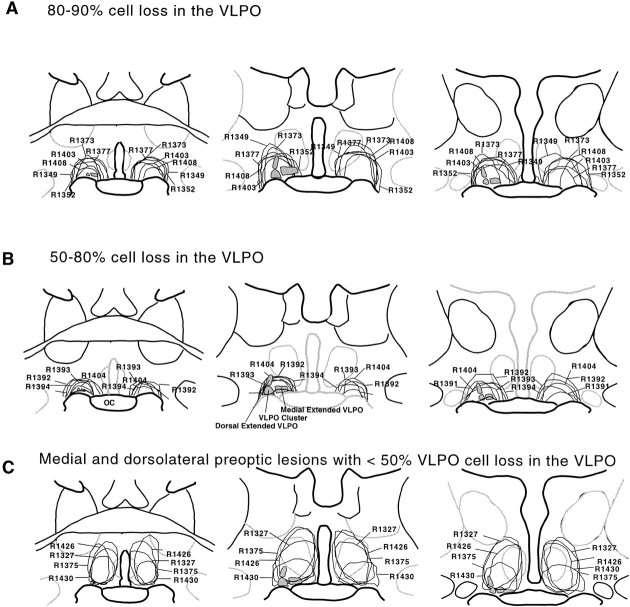
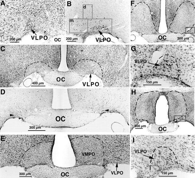
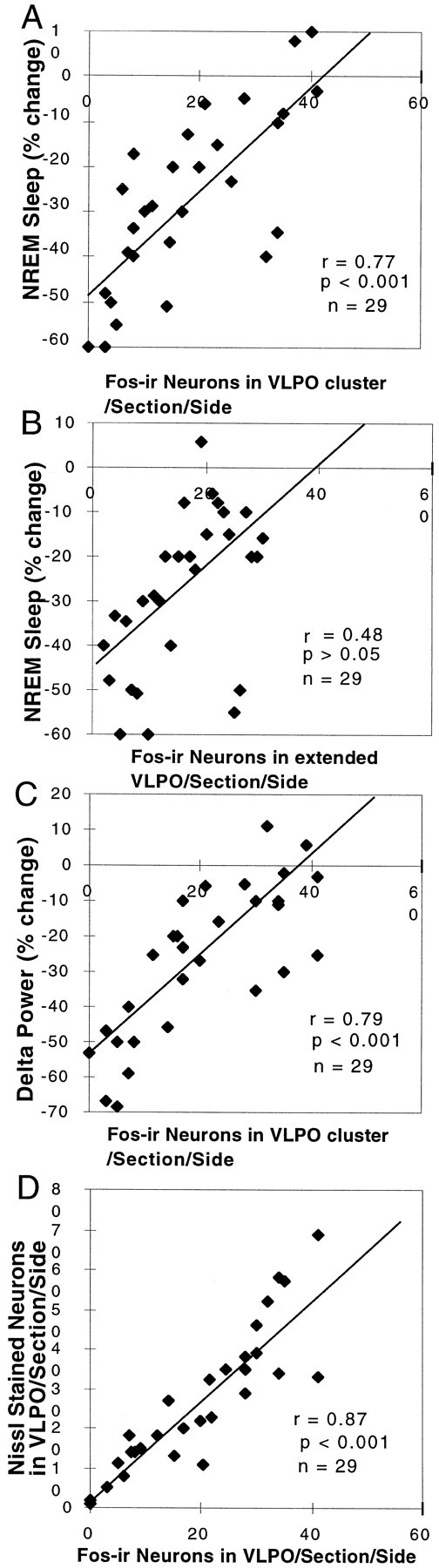
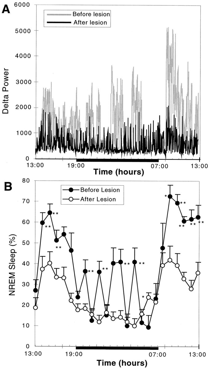
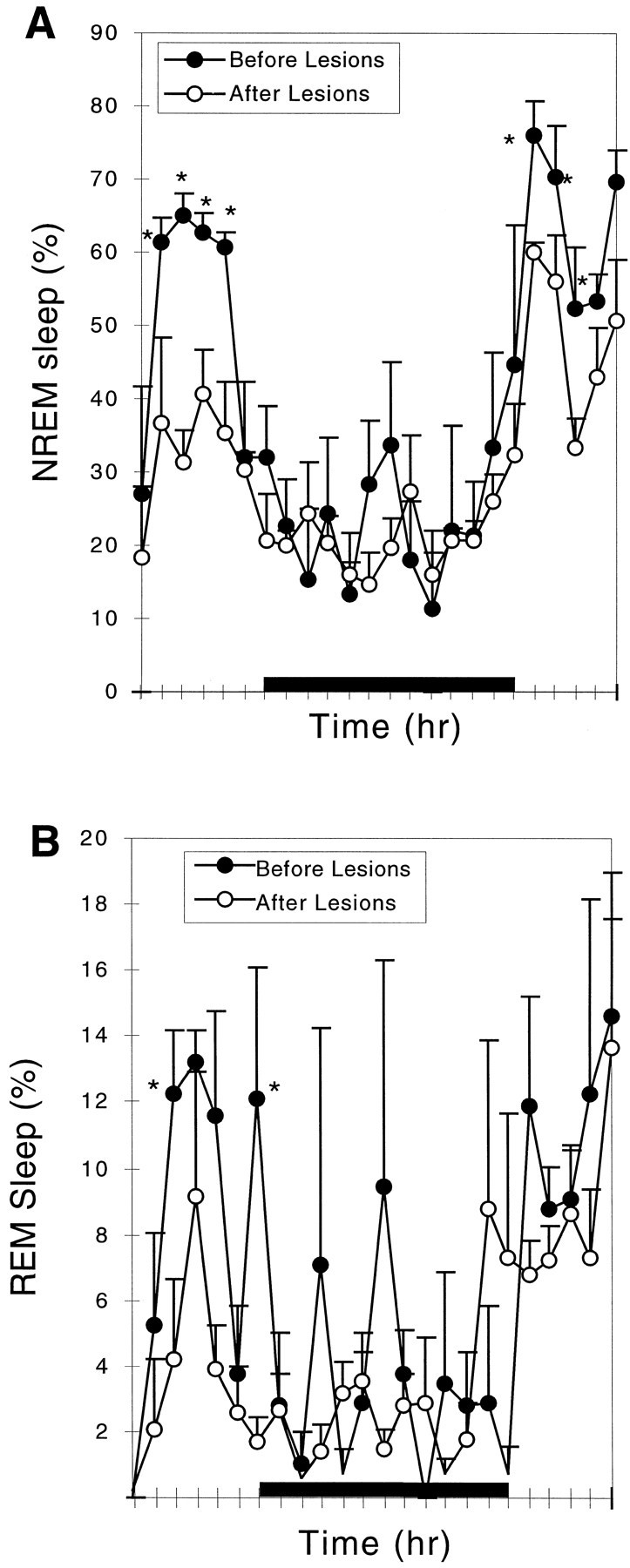
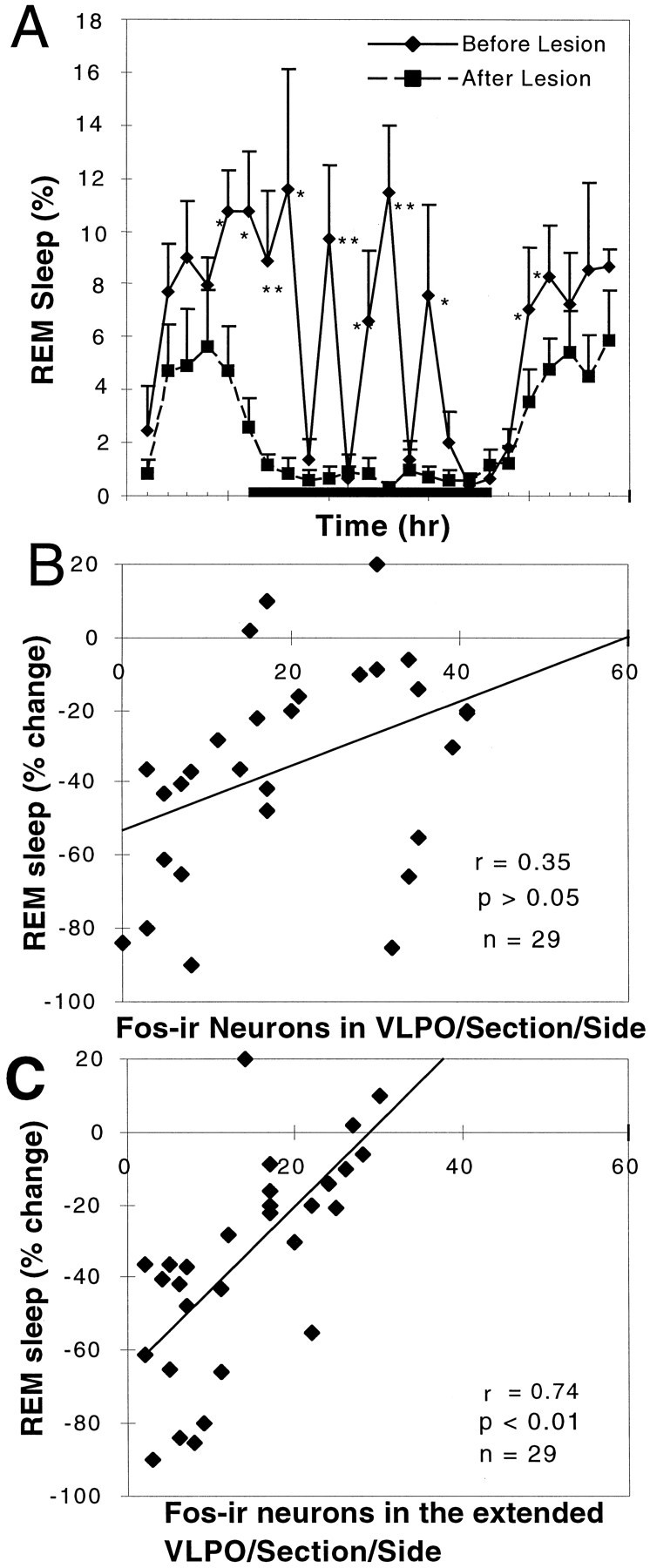
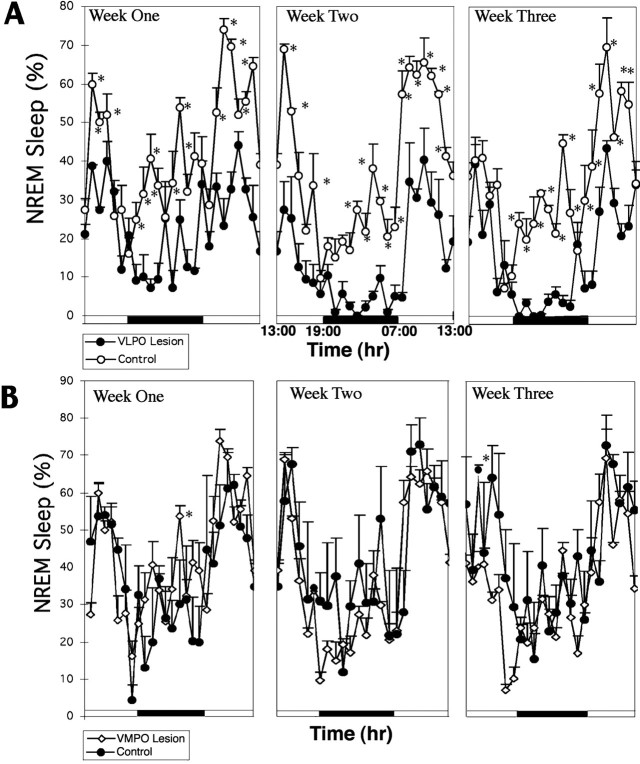
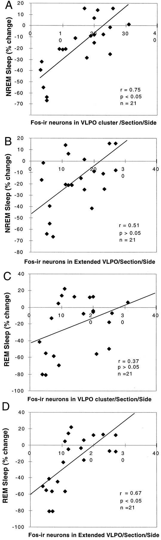
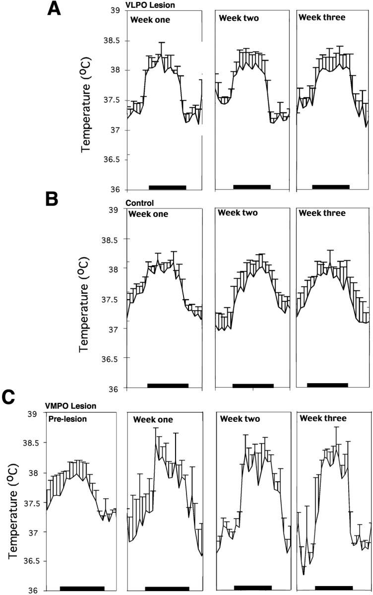
Neurons in the ventrolateral preoptic nucleus (VLPO) in rats show c-fos activation after sleep and provide GABAergic innervation of the major monoamine arousal systems, suggesting that they may be a necessary part of the brain circuitry that produces sleep. We examined the effects on sleep behavior in rats of cell-specific damage to the VLPO by microinjection of ibotenic acid. Severe lesions of the central cell cluster of the VLPO ( approximately 80-90% cell loss bilaterally) caused a 60-70% decrease in delta power and a 50-60% decrease in nonrapid-eye-movement (NREM) sleep time (p < 0.001). The number of remaining Fos-immunoreactive neurons in the VLPO cell cluster was linearly related to NREM sleep time (r = 0.77; p < 0.001) and total electroencephalogram delta power (r = 0. 79; p < 0.001) but not to rapid-eye-movement (REM) sleep (r = 0.35; p > 0.10). Lesions in the region containing scattered VLPO neurons medial or dorsal to the cell cluster caused smaller changes in NREM sleep time (24.5 or 15%, respectively) but were more closely associated with loss of REM sleep (r = 0.74; p < 0.01). The insomnia caused by bilateral VLPO lesions persisted for at least 3 weeks. Lesions of the VLPO caused no change in mean body temperature or its circadian variation; after small lesions of the ventromedial preoptic nucleus, body temperature showed normal circadian variation but a wider temperature range, and sleep behavior was not affected. These experiments delineate distinct preoptic sites with primary effects on the regulation of NREM sleep, REM sleep, and body temperature.


Figures

Fig. 1.
Fig. 1.
Camera lucida drawings illustrating the typical lesions of the VLPO cluster of 80–90% (A) and 50–80% (B) and control lesions in the medial preoptic area and dorsolateral preoptic area with <50% cell loss in the VLPO cell cluster (C), as assessed in Nissl-stained sections. A, The lesion areas in the animals that had 80–90% loss of the cells in the VLPO cluster ranged from 400 to 800 μm in diameter and included the entire extent of the VLPO. B, Because the cells in the VLPO cluster were relatively resistant to ibotenic acid compared with the cells in the surrounding regions, even when the ibotenic acid injection was placed close to the base of the hypothalamus, substantial numbers of cells in the VLPO cluster often survived (50–80% cell loss). C, Similarly when the lesion sites were placed just dorsal to the VLPO cluster, the areas around the VLPO were often devastated, but the majority of cells in the VLPO cluster survived. OC, Optic chiasm.
Fig. 2.
Fig. 2.
A series of photomicrographs illustrating lesions of the VLPO (A–D) and control lesions (E–I) by ibotenic acid injection.A, The VLPO cluster is visualized by Nissl staining as a pyramidal-shaped cell cluster along the base of the brain, just lateral to the optic chiasm. The triangularcountingbox used to quantify neurons in the VLPO cluster in Nissl preparations is shown. B, The VLPO cluster stands out as an aggregation of c-fos-immunoreactive neurons after sleep. The boxes in Brepresent those used to count cells in the VLPO cluster and the medial (m)- and dorsal (d)-extended VLPO. C, D, After bilateral ibotenic acid lesions, it is possible to quantify the remaining neurons in the VLPO cluster by Nissl staining (C; dashed lines represent borders of lesions) or by Fos immunostaining (D; thearrowheads indicate Fos-positive cells in the VLPO cluster) in animals that are perfused during the height of the sleep cycle, between 10:00 and 12:00 noon. E–I, A lesion of the ventromedial preoptic area (VMPO; E) did little if any damage to the VLPO, and lesions dorsal to the VLPO (F) or larger lesions in the medial preoptic area (H) left the VLPO cluster nearly intact, as shown by higher magnification views of the areasincluded in the boxes (G,I, respectively).
Fig. 3.
Fig. 3.
Correlation of the loss of NREM sleep and delta power with the loss of VLPO neurons. A, The relationship of the loss of Fos-IR neurons in the VLPO cluster and the percentage reduction of NREM sleep is shown. The NREM sleep reduction was calculated by the percentage change in 24 hr NREM sleep time from before to after the lesion. The number of Fos-IR neurons in the VLPO cluster was measured in rats that were perfused while sleeping between 10:00 and 12:00 noon (r = 0.77; p < 0.001). B, The relatively weak correlation (r = 0.48) of the loss of Fos-IR neurons in the extended VLPO and the percentage reduction of NREM sleep did not reach statistical significance. C, The relationship between the loss of Fos-IR neurons in the VLPO and the percentage reduction of the delta power calculated from comparing the 24 hr prelesion and postlesion EEG (r = 0.79; p < 0.001) is shown. D, The number of Nissl-stained neurons in the VLPO cluster correlates closely with the number of Fos-IR neurons in the nucleus during morning sleep (r = 0.87; p < 0.001).
Fig. 4.
Fig. 4.
The effect of severe bilateral (>80%) lesions of the VLPO cluster on delta power and NREM sleep. A, Comparison of delta power in the EEG plotted over 24 hr for a single animal with >90% cell loss in the VLPO cluster bilaterally (R1352) before lesion (gray) and 6 d after lesion (black) is shown. The blackbaralong the bottom of the graph indicates the dark cycle. Note the nearly complete loss of periods of greatest delta activity during the first part of the sleep cycle (just after lights on) and during the brief episodes of sleep during the dark cycle. B, For the entire group of seven rats with severe bilateral VLPO lesions, there was loss of nearly 55% of NREM sleep time. In particular, NREM sleep bouts during the dark cycle were almost entirely eliminated. However, circadian variation in NREM sleep time persisted. *p < 0.05; **p < 0.01.
Fig. 5.
Fig. 5.
Comparison of NREM and REM sleep before and after lesion of the medial preoptic area. The lesion caused a reduction in NREM sleep by 24.5% (A) and in REM sleep by 30% (not statistically significant; B). Loss of NREM sleep was predominantly late in the light cycle, whereas the reduction of REM sleep also involved loss of peaks during the dark cycle. *p < 0.05.
Fig. 6.
Fig. 6.
Loss of REM sleep correlates with loss of Fos-IR neurons in the extended VLPO but not in the VLPO cluster.A, There was a substantial (∼50%) loss of REM sleep in the comparison of the prelesion and postlesion behavior of the seven rats with 80–90% loss of neurons in the VLPO cluster (*p < 0.05; **p < 0.01). REM sleep episodes during the night were almost entirely eliminated.B, However, the numbers of surviving Fos-IR neurons in the VLPO cluster after sleep did not correlate well with REM sleep behavior (r = 0.35; n = 29; p > 0.05).C, In contrast, the number of Fos-positive cells in the extended VLPO (in which cell loss was equally substantial in this series) was significantly correlated with the percentage reduction of REM sleep (r = 0.74; p < 0.01;n = 29).
Fig. 7.
Fig. 7.
Change in NREM sleep time over a 3 week period after lesions of the VLPO (A) andVMPO (B). A, In four animals with 70–90% bilateral lesions of the VLPO cluster, the percentage of NREM sleep time was depressed by almost 45% in the first week, 63% in the second week, and 57% in the third week compared with that of a group of control rats that had received bilateral saline injections. As in the short-term experiments, the animals with VLPO lesions showed almost complete loss of NREM sleep during the dark cycle and substantial loss at the onset of the light cycle (*p < 0.05). There was no trend toward recovery. B, In five animals with severe bilateral lesions of the ventromedial preoptic nucleus, there was no change in NREM sleep time compared with that of saline-injected control animals.
Fig. 8.
Fig. 8.
Correlation of loss of NREM or REM sleep time with the numbers of Fos-IR neurons in the VLPO and extended VLPO 3 weeks after placement of severe bilateral lesions (70–90% cell loss in the VLPO cluster). A, B, The decrease in NREM sleep correlated closely with the loss of Fos-IR neurons in the VLPO cluster (A; r = 0.75; p< 0.05) but not with the loss of Fos-IR neurons in the extended VLPO (B; r = 0.51; p> 0.05). C, D, On the other hand, the decrease in REM sleep did not correlate with the loss of Fos-IR neurons in the VLPO cluster (C; r = 0.37;p > 0.05) but did correlate with the loss of Fos-IR neurons in the extended VLPO (D;r = 0.67; p < 0.05).
Fig. 9.
Fig. 9.
Effect of lesions of the VLPO or ventromedial preoptic nucleus on body temperature recorded over 24 hr at three weekly intervals. A, B, Severe bilateral lesions of the VLPO in four rats had no effect on the mean body temperature or its daily circadian rhythm (A) when compared with that of four control rats (B). Although the animals with VLPO lesions had a slightly greater circadian excursion of body temperature, this did not differ statistically from that of the saline-injected control animals. C, In contrast, in four animals with lesions of the ventromedial preoptic nucleus, the circadian rhythm of body temperature was unaffected, but the range was ∼2°C during the course of the day (approximately twice normal), and the peaks and troughs of body temperature both differed significantly from that of control animals (p < 0.05). Body temperature showed substantial instability in these animals, as evidenced by the significantly larger variance compared with that of the control or VLPO-lesioned animals (p < 0.05).

Similar articles

Cited by

References

    1. Alam MN, McGinty D, Szymusiak R. Neuronal discharge of preoptic/anterior hypothalamic thermosensitive neurons: relation to NREM sleep. Am J Physiol. 1995;269:R1240–R1249. - PubMed
    1. Benington JH, Heller HC. REM-sleep timing is controlled homeostatically by accumulation of REM-sleep propensity in non-REM sleep. Am J Physiol. 1994;266:R1992–R2000. - PubMed
    1. Bjorkum AA, Ha QH, Saper CB. Afferents to and efferents from the raphe nuclei to the ventrolateral preoptic nucleus. Soc Neurosci Abstr. 1999;25:625.
    1. Gaus SE, Saper CB. Sleep-active neurons in the ventrolateral preoptic area (VLPO) are galaninergic. Soc Neurosci Abstr. 1999;25:625.
    1. John J, Kumar VM. The effects of NMDA lesion of the medial preoptic neurons on sleep and other functions. Sleep. 1998;21:587–598. - PubMed

Publication types

LinkOut - more resources