. 2000 May 15;20(10):3864-73.
doi: 10.1523/JNEUROSCI.20-10-03864.2000.

Projections from the rat prefrontal cortex to the ventral tegmental area: target specificity in the synaptic associations with mesoaccumbens and mesocortical neurons

Affiliations

Projections from the rat prefrontal cortex to the ventral tegmental area: target specificity in the synaptic associations with mesoaccumbens and mesocortical neurons

D B Carr et al. J Neurosci. .

Abstract

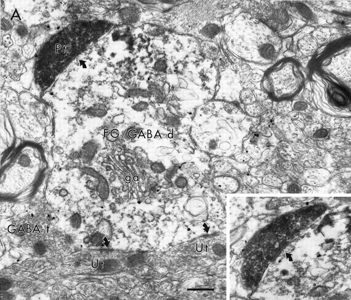
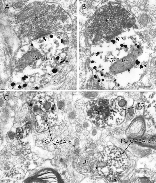
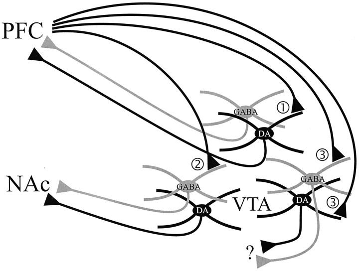
Excitatory projections from the prefrontal cortex (PFC) to the ventral tegmental area (VTA) play an important role in regulating the activity of VTA neurons and the extracellular levels of dopamine (DA) within forebrain regions. Previous investigations have demonstrated that PFC terminals synapse on the dendrites of DA and non-DA neurons in the VTA. However, the projection targets of these cells are not known. To address whether PFC afferents innervate different populations of VTA neurons that project to the nucleus accumbens (NAc) or to the PFC, a triple labeling method was used that combined peroxidase markers for anterograde and retrograde tract-tracing with pre-embedding immunogold-silver labeling for either tyrosine hydroxylase (TH) or GABA. Within the VTA, PFC terminals formed asymmetric synapses onto dendritic shafts that were immunoreactive for either TH or GABA. PFC terminals also synapsed on VTA dendrites that were retrogradely labeled from the NAc or the PFC. Dendrites retrogradely labeled from the NAc and postsynaptic to PFC afferents were sometimes immunoreactive for GABA but were never TH-labeled. Conversely, dendrites retrogradely labeled from the PFC and postsynaptic to PFC afferents were sometimes immunoreactive for TH but were never GABA-labeled. These results provide the first demonstration of PFC afferents synapsing on identified cell populations in the VTA and indicate a considerable degree of specificity in the targets of the PFC projection. The unexpected finding of selective PFC synaptic input to GABA-containing mesoaccumbens neurons and DA-containing mesocortical neurons suggests novel mechanisms through which the PFC can influence the activity of ascending DA and GABA projections.


Figures

Fig. 1.
Fig. 1.
Light micrographs of coronal sections through the rat VTA showing the overlap of the somatodendritic field of mesoaccumbens neurons and PFC afferents. A, Bright-field micrograph showing the distribution of axons and terminals after injection of BDA into the medial PFC. B, Fluorescence micrograph displaying the distribution and extent of labeling of VTA neurons after an injection of FluoroGold into the medial NAc.SNr, Substantia nigra, zona reticulata;mp, mammilary peduncle. Medial is to theright. Scale bar, 140 μm.
Fig. 2.
Fig. 2.
Electron micrographs showing synaptic contacts (curved arrows) of BDA-labeled PFC terminals (Pt) onto dendrites containing immunogold–silver labeling for TH (A, TH-d) or GABA (B, GABA-d). In B, the synapse formed by the Pt on the GABA-d is also prominent in an adjacent section (inset). Scale bar, 0.25 μm.
Fig. 3.
Fig. 3.
Electron micrograph showing the distribution of immunoperoxidase labeling for FG, retrogradely transported from the NAc, and immunogold–silver labeling for TH within the neuropil of the VTA. A dendrite (FG/TH-d) contains both peroxidase labeling for FG as well as gold–silver labeling for TH. Within the same area of neuropil are dendrites singly labeled for either FG (FG-d) or TH (TH-d). Scale bar, 1.0 μm.
Fig. 4.
Fig. 4.
Electron micrographs showing the associations of PFC terminals (Pt) with VTA dendrites retrogradely labeled from the NAc. In A, a Pt is within the same area of neuropil as a dendrite containing both immunoperoxidase labeling for FG, retrogradely transported from the NAc, as well as immunogold labeling for TH (FG/TH-d). The FG/TH-d exhibits FG labeling concentrated in a lysosome (note flocculent precipitate around the structure at the open arrow) and receives a synaptic contact from an unlabeled terminal (Ut). The Pt does not contact the FG/TH-d but instead contacts an unlabeled dendrite (Ud). The oblique synapse (curved arrow) formed by the Pt on the Ud is more evident in an adjacent section (inset). In B, a Pt forms a synaptic contact (curved arrow) onto an FG-labeled dendrite (FG-d) that does not contain gold–silver labeling for TH. In C, a Pt synapses (curved arrow) on a dendrite (PRV/GABA-d) that contains both immunoperoxidase labeling for PRV retrogradely transported from the NAc and immunogold–silver labeling for GABA. The PRV/GABA-d is adjacent to a dendrite that contains only peroxidase labeling for PRV (PRV-d). Scale bar, 0.5 μm.
Fig. 5.
Fig. 5.
Electron micrographs showing a synaptic contact (curved arrow) of a PFC terminal (Pt) on a dendrite containing both immunoperoxidase labeling for FG retrogradely transported from the NAc and immunogold–silver labeling for GABA (FG/GABA-d). The postsynaptic dendrite is proximal to the soma, [see Golgi apparatus (ga)], and receives synaptic input from the Pt (also shown in inset) and from unlabeled terminals (Ut). A GABA-containing terminal (GABA-t) lies adjacent to the dendrite. Scale bar, 0.5 μm.
Fig. 6.
Fig. 6.
Electron micrographs showing the associations of PFC terminals (Pt) with VTA dendrites retrogradely labeled from the PFC. In A and B, two different Pts form synaptic contacts (curved arrows) onto dendrites that contain both immunoperoxidase labeling for FG retrogradely transported from the PFC as well as immunogold–silver labeling for TH (FG/TH-d). In C, a Pt synapses on a dendrite (FG-d) that contains retrogradely transported FG but does not contain gold–silver labeling for GABA. Within the same area of neuropil, two other dendrites (FG/GABA-d) contain both markers. One FG/GABA-d is apposed to an unlabeled terminal (Ut). Scale bar, 0.25 μm.
Fig. 7.
Fig. 7.
Schematic drawing of the observed relationships between PFC terminals and mesoprefrontal, mesoaccumbens, and other projection neurons of the VTA. 1, A small population of PFC terminals form synaptic contacts onto DA neurons that project to the PFC. 2, Another population of PFC terminals synapse onto GABA neurons that project to the NAc. 3, The majority of PFC terminals within the VTA appear to target DA and GABA neurons that project to unknown target sites. It is also possible that PFC terminals project onto VTA neurons that contain neither DA nor GABA.

References

    1. Bassareo V, Di Chiara G. Differential influence of associative and nonassociative learning mechanisms on the responsiveness of prefrontal and accumbal dopamine transmission to food stimuli in rats fed ad libitum. J Neurosci. 1997;17:851–861. - PMC - PubMed
    1. Bean AJ, Roth RH. Extracellular dopamine and neurotensin in rat prefrontal cortex in vivo: effects of median forebrain bundle stimulation frequency, stimulation pattern, and dopamine autoreceptors. J Neurosci. 1991;11:2694–2702. - PMC - PubMed
    1. Bertolino A, Knable MB, Saunders RC, Callicott JH, Kolachana B, Mattay VS, Bachevalier J, Frank JA, Egan M, Weinberger DR. The relationship between dorsolateral prefrontal N-acetylaspartate measures and striatal dopamine activity in schizophrenia. Biol Psychiatry. 1999;45:660–667. - PubMed
    1. Card JP, Levitt P, Enquist LW. Different patterns of neuronal infection after intracerebral injection of two strains of pseudorabies virus. J Virol. 1998;72:4434–4441. - PMC - PubMed
    1. Carr DB, O'Donnell P, Card JP, Sesack SR. Dopamine terminals in the rat prefrontal cortex synapse on pyramidal cells that project to the nucleus accumbens. J Neurosci. 1999;19:11049–11060. - PMC - PubMed

Publication types

MeSH terms

LinkOut - more resources