Clinical Trial
. 2000 May;83(5):2661-75.
doi: 10.1152/jn.2000.83.5.2661.

Differences in control of limb dynamics during dominant and nondominant arm reaching

Affiliations
Clinical Trial

Differences in control of limb dynamics during dominant and nondominant arm reaching

R L Sainburg et al. J Neurophysiol. 2000 May.

Abstract

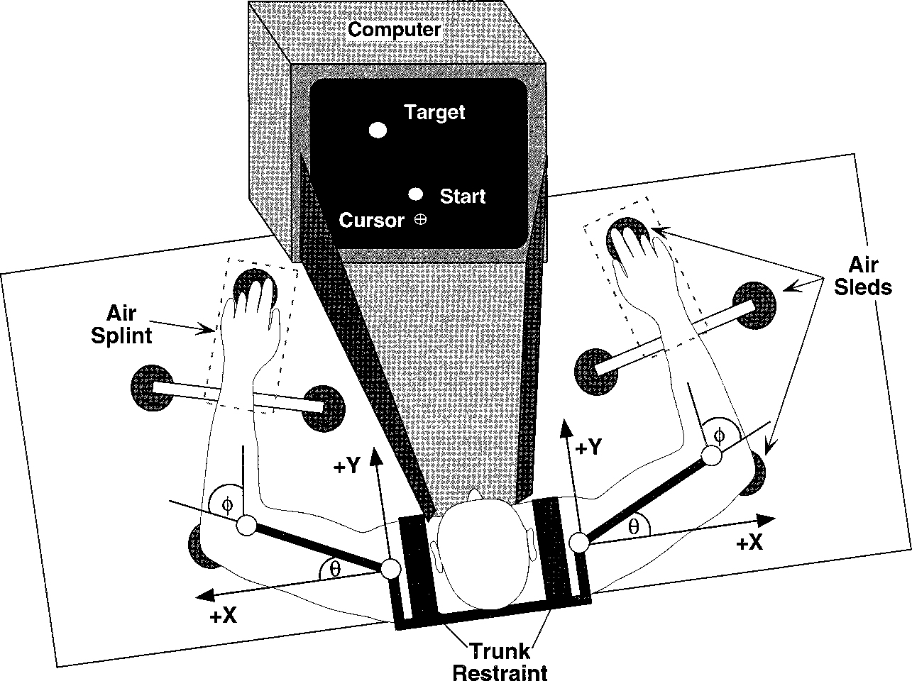
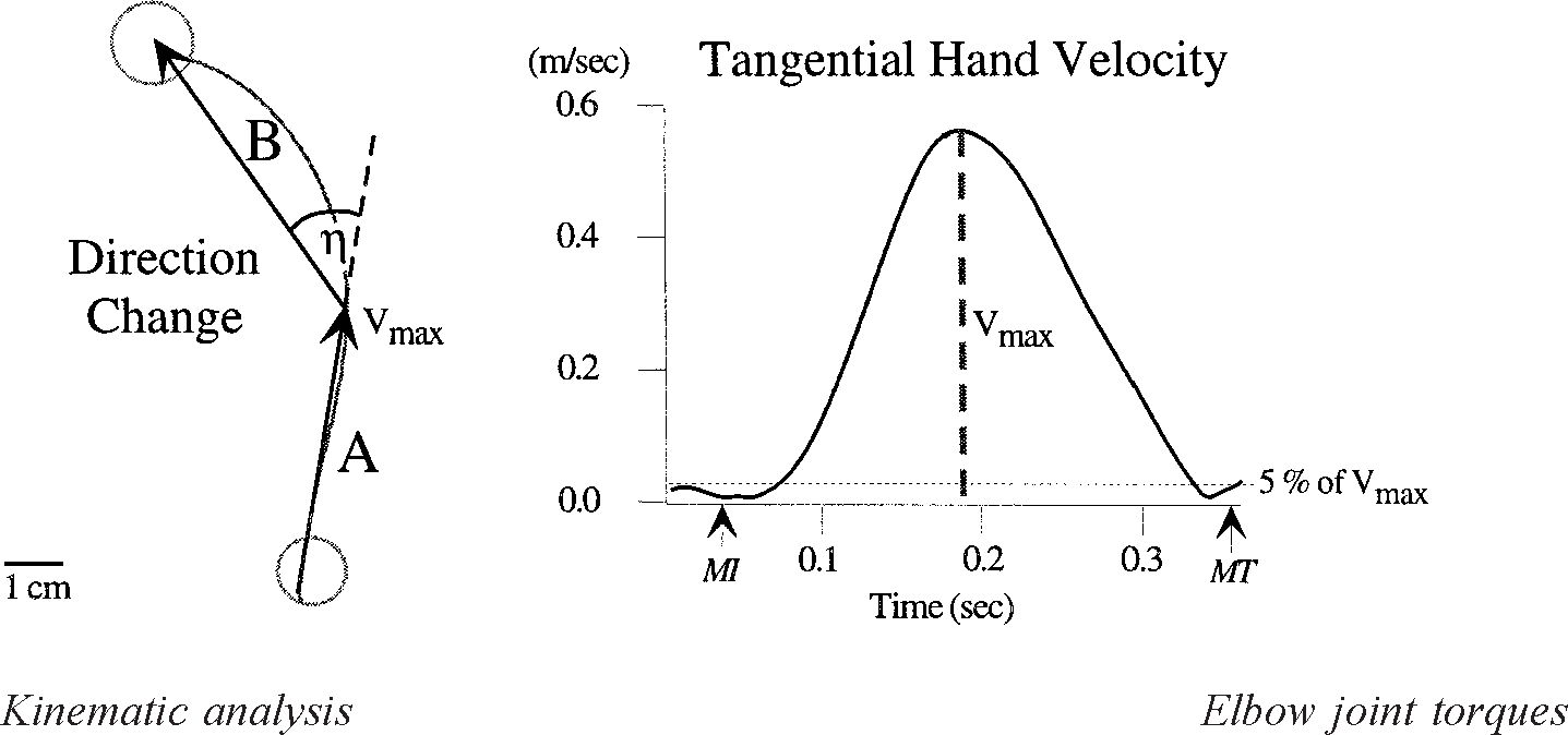
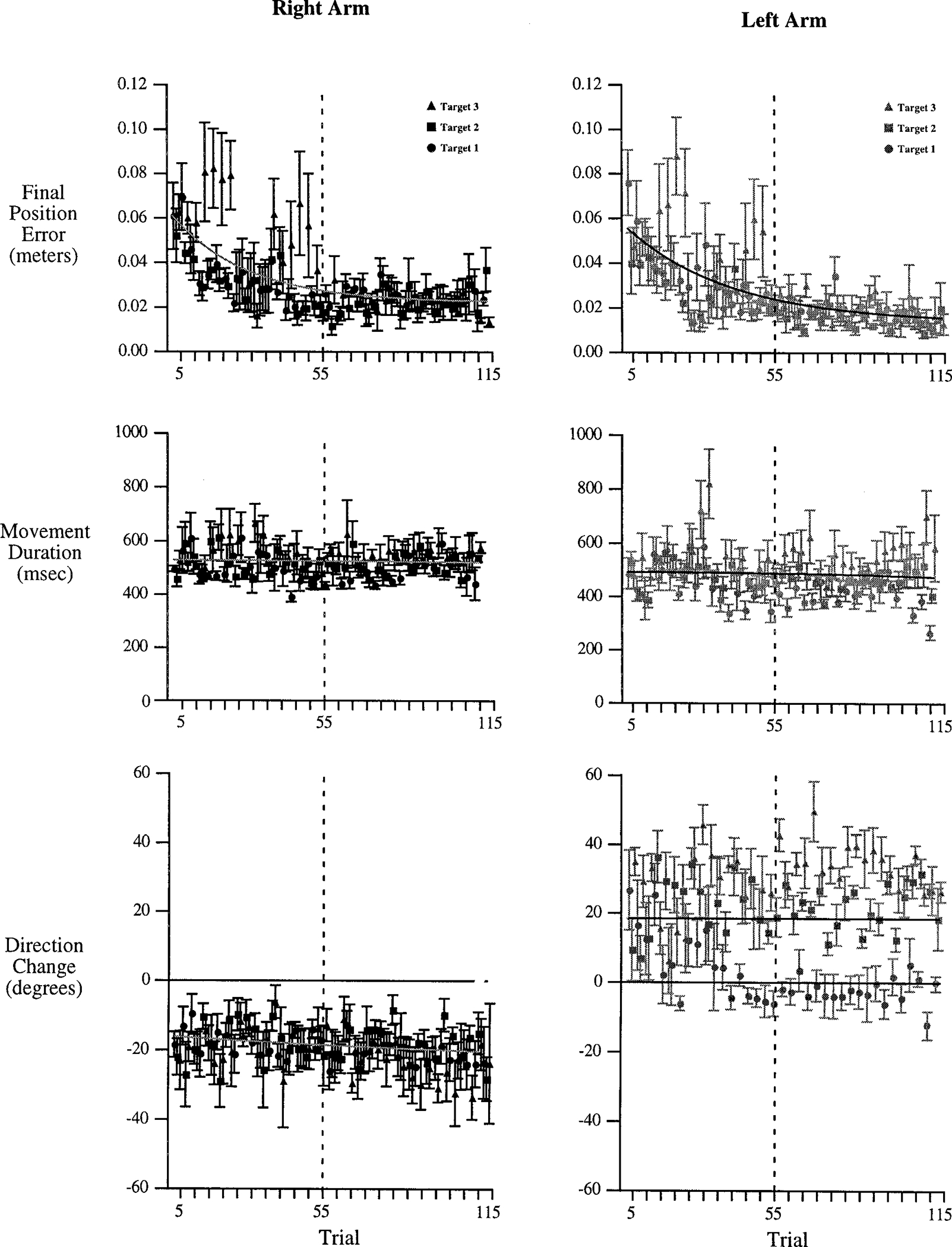
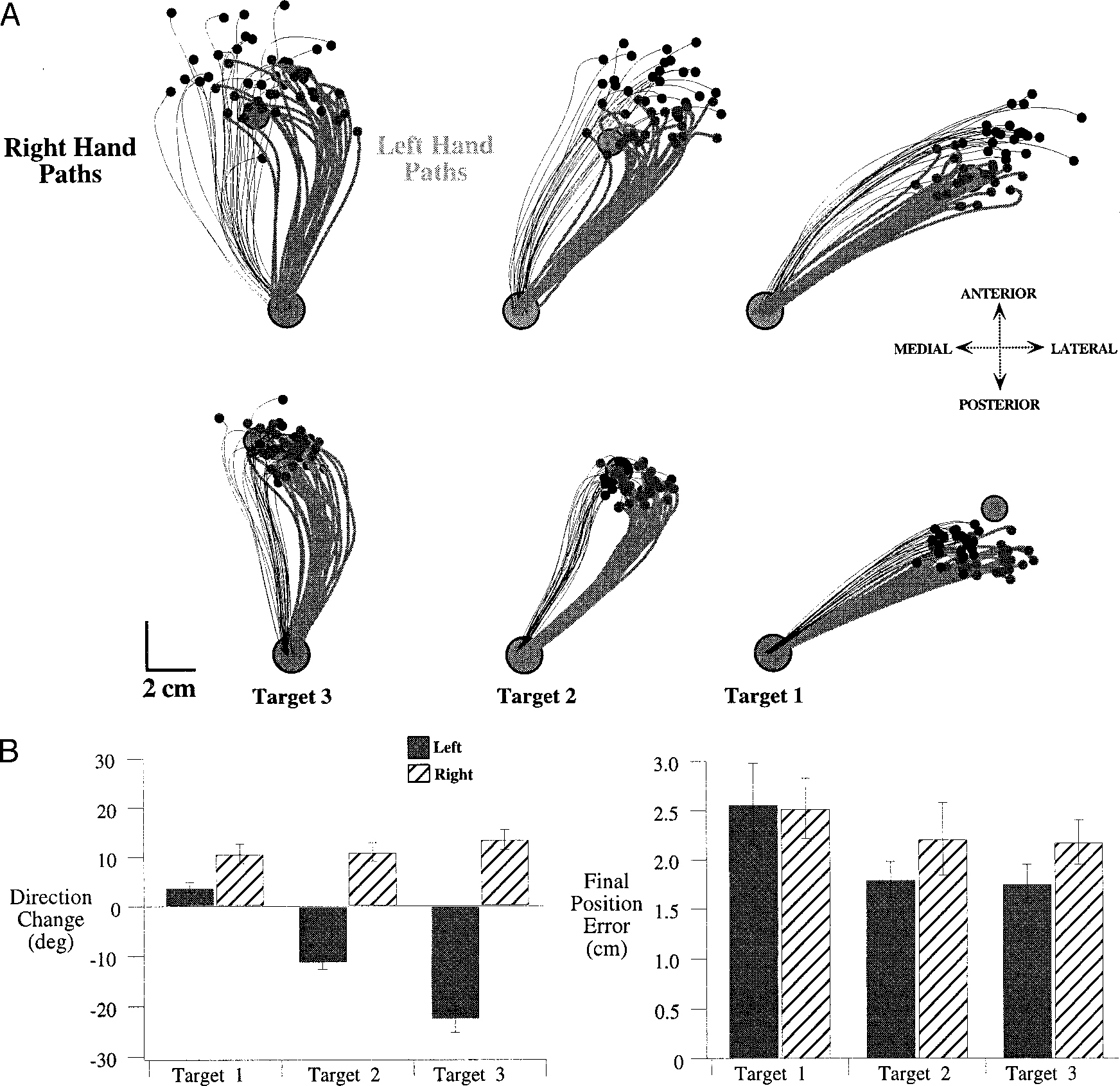
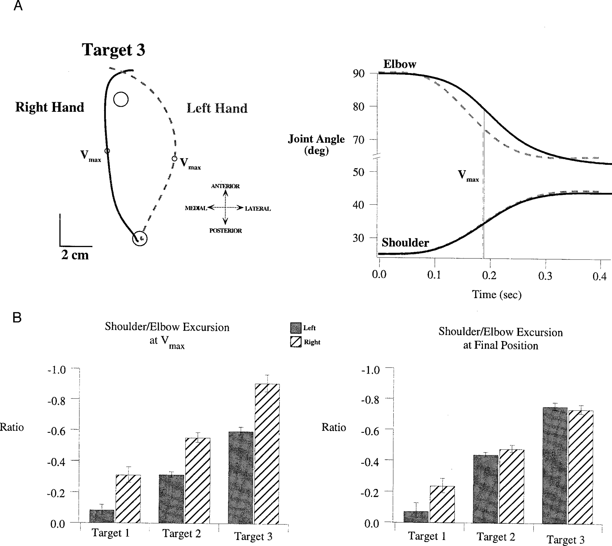
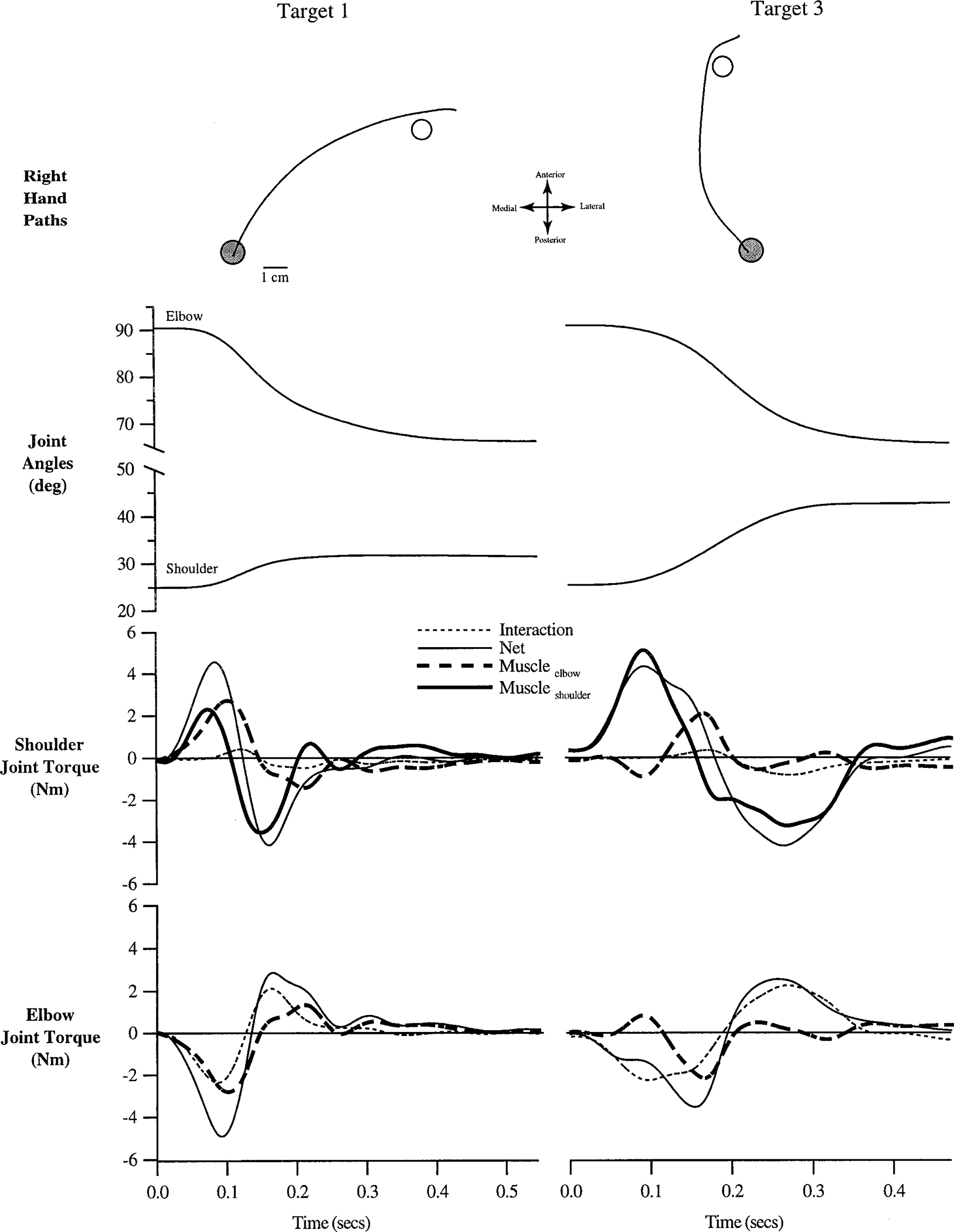
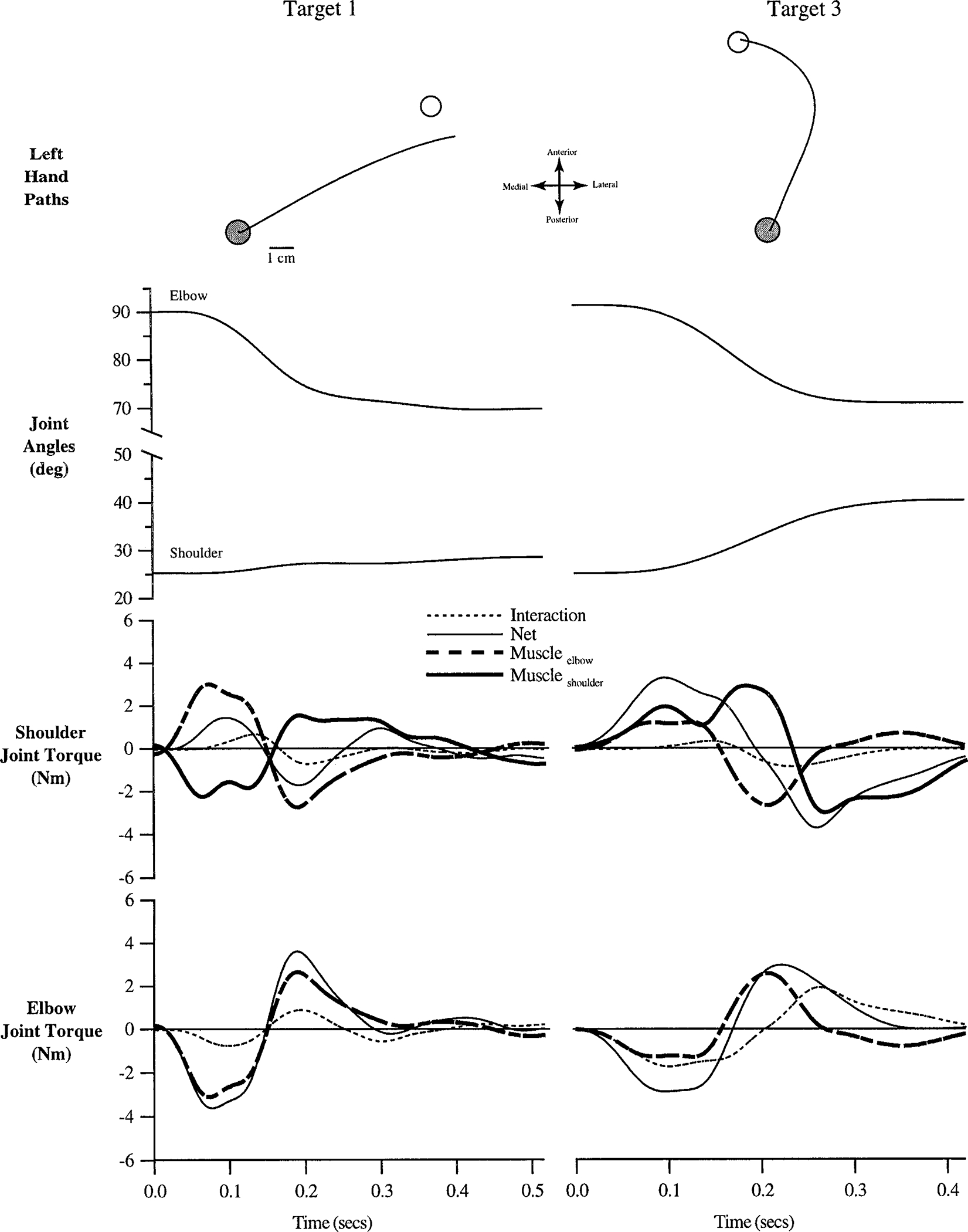
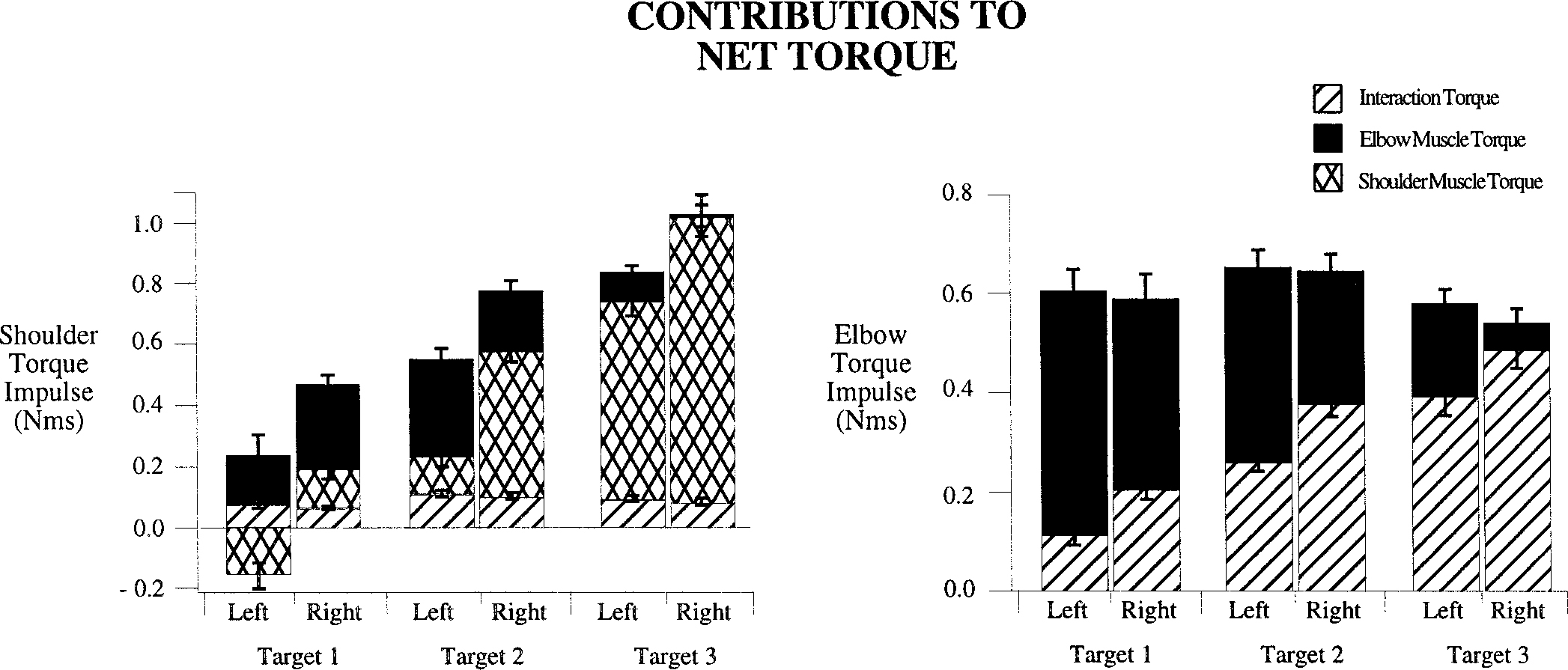
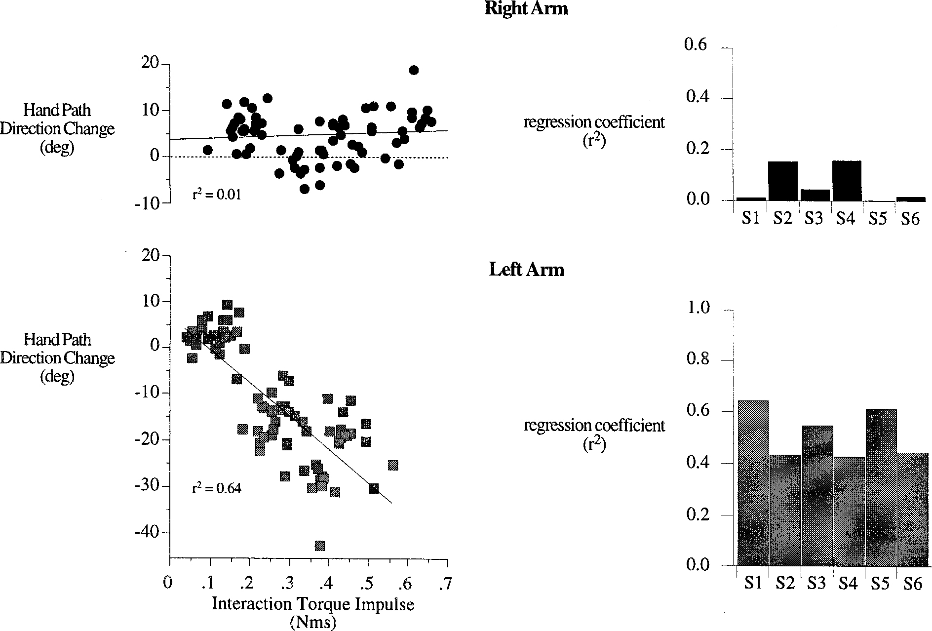
This study compares the coordination patterns employed for the left and right arms during rapid targeted reaching movements. Six right-handed subjects reached to each of three targets, designed to elicit progressively greater amplitude interaction torques at the elbow joint. All targets required the same elbow excursion (20 degrees ), but different shoulder excursions (5, 10, and 15 degrees, respectively). Movements were restricted to the shoulder and elbow and supported on a horizontal plane by a frictionless air-jet system. Subjects received visual feedback only of the final hand position with respect to the start and target locations. For motivation, points were awarded based on final position accuracy for movements completed within an interval of 400-600 ms. For all subjects, the right and left hands showed a similar time course of improvement in final position accuracy over repeated trials. After task adaptation, final position accuracy was similar for both hands; however, the hand trajectories and joint coordination patterns during the movements were systematically different. Right hand paths showed medial to lateral curvatures that were consistent in magnitude for all target directions, whereas the left hand paths had lateral to medial curvatures that increased in magnitude across the three target directions. Inverse dynamic analysis revealed substantial differences in the coordination of muscle and intersegmental torques for the left and right arms. Although left elbow muscle torque contributed largely to elbow acceleration, right arm coordination was characterized by a proximal control strategy, in which movement of both joints was primarily driven by the effects of shoulder muscles. In addition, right hand path direction changes were independent of elbow interaction torque impulse, indicating skillful coordination of muscle actions with intersegmental dynamics. In contrast, left hand path direction changes varied directly with elbow interaction torque impulse. These findings strongly suggest that distinct neural control mechanisms are employed for dominant and non dominant arm movements. However, whether interlimb differences in neural strategies are a consequence of asymmetric use of the two arms, or vice versa, is not yet understood. The implications for neural organization of voluntary movement control are discussed.


Figures

FIG. 1.
FIG. 1.
Experimental setup: X and Y represent axes of coordinate system originating at shoulder. Shoulder and elbow angles were measured as θ and ϕ, respectively. The trunk and scapulae were restrained with a butterfly style chest brace, whereas wrist and fingers were immobilized by air splints. Vision of the table and arms was blocked during the experimental sessions.
FIG. 2.
FIG. 2.
Hand path “direction-change” was quantified as the angle (η) between the 2 vectors drawn over the hand path (left). Vector A starts at movement initiation (MI) and ends at the point corresponding to maximum tangential hand velocity Vmax, identified on the tangential hand velocity plot (right). Vector B begins at Vmax and ends at movement termination (MT).
FIG. 3.
FIG. 3.
Measures of final position error (top), movement duration (middle) and direction-change (bottom). Each point represents a single trial, averaged across all 6 subjects. Trials for right (black) and left (gray) arms have been separated for comparison. The x-axis represents intralimb trial number.
FIG. 4.
FIG. 4.
A: hand paths for all trials of subject 1 (top) and subject 2 (bottom). Both right hand (thin, black) and left hand (thick, gray) paths are plotted in a “right hand” coordinate system, with the medial to lateral dimension plotted along the negative to positive x-axis. B: mean ± SE for measures of direction-change (left) and final position error (right). Values are averages of the individual subject means for each measure, taken separately for each limb and each target.
FIG. 5.
FIG. 5.
A: sample trials performed with right arm (black, solid) and left arm (gray, dashed) to target 3. Hand paths (left) are plotted in a “right hand” coordinate system, and joint displacement profiles are shown to the right of these. B: mean ± SE for measures of the shoulder excursion/elbow excursion ratio, measured at Vmax (left) and at final position (right). Values are averages of the individual subject means for each measure, taken separately for each limb and each target.
FIG. 6.
FIG. 6.
Representative trials made with the right arm to target 1 (left column) and target 3 (right column). Hand paths, joint angles, shoulder joint torques, and elbow joint torques (see methods) are shown. All time series data have been synchronized to movement initiation.
FIG. 7.
FIG. 7.
Representative trials made with the left arm to target 1 (left column) and target 3 (right column). Hand paths are plotted in a “right hand” coordinate system, with the medial to lateral dimension plotted along the negative to positive x-axis, respectively. Hand paths, joint angles, shoulder joint torques, and elbow joint torques are shown. All time series data have been synchronized to movement initiation.
FIG. 8.
FIG. 8.
Muscle and interaction torque impulse, measured as a contribution to net torque (see methods) is shown for the shoulder (left) and elbow (right) joints. Contributions of interaction torque, elbow muscle torque, and shoulder muscle torque are plotted separately in stacked bar format. Values are averages of the individual subject means for each measure, taken separately for each limb and each target.
FIG. 9.
FIG. 9.
Hand path direction-change is plotted against interaction torque impulse (left column), taken as a contribution to net torque (see methods and Fig. 8), for all trials performed with the right arm (top) and left arm (bottom) by subject 1. Simple linear regressions (solid line) were performed and r2 values are plotted in the bar plots (right) for trials performed with the right arm (top) and left arm (bottom) by each subject.

Similar articles

Cited by

References

    1. Abbott BC and Wilkie PR The relation between velocity of shortening and tension-length curve of skeletal muscle. J. Physiol. (Lond.) 120: 214–223, 1953. - PMC - PubMed
    1. Amunts K, Schlaug G, Schleicher A, Steinmetz H, Dabringhaus A, Roland PE, and Zilles K Asymmetry in the human motor cortex and handedness. Neuroimage 4: 216–222, 1996. - PubMed
    1. Annett J, Annett M, and Hudson PTW The control of movement in the preferred and non-preferred hands. Q. J. Exp. Psychol. B 31: 641–652, 1979. - PubMed
    1. Annett M Parallels between asymmetries of Planum temporale and of hand skill. Neuropsychologia 30: 951–962, 1992. - PubMed
    1. Bizzi E Motor control mechanisms. An overview. Neurologic Clinics 5: 523–528, 1987. - PubMed

Publication types

LinkOut - more resources