. 2000 Jun;20(11):3781-94.
doi: 10.1128/MCB.20.11.3781-3794.2000.

Granzyme B short-circuits the need for caspase 8 activity during granule-mediated cytotoxic T-lymphocyte killing by directly cleaving Bid

Affiliations

Granzyme B short-circuits the need for caspase 8 activity during granule-mediated cytotoxic T-lymphocyte killing by directly cleaving Bid

M Barry et al. Mol Cell Biol. 2000 Jun.

Abstract

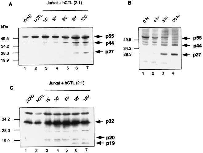
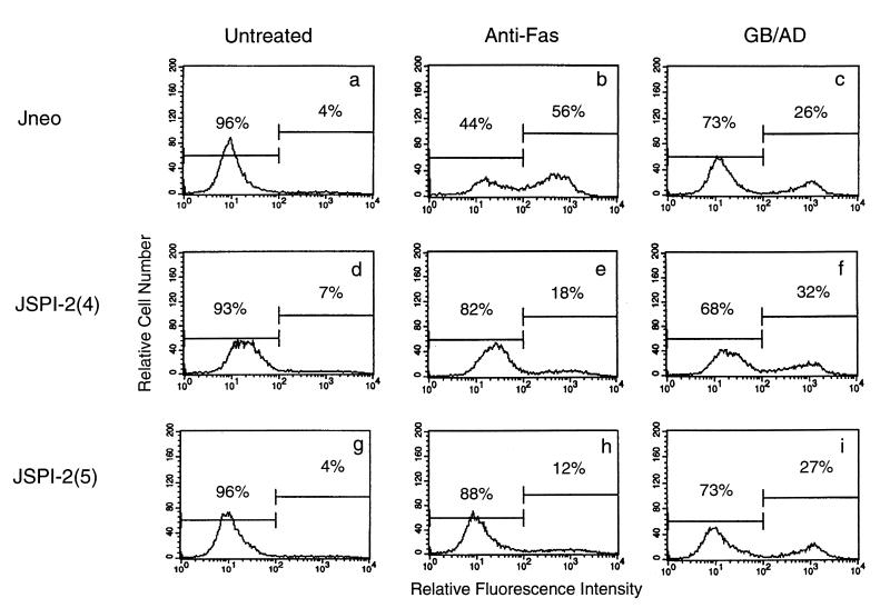
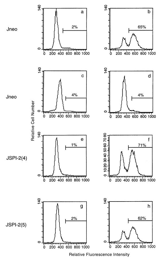
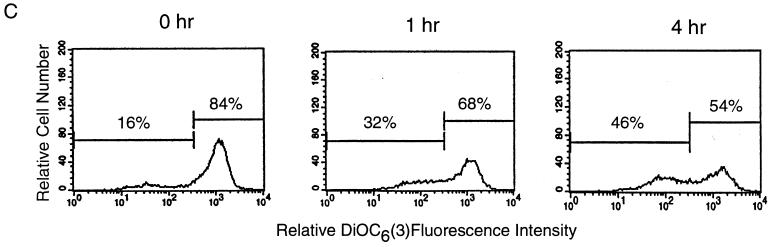
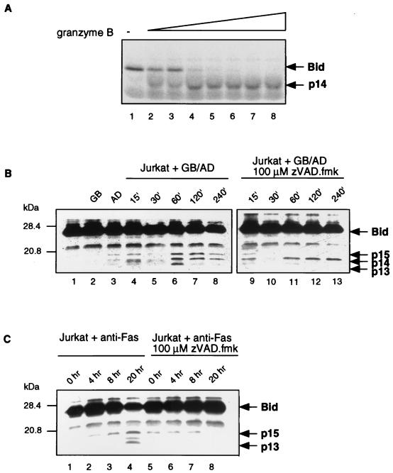
Cytotoxic T lymphocytes (CTL) can trigger an apoptotic signal through the Fas receptor or by the exocytosis of granzyme B and perforin. Caspase activation is an important component of both pathways. Granzyme B, a serine proteinase contained in granules, has been shown to proteolytically process and activate members of the caspase family in vitro. In order to gain an understanding of the contributions of caspases 8 and 3 during granule-induced apoptosis in intact cells, we have used target cells that either stably express the rabbitpox virus-encoded caspase inhibitor SPI-2 or are devoid of caspase 3. The overexpression of SPI-2 in target cells significantly inhibited DNA fragmentation, phosphatidylserine externalization, and mitochondrial disruption during Fas-mediated cell death. In contrast, SPI-2 expression in target cells provided no protection against granzyme-mediated apoptosis, mitochondrial collapse, or cytolysis, leading us to conclude that SPI-2-inhibited caspases are not an essential requirement for the granzyme pathway. Caspase 3-deficient MCF-7 cells were found to be resistant to CTL-mediated DNA fragmentation but not to CTL-mediated cytolysis and loss of the mitochondrial inner membrane potential. Furthermore, we demonstrate that granzyme B directly cleaves the proapoptotic molecule Bid, bypassing the need for caspase 8 activation of Bid. These results provide evidence for a two-pronged strategy for mediating target cell destruction and provide evidence of a direct link between granzyme B activity, Bid cleavage, and caspase 3 activation in whole cells.


Figures

FIG. 1
FIG. 1
SPI-2 expression inhibits Fas-mediated DNA fragmentation and caspase 3 activation. (A) Immunoblot analysis of SPI-2 expression in stably transfected Jurkat cells. Jurkat cells transfected with SPI-2 BMGneo [JSPI-2(4) and JSPI-2(5)] (lanes 1 and 2) express SPI-2, whereas cells transfected with the empty vector (Jneo) (lane 3) do not. (B) Jurkat cells were treated with 250 ng of anti-Fas antibody per ml for 8 h, and DNA fragmentation was monitored by TUNEL assay as described in Materials and Methods. Panels: a, untreated Jneo cells; b, Jneo cells treated with anti-Fas; c, Jneo cells treated with anti-Fas in the presence of 100 μM zIETD-fmk; d, untreated JSPI-2 (clone 4); e, JSPI-2 (clone 4) treated with anti-Fas; f, untreated JSPI-2 (clone 5); g, JSPI-2 (clone 5) treated with anti-Fas. Representative data from three experiments are shown. (C) Caspase 3 activation in untreated Jneo cells (lane 1), Jneo cells treated with anti-Fas antibody for 8 h (lane 2), untreated JSPI-2 (clone 4) cells (lane 3), JSPI-2 (clone 4) cells treated with anti-Fas (lane 4), untreated JSPI-2 (clone 5) cells (lane 5), and JSPI-2 (clone 5) cells treated with anti-Fas (lane 6) was monitored by Western blotting.
FIG. 1
FIG. 1
SPI-2 expression inhibits Fas-mediated DNA fragmentation and caspase 3 activation. (A) Immunoblot analysis of SPI-2 expression in stably transfected Jurkat cells. Jurkat cells transfected with SPI-2 BMGneo [JSPI-2(4) and JSPI-2(5)] (lanes 1 and 2) express SPI-2, whereas cells transfected with the empty vector (Jneo) (lane 3) do not. (B) Jurkat cells were treated with 250 ng of anti-Fas antibody per ml for 8 h, and DNA fragmentation was monitored by TUNEL assay as described in Materials and Methods. Panels: a, untreated Jneo cells; b, Jneo cells treated with anti-Fas; c, Jneo cells treated with anti-Fas in the presence of 100 μM zIETD-fmk; d, untreated JSPI-2 (clone 4); e, JSPI-2 (clone 4) treated with anti-Fas; f, untreated JSPI-2 (clone 5); g, JSPI-2 (clone 5) treated with anti-Fas. Representative data from three experiments are shown. (C) Caspase 3 activation in untreated Jneo cells (lane 1), Jneo cells treated with anti-Fas antibody for 8 h (lane 2), untreated JSPI-2 (clone 4) cells (lane 3), JSPI-2 (clone 4) cells treated with anti-Fas (lane 4), untreated JSPI-2 (clone 5) cells (lane 5), and JSPI-2 (clone 5) cells treated with anti-Fas (lane 6) was monitored by Western blotting.
FIG. 2
FIG. 2
Caspase 8 is processed in target cells following the addition of whole CTL or anti-Fas antibody. (A) Jurkat cells were incubated with whole hCTL at an effector-to-target ratio of 2:1. Cell lysates were generated at the times indicated and immunoblotted for caspase 8 activation. (B) Jurkat cells were treated with 250 ng of anti-Fas antibody per ml for 0, 4, 8, and 20 h, and caspase 8 activation was assessed by Western blotting analysis. (C) Jurkat cells were incubated with whole hCTL at an effector-to-target ratio of 2:1. Cell lysates were generated at the times indicated and immunoblotted for caspase 3 activation.
FIG. 3
FIG. 3
SPI-2 expression inhibits phosphatidylserine exposure in response to anti-Fas but not in response to granzyme B. Phosphatidylserine exposure was quantitated by annexin V-fluorescein isothiocyanate binding following treatment with 250 ng of anti-Fas per ml or treatment with granzyme B (1 μg/ml) and adenovirus (10 PFU/cell). (a) Untreated Jneo cells; (b) Jneo cells treated with anti-Fas; (c) Jneo cells treated with granzyme B and adenovirus; (d) untreated JSPI-2 (clone 4); (e) JSPI-2 (clone 4) cells treated with anti-Fas; (f) JSPI-2 (clone 4) cells treated with granzyme B and adenovirus; (g) untreated JSPI-2 (clone 5); (h) JSPI-2 (clone 5) cells treated with anti-Fas; (i) JSPI-2 (clone 5) cells treated with granzyme B and adenovirus. Representative data from three experiments are shown.
FIG. 4
FIG. 4
SPI-2 expression in target cells does not inhibit granzyme B-induced DNA fragmentation. Jurkat cells transfected with the empty vector or expressing SPI-2 were treated with purified granzyme B (1 μg/ml) and adenovirus (10 PFU/cell), and DNA fragmentation was assessed by the TUNEL protocol. (a) Untreated Jneo cells; (b) Jneo cells treated with granzyme B and adenovirus; (c) Jneo cells treated with granzyme B alone; (d) Jneo cells treated with adenovirus alone; (e) untreated JSPI-2 (clone 4) cells; (f) JSPI-2 (clone 4) cells treated with granzyme B and adenovirus; (g) untreated JSPI-2 (clone 5) cells; (h) JSPI-2 (clone 5) cells treated with granzyme B and adenovirus. Representative data from three experiments are shown.
FIG. 5
FIG. 5
SPI-2 expression does not provide protection against whole CTL-mediated DNA fragmentation or membrane damage. Labeled target cells were incubated with whole CTL at a range of effector-to-target ratios. (A) 51Cr release was measured after 4 h. (B) [3H]thymidine release was measured after 2 h. The means and standard deviations from triplicate samples are shown.
FIG. 6
FIG. 6
SPI-2 expression inhibits ΔΨm and ROS production during Fas-mediated cell death but not during granzyme-mediated cell death. Jurkat cells were treated either with anti-Fas or with granzyme B and adenovirus (GB/AD). Mitochondrial transmembrane potential was determined using 40 nM DiOC6(3), and the production of ROS was assessed with 2 μM hydroethidine (HE). (a) Untreated Jneo cells; (b) Jneo cells treated with the membrane uncoupler mClCCP; (c) Jneo cells exposed to anti-Fas for 8 h; (d) Jneo cells treated with purified granzyme B and adenovirus for 2 h; (e) untreated JSPI-2 (clone 4) cells; (f) JSPI-2 (clone 4) cells treated with the membrane uncoupler mClCCP; (g) JSPI-2 (clone 4) cells exposed to anti-Fas for 8 h; (h) JSPI-2 (clone 4) cells treated with purified granzyme B and adenovirus for 2 h; (i) untreated JSPI-2 (clone 5) cells; (j) JSPI-2 (clone 5) cells treated with the membrane uncoupler mClCCP; (k) JSPI-2 (clone 5) cells exposed to anti-Fas for 8 h; (l) JSPI-2 (clone 5) cells treated with purified granzyme B and adenovirus for 2 h. Representative data from three experiments are shown.
FIG. 7
FIG. 7
Caspase 3 is normally activated in SPI-2 expressing cells. Cells were treated with whole CTL at an effector-to-target ratio of 2:1, and caspase 3 processing in both SPI-2-expressing cells (lanes 1 and 3 to 7) and Jneo cells (lanes 8 and 9) was monitored by Western blotting.
FIG. 8
FIG. 8
Caspase 3 activation is a necessary component for granule-mediated DNA fragmentation. MCF-7 cells lacking caspase 3 are unable to undergo DNA fragmentation after treatment with whole CTL but still undergo CTL-mediated membrane damage. (A) MCF-7 cells and Jurkat cells were labeled with [3H]thymidine. Labeled cells were incubated with a range of effectors-to-target ratios in the presence of 2 μg of concanavalin A per ml, and [3H]thymidine release was measured after 2 h. Data from triplicate samples are shown. (B) MCF-7 cells were labeled with 51Cr and incubated with whole CTL at a range of effector-to-target ratios in the presence and absence of 5 mM EGTA. 51Cr release was measured after 4 h. The means and standard deviations from triplicate samples are shown. (C) MCF-7 cells were treated with hCTL at an effector-to-target ratio of 2.5:1. Mitochondrial transmembrane potential was determined using 40 nM DiOC6(3). Representative data from three independent experiments are shown.
FIG. 8
FIG. 8
Caspase 3 activation is a necessary component for granule-mediated DNA fragmentation. MCF-7 cells lacking caspase 3 are unable to undergo DNA fragmentation after treatment with whole CTL but still undergo CTL-mediated membrane damage. (A) MCF-7 cells and Jurkat cells were labeled with [3H]thymidine. Labeled cells were incubated with a range of effectors-to-target ratios in the presence of 2 μg of concanavalin A per ml, and [3H]thymidine release was measured after 2 h. Data from triplicate samples are shown. (B) MCF-7 cells were labeled with 51Cr and incubated with whole CTL at a range of effector-to-target ratios in the presence and absence of 5 mM EGTA. 51Cr release was measured after 4 h. The means and standard deviations from triplicate samples are shown. (C) MCF-7 cells were treated with hCTL at an effector-to-target ratio of 2.5:1. Mitochondrial transmembrane potential was determined using 40 nM DiOC6(3). Representative data from three independent experiments are shown.
FIG. 9
FIG. 9
Granzyme B is responsible for cleaving Bid. (A) Granzyme B cleaves Bid in vitro. Bid was translated in the presence of [35S]methionine, and purified granzyme B was added in increasing amounts (0, 0.001, 0.0025, 0.01, 0.025, 0.1, 0.25, and 1.0 μg). (B) Bid is cleaved in intact cells by granzyme B. Jurkat cells were untreated (lane 1), treated with granzyme B (GB) (lane 2), treated with adenovirus (AD) (lane 3), or treated with adenovirus and granzyme B simultaneously for 15, 30, 60, 120, and 240 min (lanes 4 to 8, respectively). Jurkat cells were pretreated with 100 μM zVAD-fmk prior to the simultaneous treatment with granzyme B and adenovirus for 15, 30, 60, 120, and 240 min (lanes 9 to 13, respectively). (C) Fas activation results in Bid cleavage. Jurkat cells were untreated (lane 1), treated with anti-Fas antibody for 4, 8, and 20 h (lanes 2 to 4, respectively), or pretreated with 100 μM zVAD-fmk prior to the addition of anti-Fas antibody (lanes 5 to 8).

Similar articles

Cited by

References

    1. Atkinson E A, Barry M, Darmon A J, Shostak I, Turner P C, Moyer R W, Bleackley R C. Cytotoxic T lymphocyte-assisted suicide. J Biol Chem. 1998;273:21261–21266. - PubMed
    1. Atkinson E A, Bleackley R C. Mechanisms of lysis by cytotoxic T cells. Crit Rev Immunol. 1995;15:359–384. - PubMed
    1. Boldin M P, Goncharov T M, Goltsev Y V, Wallach D. Involvement of MACH, a novel MORT1/FADD-interacting protease, in Fas/APO-1 and TNF receptor-induced cell death. Cell. 1996;85:803–815. - PubMed
    1. Caputo A, James M N G, Powers J C, Hudig D, Bleackley R C. Conversion of the substrate specificity of mouse proteinase granzyme B. Nat Struct Biol. 1994;1:364–367. - PubMed
    1. Caputo A, Parrish J C, James M N G, Powers J C, Bleackley R C. Electrostatic reversal of serine proteinase substrate specificity. Proteins. 1999;35:415–424. - PubMed

Publication types

MeSH terms