. 2000 Jun;20(11):3807-16.
doi: 10.1128/MCB.20.11.3807-3816.2000.

Three yeast proteins related to the human candidate tumor suppressor p33(ING1) are associated with histone acetyltransferase activities

Affiliations

Three yeast proteins related to the human candidate tumor suppressor p33(ING1) are associated with histone acetyltransferase activities

R Loewith et al. Mol Cell Biol. 2000 Jun.

Abstract

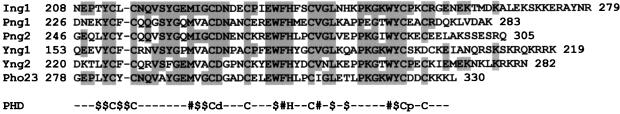
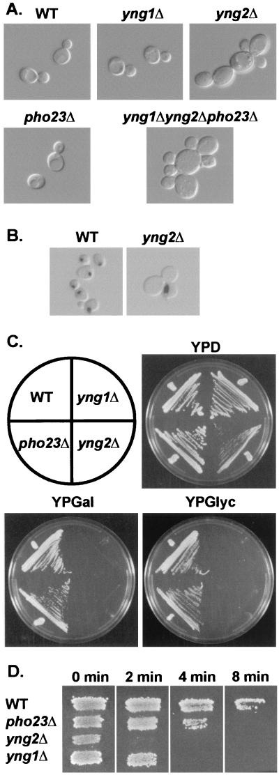
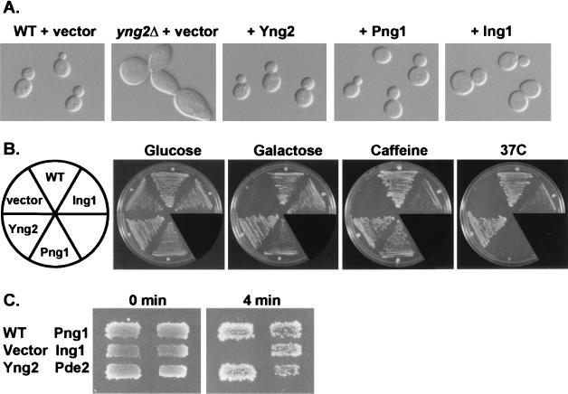
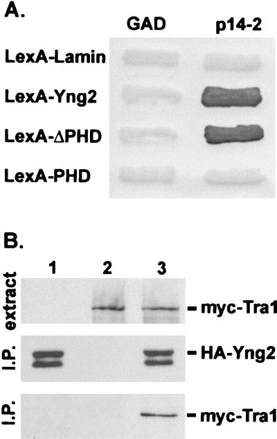
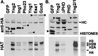
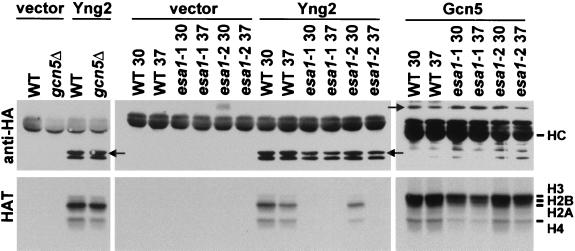
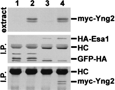
Three Saccharomyces cerevisiae proteins (Yng1/YOR064c, Yng2/YHR090c, and Pho23) and two Schizosaccharomyces pombe proteins (Png1/CAA15917 and Png2/CAA21250) share significant sequence identity with the human candidate tumor suppressor p33(ING1) in their C-terminal regions. The homologous regions contain PHD finger domains which have been implicated in chromatin-mediated transcriptional regulation. We show that GFP-Yng2, like human Ing1, is localized in the nucleus. Deletion of YNG2 results in several phenotypes, including an abnormal multibudded morphology, an inability to utilize nonfermentable carbon sources, heat shock sensitivity, slow growth, temperature sensitivity, and sensitivity to caffeine. These phenotypes are suppressed by expression of either human Ing1 or S. pombe Png1, suggesting that the yeast and human proteins are functionally conserved. Yng1- and Pho23-deficient cells also share some of these phenotypes. We demonstrated by yeast two-hybrid and coimmunoprecipitation tests that Yng2 interacts with Tra1, a component of histone acetyltransferase (HAT) complexes. We further demonstrated by coimmunoprecipitation that HA-Yng1, HA-Yng2, HA-Pho23, and HA-Ing1 are associated with HAT activities in yeast. Genetic and biochemical evidence indicate that the Yng2-associated HAT is Esa1, suggesting that Yng2 is a component of the NuA4 HAT complex. These studies suggest that the yeast Ing1-related proteins are involved in chromatin remodeling. They further suggest that these functions may be conserved in mammals and provide a possible mechanism for the human Ing1 candidate tumor suppressor.


Figures

FIG. 1
FIG. 1
Alignment of PHD finger domains. The C-terminal ends of human Ing1 (AAC00501), S. pombe Png1 (CAA15917) and Png2 (CAA21250), and S. cerevisiae Yng1 (P50947), Yng2 (P38806), and Pho23 (S66947) were aligned to fit the PHD finger consensus sequence (1). The symbols $ and # indicate semiconserved and highly conserved hydrophobic residues, respectively. Regions of protein sequence identity with Ing1 are shaded. Numbers at the right of each sequence indicate the length of each protein.
FIG. 2
FIG. 2
Deletion of YNG1, YNG2, and PHO23 result in various phenotypes. (A) Normal wild-type (WT) (JC1), yng1Δ (RL3-32), yng2Δ (RL3-53), pho23Δ (RL3-17), and triple mutant yngyngpho23Δ (RL5-36) cells were grown in yeast extract-peptone-dextrose (YPD) at 30°C and examined by differential interference contrast (DIC) microscopy. (B) Localization of DNA in normal WT (JC1) and yng2Δ cells (RL3-53) cells grown in YPD was examined by DAPI staining (Materials and Methods). DIC and fluorescent images were overlaid to produce the images shown. (C) The carbon source requirements of normal WT (JC1), yng1Δ (RL3-32), yng2Δ (RL3-53), and pho23Δ (RL3-17) cells were determined by growth on either glucose (YPD), galactose (YPGal), or glycerol (YPGlyc) as the sole carbon source at 30°C for 3 to 5 days. (D) Normal WT (JC1), pho23Δ (RL3-17), yng2Δ (RL3-53), and yng1Δ (RL3-32) were tested for heat shock sensitivity by replica plating onto prewarmed YPD plates and incubated at 55°C for 0, 2, 4, or 8 min. Plates were then allowed to cool to room temperature before incubation at 30°C for 3 days.
FIG. 3
FIG. 3
Expression of human Ing1 or S. pombe Png1 rescues yng2 null phenotypes. (A) Normal wild-type (WT) (JC1) or yng2Δ (RL3-53) cells harboring a control vector, or yng2Δ cells expressing either Yng2, Png1, or Ing1 were grown at 30°C in SC-L synthetic medium and examined by differential interference contrast microscopy. Plasmids used to express proteins were pAD4.H (+vector), pADHA-Yng2 (+Yng2), pADHA-Png1 (+Png1), and pADHA-Ing1 (+Ing1). (B) These strains were also tested for growth under different conditions. Cells were grown on synthetic complete (SC) medium (Glucose) at 30°C, SC containing galactose (Galactose) instead of glucose as the sole carbon source at 30°C, SC containing 3 mM caffeine (Caffeine) at 30°C, or SC at 37°C (37C). (C) These strains and yng2Δ (RL3-53) overexpressing PDE2 (pYepPDE2) were also tested for hypersensitivity to heat shock at 53°C for 0 or 4 min (see Fig. 2 legend).
FIG. 4
FIG. 4
Yng2 is localized in the nucleus. Normal JC1 cells expressing GFP (left panels) or GFP-Yng2 (right panels) were grown in HC medium, harvested, and briefly fixed before being mounted in medium containing DAPI (Materials and Methods). Cells were visualized by differential interference contrast microscopy (DIC [top row]), DNA was visualized using UV optics (DAPI [middle row]), and GFP localization was visualized using fluorescein isothiocyanate optics (GFP [bottom row]). Images in each column are of the same field of cells. Plasmids used to express proteins were pADGFPHA (GFP) and pADGFPHA-Yng2 (GFP-Yng2).
FIG. 5
FIG. 5
Yng2 is associated with Tra1. (A) The Gal activation domain (GAD) and a GAD fusion with Tra1 (residues 1678 to 1740) (p14-2) were tested for their ability to interact with LexA fusions with lamin, Yng2, the N-terminal domain (residues 1 to 222) of Yng2 (LexA-ΔPHD), or the C-terminal domain (residues 222 to 282) of Yng2 (LexA-PHD) by the yeast two-hybrid test (Materials and Methods). Each patch represents an independent transformant of the yeast two-hybrid tester strain L40 expressing the indicated proteins. Interaction between fusion proteins was assayed by their ability to induce expression of β-galactosidase by a filter assay (10). (B) Extracts from JC1 cells expressing HA-Yng2 (lane 1), myc-Tra1 (lane 2), or HA-Yng2 and myc-Tra1 (lane 3) were assayed for expression of myc-tagged proteins by Western blot analysis using anti-myc (monoclonal antibody 9E10) antibody (top panel). Ten milligrams of total protein from each extract was immunoprecipitated (I.P.) with anti-HA (monoclonal antibody 12CA5) antibody; half of the immunoprecipitate was probed with anti-HA antibody (middle panel), and half was probed with anti-myc antibody (bottom panel). Plasmids used to express proteins were pADHA-Yng2 and pUAD6 (lane 1), pAD4.H and pADmyc-Tra1 (lane 2), or pADHA-Yng2 and pADmyc-Tra1 (lane 3).
FIG. 6
FIG. 6
Yng1, Yng2, and Pho23 are associated with HAT activities. (A) Extracts from JC1 cells expressing GFP-HA (GFP), HA-Yng2 (Yng2), HA-Ing1 (Ing1), HA-Pho23 (Pho23), HA-Yng1 (Yng1), GFP-HA-Esa1 (Esa1), or GFP-HA-Gcn5 (Gcn5) were immunoprecipitated with anti-HA (12CA5) antibody. Immunoprecipitates were split, and half of each sample was examined by Western blot analysis using anti-HA antibody (top), and half was assayed for HAT activity (bottom panels) (see Materials and Methods). The left lanes (GFP, Yng2, Ing1, Pho23, and Yng1) and right lanes (Gcn5 and Esa1) of the HAT assay were run on the same gel, but they were exposed to film for 15 and 3 days, respectively. (B) Extracts from JC1 cells expressing GFP-HA (GFP), or GFP-HA fusions with either Yng2 (Yng2), the N-terminal domain (residues 1 to 222) of Yng2 (ΔPHD), the C-terminal domain (residues 222 to 282) of Yng2 (PHD), or Yng2/1 (Yng2 residues 1 to 222 fused to Yng1 residues 155 to 219) were immunoprecipitated with anti-HA antibody. Immunoprecipitates were examined by a Western blot using anti-HA antibody (top), or HAT assays (bottom). Plasmids used to express proteins were pADGFPHA (GFP), pADHA-Yng2 (Yng2), pADHA-Ing1 (Ing1), pADHA-Pho23 (Pho23), pADHA-Yng1 (Yng1), pADGFPHA-Esa1 (Esa1), and pADGFPHA-Gcn5 (Gcn5) (in panel A) and pADGFPHA (GFP), pADGFPHA-Yng2 (Yng2), pADGFPHA-Yng2ΔPHD (ΔPHD), pADGFPHA-Yng2PHD (PHD), and pADGFPHA-Yng2/1 (Yng2/1) (in panel B). Arrows denote migration of relevant proteins. HC denotes antibody heavy chain. The panel on the lower right shows a Coomassie-stained lane from the HAT gel and the migration of histones H3, H2B, H2A, and H4.
FIG. 7
FIG. 7
yng2Δ and esa1(Ts) cells exhibit similar H4 acetylation deficiency. Whole-cell extracts from wild-type strain FY105 (WT), gcn5Δ, wild-type strain LPY3498 grown at 30°C (WT 30) or 37°C (WT 37), esa1(Ts) strain LPY3291 grown at 30°C (esa1 30) or 37°C (esa1 37), wild-type strain JC1, yng1Δ, yng2Δ, and pho23Δ cells were separated on an SDS–18% polyacrylamide gel and analyzed by Western blotting using antibodies (Upstate Biochemical) that specifically recognize acetylated H3 (H3), acetylated lysine (H4; histone H4 region of gel shown), and H4 acetylated at lysine residues 12 (H4K12), 8 (H4K8), and 5 (H4K5), or stained with Coomassie brilliant blue (total protein).
FIG. 8
FIG. 8
Yng2-associated HAT activity is deficient in esa1(Ts) cells. Extracts from cells containing a control plasmid pAD4.H (vector), or expressing HA-Yng2 (Yng2) or GFP-HA-Gcn5 (Gcn5) were immunoprecipitated with anti-HA. Immunoprecipitates were split, and half of each sample was examined by Western blot analysis using anti-HA antibody (top), and half was assayed for HAT activity (bottom). Strains used include FY105 (WT), gcn5Δ, LPY3498 grown at 30°C (WT 30) or 37°C (WT 37), LPY3291 grown at 30°C (esa1-1 30) or 37°C (esa1-1 37), and LPY3500 grown at 30°C (esa1-2 30) or 37°C (esa1-2 37).
FIG. 9
FIG. 9
Yng2 and Esa1 form a complex in vivo. Extracts from JC1 cells expressing GFP-HA (lane 1), GFP-HA and myc-Yng2 (lane 2), GFP-HA-Esa1 (HA-Esa1) (lane 3), or GFP-HA-Esa1 and myc-Yng2 (lane 4) were assayed for expression of myc-tagged proteins by Western blot analysis using anti-myc (mAb 9E10) antibody (top panel). 10 mg of total protein from each extract was immunoprecipitated with anti-HA (monoclonal antibody 12CA5) antibody; half of the immunoprecipitate was probed with anti-HA antibody (middle panel), and half was probed with anti-myc antibody (bottom panel). myc-Yng2 was only precipitated when coexpressed with GFP-HA-Esa1. Plasmids used to express proteins were pADGFPHA and pUAD6 (lane 1), pADGFPHA and pADmyc-Yng2 (lane 2), pADGFPHA-Esa1 and pUAD6 (lane 3), or pADGFPHA-Esa1 and pADmyc-Yng2 (lane 4).

Similar articles

Cited by

References

    1. Aasland R, Gibson T J, Stewart A F. The PHD finger: implications for chromatin-mediated transcriptional regulation. Trends Biochem Sci. 1995;20:56–59. - PubMed
    1. Adams A, Gottschling D E, Kaiser C A, Stearns T. Methods in yeast genetics, 1997 ed. Plainview, N.Y: Cold Spring Harbor Laboratory Press; 1998.
    1. Allard S, Utley R T, Savard J, Clarke A, Grant P, Brandl C J, Pillus L, Workman J L, Cote J. NuA4, an essential transcription adaptor/histone H4 acetyltransferase complex containing Esa1p and the ATM-related cofactor Tra1p. EMBO J. 1999;18:5108–5119. - PMC - PubMed
    1. Archer S Y, Hodin R A. Histone acetylation and cancer. Curr Opin Genet Dev. 1999;9:171–174. - PubMed
    1. Bannister A J, Kouzarides T. The CBP co-activator is a histone acetyltransferase. Nature. 1996;384:641–643. - PubMed

Publication types

MeSH terms