. 2000 Jun;20(11):3817-30.
doi: 10.1128/MCB.20.11.3817-3830.2000.

Class II phosphoinositide 3-kinases are downstream targets of activated polypeptide growth factor receptors

Affiliations

Class II phosphoinositide 3-kinases are downstream targets of activated polypeptide growth factor receptors

A Arcaro et al. Mol Cell Biol. 2000 Jun.

Abstract

The class II phosphoinositide 3-kinases (PI3K) PI3K-C2alpha and PI3K-C2beta are two recently identified members of the large PI3K family. Both enzymes are characterized by the presence of a C2 domain at the carboxy terminus and, in vitro, preferentially utilize phosphatidylinositol and phosphatidylinositol 4-monophosphate as lipid substrates. Little is understood about how the catalytic activity of either enzyme is regulated in vivo. In this study, we demonstrate that PI3K-C2alpha and PI3K-C2beta represent two downstream targets of the activated epidermal growth factor (EGF) receptor in human carcinoma-derived A431 cells. Stimulation of quiescent cultures with EGF resulted in the rapid recruitment of both enzymes to a phosphotyrosine signaling complex that contained the EGF receptor and Erb-B2. Ligand addition also induced the appearance of a second, more slowly migrating band of PI3K-C2alpha and PI3K-C2beta immunoreactivity on sodium dodecyl sulfate-polyacrylamide gel electrophoresis. Since both PI3K enzymes can utilize Ca(2+) as an essential divalent cation in lipid kinase assays and since the catalytic activity of PI3K-C2alpha is refractory to the inhibitor wortmannin, these properties were used to confirm the recruitment of each PI3K isozyme to the activated EGF receptor complex. To examine this interaction in greater detail, PI3K-C2beta was chosen for further investigation. EGF and platelet-derived growth factor also stimulated the association of PI3K-C2beta with their respective receptors in other cells, including epithelial cells and fibroblasts. The use of EGF receptor mutants and phosphopeptides derived from the EGF receptor and Erb-B2 demonstrated that the interaction with recombinant PI3K-C2beta occurs through E(p)YL/I phosphotyrosine motifs. The N-terminal region of PI3K-C2beta was found to selectively interact with the EGF receptor in vitro, suggesting that it mediates the association of this PI3K with the receptor. However, the mechanism of this interaction remains unclear. We conclude that class II PI3K enzymes may contribute to the generation of 3' phosphoinositides following the activation of polypeptide growth factor receptors in vivo and thus mediate certain aspects of their biological activity.


Figures

FIG. 1
FIG. 1
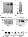
Both PI3K-C2α and PI3K-C2β are immunoprecipitated by antiphosphotyrosine antibody from lysates of EGF-stimulated cultures. (A) Confluent cultures of A431 and HEK293 cells were lysed with Laemmli sample buffer on ice. These lysates were boiled, fractionated by SDS-PAGE, and Western blotted with either anti–PI3K-C2α or anti–PI3K-C2β antiserum. (B to E) Confluent and quiescent cultures of A431 cells were stimulated with EGF (100 nM) for the indicated times. Cells were lysed with buffer containing Triton X-100 at 4°C, and the supernatants were clarified by centrifugation (13,000 × g). Antiphosphotyrosine antibody (PY20) was added (4 h, 4°C), and the resulting immune complexes were collected on protein G-Sepharose beads. Following extraction with sample buffer, proteins were fractionated by SDS-PAGE and Western blotted with either anti–PI3K-C2α antiserum (B), anti–PI3K-C2β antiserum (C), anti-EGFR antibody (R1) (D), or antiphosphotyrosine antibody (PY99) (E). Numbers at left indicate the apparent molecular mass.
FIG. 2
FIG. 2
PI3K-C2α and PI3K-C2β coimmunoprecipitate with EGFR and ErbB-2 following EGF stimulation. Quiescent and confluent cultures of A431 cells were incubated in the absence (−) or presence (+) of 100 nM EGF for 10 min. Clarified lysates were prepared and incubated (4 h, 4°C) with either protein G-Sepharose beads alone (beads) or together with either anti-EGFR (R1), anti–ErbB-2 (Ab2), anti–ErbB-3 (C17), or anti–ErbB-4 (Ab-1) antibody. The resultant immune complexes were washed and, following extraction, associated proteins were fractionated by SDS-PAGE before Western blotting with either anti–PI3K-C2α antiserum (A), anti–PI3K-C2β antiserum (B), or antiphosphotyrosine antibody (PY99) (C).
FIG. 3
FIG. 3
Cation specificity of p110α, PI3K-C2α, and PI3K-C2β and their sensitivity to inhibition by wortmannin. (A and B) Recombinant p85α-p110α, PI3K-C2α, and PI3K-C2β were assayed for lipid kinase activity using either PtdIns (A) or PtdIns(4)P (B) in the absence (−) or presence of either Ca2+, Mg2+, or Mn2+. Reaction products were extracted, fractionated by TLC, and examined by autoradiography. (C) p85α-p110α, PI3K-C2α, and PI3K-C2β were also examined for their ability to phosphorylate PtdIns in the presence of either Ca2+ or Mg2+ as the divalent cation and in the absence (−) or presence (+) of wortmannin (50 nM).
FIG. 4
FIG. 4
Ca2+-dependent PI3K activity associates with the EGFR following ligand addition. Confluent and quiescent A431 cultures were stimulated with EGF (100 nM) for the times indicated, and lysates were prepared. These were incubated with anti-EGFR antibody (R1) for 4 h at 4°C, and the immune complexes were collected with protein G-Sepharose beads. After washing, these immunoprecipitates were used for lipid kinase assays with PtdIns in the presence of Ca2+ (top panel) or Ca2+ and 50 nM wortmannin (bottom panel). Reactions were terminated, and radiolabeled phospholipids were extracted, separated by TLC, and examined by autoradiography. For reference, a mixture of PtdIns(3)P and PtdIns(4)P was also separated and served as a control (arrows). The slight shift in mobility observed is an artifact of the TLC run.
FIG. 5
FIG. 5
Differential regulation of PI3K-C2β and p85α by EGF and insulin. (A and B) HEK293 cells were stimulated with EGF (100 nM) or insulin (Ins) (1 μg/ml) for 5 min. Lysates were incubated with antiphosphotyrosine antibody (α-PY), and the resulting immunoprecipitates were fractionated by SDS-PAGE. Proteins were transferred to polyvinylidene difluoride membranes and Western blotted with either anti–PI3K-C2β antiserum (A) or anti-p85α antibody (B). (C and D) Cos 7 cells were also stimulated with either EGF or insulin, lysed, and incubated with either antiphosphotyrosine antibody (α-PY), anti–PI3K-C2β antiserum, or anti-p85α antibody. After SDS-PAGE, the proteins were Western blotted with either anti-PI3K-C2β antiserum (C) or anti-p85α antibody (D). The positions of PI3K-C2β (p180) and p85α are indicated. MW, molecular weight (in thousands).
FIG. 6
FIG. 6
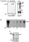
PI3K-C2β is present in antiphosphotyrosine antibody immunoprecipitates of PDGF-stimulated fibroblasts. (A and B) NIH 3T3 cells were stimulated with PDGF (10 nM), and lysates were immunoprecipitated with mouse immunoglobulin antibody (Ctr), antiphosphotyrosine antibody (α-PY), or anti–PI3K-C2β antiserum. The samples were assayed for PI3K in the presence of Ca2+, and the radioactive phosphoinositide products were separated by TLC (A). Antiphosphotyrosine antibody immunoprecipitates from quiescent or PDGF-stimulated cultures were also fractionated by SDS-PAGE and Western blotted with anti–PI3K-C2β antiserum (B). (C and D) Cultures of cells stably expressing epitope-tagged PI3K-C2β were stimulated with PDGF for various times as indicated, and their lysates were immunoprecipitated with anti-Glu tag monoclonal antibody. After SDS-PAGE, immunoprecipitated proteins were Western blotted with antiphosphotyrosine antibody (C), stripped, and reprobed with anti–PI3K-C2β antiserum (D). (E) Lysates from NIH 3T3 cells stimulated with PDGF were also immunoprecipitated with anti-Glu tag antibody, antiphosphotyrosine antibody, or anti-PDGFR antibody. The resultant immune complexes were analyzed for phospholipid kinase activity in the presence of Ca2+. Radiolabeled phosphoinositide products were separated by TLC. MW, molecular weight.
FIG. 7
FIG. 7
Interaction of PI3K-C2β with the EGFR and PDGFR in vitro. (A) Immobilized EGFR immunoprecipitated from A431 cells (R1) was autophosphorylated in vitro (R1P) and incubated with a cell lysate from HEK293 cells that had been transfected with either Glu-tagged PI3K-C2β or an empty expression vector. Lysates from transfected cells were also immunoprecipitated with anti-Glu tag antibodies (α-Glu) or mouse immunoglobulins (contr). Each affinity complex was fractionated by SDS-PAGE and Western blotted with anti–PI3K-C2β antiserum. Aliquots of unphosphorylated (R1) or autophosphorylated (R1P) EGFR were also Western blotted in parallel with monoclonal antiphosphotyrosine antibody. (B) Recombinant epitope-tagged PI3K-C2β was purified from infected Sf9 cells and incubated with anti–PI3K-C2β antiserum (α-C2β), Actigel beads (Act), Actigel beads coupled to phosphotyrosine (Act PY), or immobilized purified EGFR (R1) that had been autophosphorylated in vitro (R1P). The samples were assayed for phospholipid kinase activity with PtdIns in the presence of Ca2+. The radiolabeled phospholipid products were analyzed by TLC. (C) Purified epitope-tagged PI3K-C2β was also incubated with either anti-Glu tag antibody–protein G (control) (ctr), immobilized PDGFR immunoprecipitated from resting NIH 3T3 cells (R), or receptor that had been autophosphorylated in vitro (RP). The samples were fractionated by SDS-PAGE and Western blotted with anti–PI3K-C2β antiserum. The position of PI3K-C2β (p180) is shown. MW, molecular weight.
FIG. 8
FIG. 8
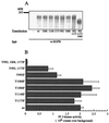
Interactions of EGFR mutants with PI3K-C2β. (A) A mammalian expression vector containing cDNA encoding various human EGFR mutants was transfected into HEK293 cells. Each recombinant EGFR mutant was immunoprecipitated with anti-EGFR antibody and tested for its ability to interact in vitro with recombinant PI3K-C2β isolated from HEK293 cell lysates. Quantification of each mutant receptor present in the immunoprecipitates was done by SDS-PAGE and Coomassie blue staining. The control sample (−) represents EGFR immunoprecipitation from nontransfected cells. (B) The samples were assayed for PI3K activity in the presence of Ca2+. Radiolabeled phospholipid products were analyzed by TLC and quantified by PhosphorImager analysis. Background binding to endogenous EGFR was subtracted from all values, and data are presented as mean ± standard error from six independent experiments. MW, molecular weight; wt, wild type.
FIG. 9
FIG. 9
Phosphopeptides containing an E(p)YL sequence bind PI3K-C2β. (A) Lysates from HEK293 cells transfected with either empty vector (−) or wild-type EGFR (+) were immunoprecipitated with anti-EGFR antibodies (R). The isolated EGFR was autophosphorylated in vitro and incubated with lysates of HEK293 cells transfected with either vector (−) or myc-tagged PI3K-C2β in the absence or presence of phosphopeptides corresponding to the EGFR at residues (p)992, (p)Y1068, and (p)Y1148. The receptors were isolated, and the associated proteins were fractionated by SDS-PAGE and Western blotted with anti–PI3K-C2β antiserum. p180 represents PI3K-C2β. (B) Immobilized peptides corresponding to ErbB-2 Y1196, (p)Y1196, or phosphotyrosine were incubated with purified recombinant PI3K-C2β that had been expressed in Sf9 cells. Following incubation, each sample was washed and assayed for PI3K activity in the presence of Ca2+. Radiolabeled phospholipid products were fractionated by TLC, and the spots were quantified by PhosphorImager analysis. Data are presented as mean ± standard error from four independent experiments.
FIG. 10
FIG. 10
The N-terminal region of PI3K-C2β interacts with the EGFR. (A) Purified recombinant domains corresponding to the PI3K-C2β N terminus (NT), C2 domain (C2), or putative PTB domain were produced in E. coli as GST fusion proteins. The immobilized domains and GST were incubated with lysates from quiescent or EGF-stimulated A431 cells. The fusion proteins were isolated, and the associated proteins were fractionated by SDS-PAGE and Western blotted with anti-EGFR antiserum. (B) Recombinant EGFR was purified from transfected HEK293 cells by immunoprecipitation with immobilized anti-EGFR antibody and autophosphorylated in vitro (R). The receptor was then incubated with either soluble PI3K-C2β N-terminal fragment (NT) or buffer alone. Samples were fractionated by SDS-PAGE and Western blotted with antiserum directed against the PI3K-C2β N terminus. Protein G (−) and immobilized anti-EGFR antibody alone (Ig) served as controls and were assayed in parallel. (C) Purified soluble GST or purified soluble PI3K-C2β N-terminal fragment (NT) was added to the EGFR PI3K-C2β association assay described in the legend to Fig. 9A. The resultant blots were probed with anti–PI3K-C2β antiserum.

Similar articles

Cited by

References

    1. Alessi D R, James S R, Downes C P, Holmes A B, Gaffney P R J, Reese C B, Cohen P. Characterization of a 3-phosphoinositide-dependent protein kinase which phosphorylates and activates protein kinase Bα. Curr Biol. 1997;7:261–269. - PubMed
    1. Arcaro A, Volinia S, Zvelebil M J, Stein R, Watton S J, Layton M J, Gout I, Ahmadi K, Downward J, Waterfield M D. Human PI3-kinase C2β—the role of calcium and the C2 domain in enzyme activity. J Biol Chem. 1998;273:33082–33091. - PubMed
    1. Arcaro A, Wymann M P. Wortmannin is a potent phosphatidylinositol 3-kinase inhibitor: the role of phosphatidylinositol 3,4,5-trisphosphate in neutrophil responses. Biochem J. 1993;296:297–301. - PMC - PubMed
    1. Bondeva T, Pirola L, Bulgarelli-Leva G, Rubio I, Wetzker R, Wymann M. Bifurcation of lipid and protein kinase signals of PI3Kγ to the protein kinases PBK and MAPK. Science. 1998;282:293–296. - PubMed
    1. Brown R A, Domin J, Arcaro A, Waterfield M D, Shepherd P R. Insulin activates the alpha isoform of class II phosphoinositide 3-kinase. J Biol Chem. 1999;274:14529–14532. - PubMed

Publication types

MeSH terms