. 2000 Jun;20(11):3887-95.
doi: 10.1128/MCB.20.11.3887-3895.2000.

Rck2 kinase is a substrate for the osmotic stress-activated mitogen-activated protein kinase Hog1

Affiliations

Rck2 kinase is a substrate for the osmotic stress-activated mitogen-activated protein kinase Hog1

E Bilsland-Marchesan et al. Mol Cell Biol. 2000 Jun.

Abstract

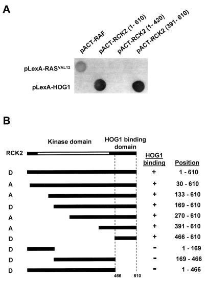
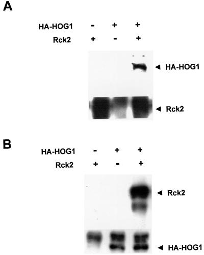
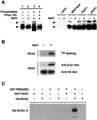
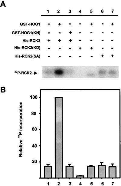
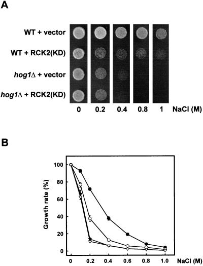
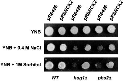
Exposure of yeast cells to increases in extracellular osmolarity activates the Hog1 mitogen-activated protein kinase (MAPK). Activation of Hog1 MAPK results in induction of a set of osmoadaptive responses, which allow cells to survive in high-osmolarity environments. Little is known about how the MAPK activation results in induction of these responses, mainly because no direct substrates for Hog1 have been reported. We conducted a two-hybrid screening using Hog1 as a bait to identify substrates for the MAPK, and the Rck2 protein kinase was identified as an interactor for Hog1. Both two-hybrid analyses and coprecipitation assays demonstrated that Hog1 binds strongly to the C-terminal region of Rck2. Upon osmotic stress, Rck2 was phosphorylated in vivo in a Hog1-dependent manner. Furthermore, purified Hog1 was able to phosphorylate Rck2 when activated both in vivo and in vitro. Rck2 phosphorylation occurred specifically at Ser519, a residue located within the C-terminal putative autoinhibitory domain. Interestingly, phosphorylation at Ser519 by Hog1 resulted in an increase of Rck2 kinase activity. Overexpression of Rck2 partially suppressed the osmosensitive phenotype of hog1Delta and pbs2Delta cells, suggesting that Rck2 is acting downstream of Hog1. Consistently, growth arrest caused by hyperactivation of the Hog1 MAPK was abolished by deletion of the RCK2 gene. Furthermore, overexpression of a catalytically impaired (presumably dominant inhibitory) Rck2 kinase resulted in a decrease of osmotolerance in wild-type cells but not in hog1Delta cells. Taken together, our data suggest that Rck2 acts downstream of Hog1, controlling a subset of the responses induced by the MAPK upon osmotic stress.


Figures

FIG. 1
FIG. 1
Identification by two-hybrid analysis of the HOG1 binding domain in Rck2. (A) Interactions of various Rck2 fragments fused to the GAL4 activator domain with the full-length Hog1 fused to the LexA DB were examined. Representative filter β-galactosidase assays demonstrating interactions between Hog1 and Rck2 are shown. Positions of the Rck2 fragments included in the constructs are indicated in parentheses. Proteins encoded by the control plasmids pLexA-RASV12 and pACT-RAF, which are known to interact with each other (37), are shown for comparison. (B) Summary of the two-hybrid interaction analysis between Rck2 and Hog1. The Rck2 segments included in the AD (denoted as A) or DB (denoted as D) constructs are schematically shown on the left, with their precise amino acid positions indicated on the right. The presence or absence of interaction is depicted by a plus or minus sign, respectively.
FIG. 2
FIG. 2
In vivo association of Hog1 and Rck2 proteins. rck2Δ yeast strain EBΔR2Wa was transformed with a plasmid expressing HA-HOG1 and a multicopy plasmid carrying RCK2. (A) Hog1 coprecipitates with Rck2. Rck2 was immunoprecipitated using specific antibodies against this kinase (lower panel), and the presence of HA-HOG1 in the precipitates was determined with an anti-HA antibody (upper panel) (B) Rck2 coprecipitates with Hog1. HA-HOG1 was immunoprecipitated using anti-HA antibody (lower panel), and the presence of Rck2 was determined with specific antibodies against Rck2 (upper panel).
FIG. 3
FIG. 3
In vivo and in vitro phosphorylation of Rck2 by Hog1. (A) Dependence of the HOG pathway for the in vivo osmotic stress-induced phosphorylation of Rck2. Wild-type cells were grown in sc medium and subjected (+) or not (−) to a brief osmotic shock (0.4 M NaCl, 10 min). Cell extracts were prepared and treated with (+) or without (−) λ phosphatase in the presence (+) or absence (−) of a mixture of phosphatase inhibitors as described in Materials and Methods (left panel). Rck2 was detected by immunoblotting using anti-Rck2 antibodies (arrowheads), and its phosphorylation state was monitored by noting changes in the electrophoretic mobility of the protein. Results from several strains subjected (+) or not (−) to brief osmotic stress (0.4 M NaCl, 5 min) are shown in the right panel. Relevant genotypes are depicted. Extracts from rck2Δ cells are included in order to monitor the specificity of the anti-Rck2 antibodies. (B) In vivo-activated Hog1 phosphorylates Rck2. HA-HOG1 was immunoprecipitated by using anti-HA monoclonal antibody from yeast cells before (−) or after (+) the addition of NaCl to a final concentration of 0.4 M. The presence of HA-HOG1 in the precipitates was detected with an anti-HA antibody, and Hog1 activation was monitored by immunoblot analysis using a monoclonal antibody specific to phosphotyrosine (4G10) (middle panel). After immunoprecipitation, HA-HOG1 was incubated with purified His-tagged RCK2(KD) in the presence of kinase buffer and radioactive ATP. Phosphorylated proteins were separated by SDS-PAGE and detected by autoradiography. (C) In vitro phosphorylation of Rck2 by Hog1. Recombinant tagged proteins were purified from E. coli as described in Materials and Methods. Hog1 and the constitutively activated Pbs2 allele [PBS2(EE)] were incubated in the presence of kinase buffer and ATP. Rck2 was then added (when indicated) in the presence of radioactive ATP. Phosphorylated proteins were resolved by SDS-PAGE and detected by autoradiography. The position of tagged Rck2 is indicated on the left.
FIG. 4
FIG. 4
Hog1 phosphorylates Ser519 at the regulatory domain of Rck2. (A) Phosphorylation of different fragments of Rck2 by Hog1. Various Rck2 fragments were tested for their ability to be phosphorylated by an in vitro-activated Hog1 (as described in Materials and Methods). After the in vitro kinase assay, phosphorylated proteins were resolved by SDS-PAGE and detected by autoradiography. Positions of the Rck2 fragments included in the constructs are indicated in parentheses. Proteins were His tagged and contained a Lys201→Met mutation which results in catalytically inactive enzymes, to avoid autophosphorylation. (B) Mutation of Rck2 Ser519→Ala abolishes Hog1 phosphorylation. The full-length RCK2(KD) and its Ser519 mutant form were tested for Hog1 phosphorylation as described for panel A. After phosphorylation, proteins were resolved by SDS-PAGE and transferred to a nylon membrane. Phosphorylated proteins were detected by autoradiography (upper panel). His-tagged Rck2 proteins were detected by immunoblotting by using the anti-His monoclonal antibody BMG-His-1 (lower panel).
FIG. 5
FIG. 5
Induction of Rck2 activity by Hog1 phosphorylation. (A) Purified wild-type Rck2 protein or the indicated mutant forms of Rck2 were incubated with (+) or without (−) wild-type GST-HOG1 or the catalytically inactive mutant version GST-HOG1(KN), in the presence of kinase buffer and cold ATP. After 15 min at 30°C, HOG1 was removed by affinity chromatography and Rck2 was further incubated in the presence of kinase buffer and radioactive ATP. Phosphorylated proteins were separated by SDS-PAGE and detected by autoradiography. The position of Rck2 is indicated on the left. A representative experiment is shown. (B) The intensity of autophosphorylation was quantified with a phosphorimager (Fuji BAS1000). Samples from three independent experiments were measured, and the intensity of each band was normalized to that of lane 2. Error bars indicate standard errors of the means.
FIG. 6
FIG. 6
Cellular osmosensitivity induced by overexpression of a catalytically inactive RCK2(KD) allele. (A) Wild-type (WT) and hog1Δ cells transformed with an empty vector (pCM262) or a plasmid expressing the catalytically inactive RCK2(KD) (pCMkdR2) were grown on YPD or YPD containing NaCl at different concentrations, in the absence of doxycycline (allowing full expression from the Tet promoter). Growth in plates was scored after 3 days at 30°C. (B) Wild-type cells transformed with pCM262 (●) or pCMkdR2 (○) and hog1Δ cells transformed with pCM262 (▾) or pCMkdR2 (▿) were grown in liquid medium in the presence of different concentrations of NaCl, and the effect of stress on cell growth was determined as described in Materials and Methods. Results are means ± standard errors of the means from three independent experiments.
FIG. 7
FIG. 7
Suppression of hog1Δ and pbs2Δ cell osmosensitivity by RCK2 overexpression. Yeast cells deficient in the HOG1 or PBS2 gene were transformed with either an empty vector (pRS426) or a multicopy plasmid carrying wild-type (WT) RCK2 (pRSRCK2). Cells were grown in YPD, YPD containing NaCl at a concentration of 0.4 M, or sorbitol at 1 M. Growth was scored after 3 days at 30°C.
FIG. 8
FIG. 8
Deletion of the RCK2 gene suppresses the lethality of the PBS2DD mutation in yeast. The wild-type and rck2Δ yeast strains were transformed with either pGal control vector or pGal:PBS2DD. Yeast cells were grown at 30°C on sc plates containing glucose or galactose. Growth was scored after 4 days.

Similar articles

Cited by

References

    1. Albertyn J, Hohmann S, Thevelein J M, Prior B A. GPD1, which encodes glycerol-3-phosphate dehydrogenase, is essential for growth under osmotic stress in Saccharomyces cerevisiae, and its expression is regulated by the high-osmolarity glycerol response pathway. Mol Cell Biol. 1994;14:4135–4144. - PMC - PubMed
    1. Bartel P L, Chien C, Sternglanz R, Fields S. Using the two-hybrid system to detect protein-protein interactions. In: Hartley D, editor. Cellular interactions in development: a practical approach. Oxford, United Kingdom: Oxford University Press; 1993. pp. 153–179.
    1. Bellí G, Garí E, Piedrafita L, Aldea M, Herrero E. An activator/repressor dual system allows tight tetracycline-regulated gene expression in budding yeast. Nucleic Acids Res. 1998;26:942–947. - PMC - PubMed
    1. Bendixen C, Gangloff S, Rothstein R. A yeast mating-selection scheme for detection of protein-protein interactions. Nucleic Acids Res. 1994;22:1778–1779. - PMC - PubMed
    1. Boguslawski G. PBS2, a yeast gene encoding a putative protein kinase, interacts with the RAS2 pathway and affects osmotic sensitivity of Saccharomyces cerevisiae. J Gen Microbiol. 1992;138:2425–2432. - PubMed

Publication types

MeSH terms

Substances