. 2000 Jun;20(11):3951-64.
doi: 10.1128/MCB.20.11.3951-3964.2000.

p38 and extracellular signal-regulated kinases regulate the myogenic program at multiple steps

Affiliations

p38 and extracellular signal-regulated kinases regulate the myogenic program at multiple steps

Z Wu et al. Mol Cell Biol. 2000 Jun.

Abstract

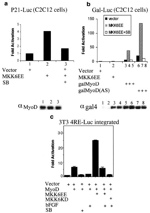
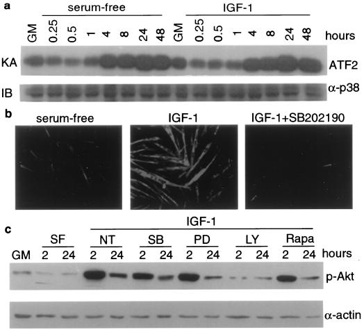
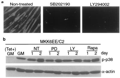
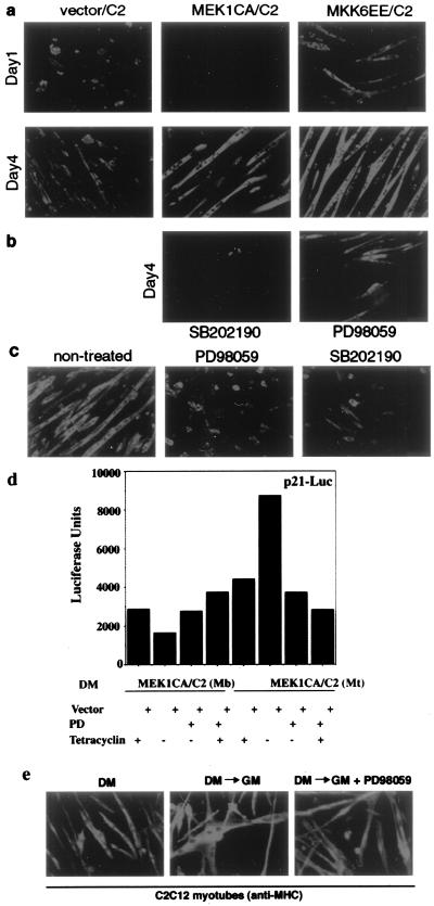
The extracellular signals which regulate the myogenic program are transduced to the nucleus by mitogen-activated protein kinases (MAPKs). We have investigated the role of two MAPKs, p38 and extracellular signal-regulated kinase (ERK), whose activities undergo significant changes during muscle differentiation. p38 is rapidly activated in myocytes induced to differentiate. This activation differs from those triggered by stress and cytokines, because it is not linked to Jun-N-terminal kinase stimulation and is maintained during the whole process of myotube formation. Moreover, p38 activation is independent of a parallel promyogenic pathway stimulated by insulin-like growth factor 1. Inhibition of p38 prevents the differentiation program in myogenic cell lines and human primary myocytes. Conversely, deliberate activation of endogenous p38 stimulates muscle differentiation even in the presence of antimyogenic cues. Much evidence indicates that p38 is an activator of MyoD: (i) p38 kinase activity is required for the expression of MyoD-responsive genes, (ii) enforced induction of p38 stimulates the transcriptional activity of a Gal4-MyoD fusion protein and allows efficient activation of chromatin-integrated reporters by MyoD, and (iii) MyoD-dependent myogenic conversion is reduced in mouse embryonic fibroblasts derived from p38alpha(-/-) embryos. Activation of p38 also enhances the transcriptional activities of myocyte enhancer binding factor 2A (MEF2A) and MEF2C by direct phosphorylation. With MEF2C, selective phosphorylation of one residue (Thr293) is a tissue-specific activating signal in differentiating myocytes. Finally, ERK shows a biphasic activation profile, with peaks of activity in undifferentiated myoblasts and postmitotic myotubes. Importantly, activation of ERK is inhibitory toward myogenic transcription in myoblasts but contributes to the activation of myogenic transcription and regulates postmitotic responses (i.e., hypertrophic growth) in myotubes.


Figures

FIG. 1
FIG. 1
p38 activity increases upon muscle differentiation prior to induction of myogenin. (a) C2C12 myoblasts were cultured in GM until they were 90 to 95% confluent and then shifted to DM. Lysates were prepared from subconfluent cells in GM or cells kept for 1 to 3 days in DM as indicated. p38 was immunoprecipitated and its activity was measured by an immune complex kinase assay (KA) using AFT2 as a substrate. The amount of p38 was assessed by immunoblotting (IB). α-p38, anti-p38 antibody. (b) Equal amounts of whole-cell lysates were separated by SDS-PAGE in duplicate. Activation of p38 in C2C12 cell lysates was determined by immunoblotting with antibodies specific to phosphorylated and activated p38 (α-pP38) or total p38. (c) JNK activity and expression in C2C12 cell lysates were determined by an immune complex kinase assay using c-Jun as a substrate and by immunoblotting, respectively. α-JNK, anti-JNK antibody. (d) Samples (30 μg) of C2C12 extracts were analyzed by immunoblotting with anti-phospho-ERK antibody (α-pERK). The same membrane was stripped and reprobed with antibody against total ERK (α-ERK) to monitor the amounts of loaded proteins. (e) A coculture of undifferentiated C2C12 myoblasts and differentiated myotubes was prepared by mixing a population of myoblasts growing in GM and myoblasts previously cultured in DM for 24 h. This mixed population was cultured in GM for 1 day. Thereafter, cells were fixed and analyzed by immunohistochemistry with antibody specific to pP38. (f) Subconfluent C2C12 cells in GM were switched to DM and were collected at the indicated times (in hours). p38 activity was examined by an immune complex kinase assay, and expression of myogenin was determined by immunoblotting. α-Myog, anti-myogenin antibody.
FIG. 2
FIG. 2
p38 activity is required for myogenic differentiation. (a) The indicated cell lines were transfected with the myogenic reporter 4RE-Luc (together with MyoD in the case of 10T1/2 cells). The indicated concentrations of SB202190 (SB) were added immediately after transfection, and the cells were switched to DM for 36 h, after which they were collected to measure luciferase (Luc.) activity. (b) C2C12 cells in GM were switched to DM for 2 days with (5 μM) or without SB202190. Cell extracts were analyzed by immunoblotting for the presence of myogenic markers. α-Myog, α-p21, α-MHC, α-MyoD, and α-p38, anti-myogenin, anti-p21, anti-MHC, anti-MyoD, and anti-p38 antibodies, respectively. (c) Subconfluent C2C12 cells or primary human myoblasts were placed in DM with (5 μM) or without SB202190. At the indicated times, cells were fixed and immunostained with anti-MHC antibody to visualize differentiated cells. C2C12 cells were examined by immunohistochemistry and bright-field microscopy, and primary human myoblasts were examined by indirect immunofluorescence.
FIG. 3
FIG. 3
Deliberate p38 activation induces myotube formation and expression of muscle differentiation markers. (a) Different muscle-specific reporters (4RE-Luc or MCK-Luc) were cotransfected with expression vectors encoding activated MKKs into either C2C12 or MyoD-expressing 10T1/2 cells. After growing in GM for 1 day, cells were shifted to DM for another 36 h and then harvested to measure luciferase activity. All determinations were done in duplicate, and the data shown are representative of three independent experiments. Fold activation is the ratio of luciferase activity in MKK-transfected cells to cells transfected with the empty vector. SB, SB202190. (b) C2C12 cells in GM were cotransfected with a construct encoding a β-galactosidase protein fused to a nuclear localization signal controlled by the MLC promoter. (see the text) and either an empty vector or a JNKK2CA or MKK6EE expression vector. After 1 day in DM without or with SB202190, the cells were fixed and stained with X-Gal (5-bromo-4-chloro-3-indolyl-β-d-galactopyranoside). In MKK6EE-transfected cells cultured without SB202190, a blue color appeared 12 h earlier than in the other samples. (c) Conditions were the same as those described above except that the cells were cultured in GM after transfection. (d) Subconfluent (sub) C2C12 clones stably transfected with either an empty vector or an MKK6EE expression vector were cultured in GM. The cells were lysed at the times indicated after reaching confluence and analyzed by immunoblotting for expression of MyoD, MHC, and myogenin. αMyoD, αMHC, and α-myog, anti-MyoD, anti-MHC, and anti-myogenin antibodies, respectively. (e) C2C12 cells stably expressing MKK6EE under the control of a tetracycline (Tet)-regulated promoter were cultured in GM until confluent. Tetracycline was either left in the culture medium or removed. After 48 h the cells were fixed, stained with anti-MHC antibody, and visualized by indirect immunofluorescence.
FIG. 4
FIG. 4
p38 phosphorylates MEF2C and increases its transcriptional activity during muscle differentiation. (a) 10T1/2 cells were cotransfected with a Gal4-Luc reporter and expression vectors for wild type (wt) and mutant (mt) Gal4-MEF2 fusion proteins [MEF2A(A312/A319) and MEF2C(A293)] along with either an empty vector or an MKK6EE expression vector. Fold activation is the ratio of the luciferase activity in cells transfected with the activator (Gal4-MEF2) to that of cells transfected with the reporter but no activators. The data are representative of three independent experiments. SB, SB202190. (b) C2C12 cells were transfected with either wild-type or mutant Gal4-MEF2C vectors along with either an empty vector or an MKK6EE expression vector. After in vivo labeling with 32P, Gal4-MEF2C was immunoprecipitated, separated by SDS-PAGE, and visualized by autoradiography. The level of Gal4-MEF2C expression was determined by immunoblotting (IB) with anti-Gal4 antibody (α-gal4). (c, upper panels) C2C12 cells kept in either GM or DM (2 days) were metabolically labeled with 32P in the absence or presence of SB202190 as indicated. Endogenous MEF2C was immunoprecipitated, the immune complexes were separated by SDS-PAGE, and the MEF2C band was excised and analyzed by two-dimensional tryptic phosphopeptide mapping. (lower panels), M2-tagged wild-type MEF2C or mutant MEF2C(A293) were transfected into C2C12 cells and were kept in DM for 2 days before being subjected to metabolic labeling as described for panel a. Tagged MEF2C proteins were immunoprecipitated and subjected to two-dimensional mapping. o, the origin of the chromatogram. The phosphopeptide containing T293 is indicated by an arrow.
FIG. 5
FIG. 5
p38 activation enhances MyoD transcriptional activity. (a) A p21-Luc reporter lacking p53 binding sites was transfected into C2C12 cells along with either an empty vector or an MKK6EE expression vector in the presence or absence of SB202190 (SB) as indicated. After growing in GM for 1 day, cells were shifted to DM for another 36 h and then harvested to measure luciferase activity. Fold activation is the luciferase activity in MKK6EE-transfected cells relative to that in empty-vector-transfected cells. The levels of endogenous MyoD in each sample were monitored by Western blotting using the monoclonal MyoD antibody (αMyoD) 5.8 and are presented in the gel below the graph. (b) A Gal4-Luc reporter and various Gal4-MyoD activator constructs (galMyoD) (58) were cotransfected into C2C12 cells along with either an empty vector or an MKK6EE expression vector. After growing in GM for 1 day, cells were shifted to DM for another 36 h and then harvested to measure luciferase activity. galMyoD(AS), Gal4 DNA binding domain fused to a mutant MyoD whose basic region is replaced by that of the Drosophila bHLH protein Achaete-scute. The levels of wild-type galMyoD and galMyoD(AS) proteins were monitored by Western blotting using antibody against Gal4 and are presented in the gel below the graph. The sample order in the Western blot is the same as that in the bar graph. αgal4, anti-Gal4 antibody. (c) 3T3 clones with a chromosome-integrated MyoD-responsive reporter (3T3 4RE-Luc) were transfected with the indicated plasmids. After growing in GM for 1 day, cells were shifted to DM, in the presence or absence of bFGF (25 ng/ml) and SB202190, for an additional 36 h and then harvested to measure luciferase activity. All transfections and measurements were done in duplicate, and the results shown are representative of two independent experiments.
FIG. 6
FIG. 6
Inhibition of p38 blocks IGF-induced myogenic differentiation without affecting the IGF-PI3K-Akt pathway. Nearly confluent C2C12 cells were shifted to serum-free medium in either the absence or presence of IGF1 (50 μg/ml). (a) Cells were harvested at the indicated times, and 100-μg samples of cell lysates were used for a p38 immune complex kinase assay (KA) using AFT2 as a substrate. The level of p38 was determined by immunoblotting (IB) with anti-p38 antibody (α-p38). (b) C2C12 cells were cultured in serum-free medium without or with IGF1 (50 ng/ml). One of the cultures was also treated with SB202190, as indicated. Cells were fixed 24 h after treatment and subjected to immunofluorescence analysis with anti-MHC antibody. (c) Cells were either left untreated or pretreated with different kinase inhibitors for 30 min before IGF1 was added. Whole-cell extracts were analyzed by immunoblotting with antibodies against phospho-Akt (p-Akt) or α-actin. NT, nontreated; SF, serum free; SB, SB202190 (10 μM); PD, PD98059 (25 μM); LY, LY294002 (25 μM); Rapa, rapamycin (10 ng/ml).
FIG. 7
FIG. 7
Inhibition of PI3K blocks MKK6EE-induced myogenic differentiation without affecting p38 activity. Tetracycline (Tet)-inducible MKK6EE/C2 cells were cultured in the presence of tetracycline (2 μg/ml) until they were 70% confluent. Tetracycline was then removed to induce MKK6EE expression. Cells were cultured in tetracycline-free medium for 24 h and then shifted to DM. (a) Cells were treated with either the vehicle (dimethyl sulfoxide) or different kinase inhibitors for 36 h in DM. (b) Whole-cell extracts were analyzed by immunoblotting with antibodies against phospho-p38 (p-p38) and total p38. The final concentrations of inhibitors used were 10 μM for SB202190, 25 μM for PD98059 (PD), 25 μM for LY294002 (LY), and 10 ng/ml for rapamycin (Rapa). NT, nontreated.
FIG. 8
FIG. 8
Dual roles of ERK during myogenesis. (a) Nearly confluent C2C12 cells stably transfected with either an empty vector, an MEK1(CA) expression vector, or an MKK6EE expression vector were first induced by tetracycline removal from the culture medium for 1 day and then shifted to DM for various times as indicated. Cells were fixed and stained for the expression of MHC. (b) Conditions were the same as those described for panel a except that SB202190 (final concentration, 10 μM) was added to MEK1CA/C2 cells and PD98059 (final concentration, 25 μM) was added to MKK6EE/C2 cells at the time when cells were shifted to DM and inhibitors were kept for 4 days. Culture medium was replaced every 24 h with freshly added inhibitors. (c) C2C12 myotubes were exposed to either SB202190 (final concentration, 10 μM) or PD98059 (final concentration, 25 μM) for 36 h and then fixed and stained for the expression of MHC. (d) The MEK1CA/C2 cell line was transfected with the p53-site-deleted p21-Luc reporter. The ERK pathway was either stimulated by tetracycline removal or inhibited by PD98059 treatment of myoblasts (MEK1CA/C2 Mb) induced to differentiate by being cultured in DM and of myotubes (MEK1CA/C2 MT). Cells were harvested to measure luciferase activity. All determinations were done in duplicate, and the data shown are representative of three independent experiments. Fold activation is the ratio of luciferase activity in MKK-transfected cells to cells transfected with an empty vector. (e) C2C12 cells were allowed to differentiate into multinucleated myotubes by culturing them in DM for 3 days (left panel) and then restimulated by 20% FBS in the absence (middle image) or in the presence (right panel) of the ERK inhibitor PD98059. Cells were fixed at the indicated times and examined for MHC expression by indirect immunofluorescence. DM→GM, serum restimulation.

Similar articles

Cited by

References

    1. Alemà S, Tatò F. Oncogenes and muscle differentiation: multiple mechanism of interference. Semin Cancer Biol. 1994;5:147–156. - PubMed
    1. Anderson J E. Murray L. Barr Award lecture. Studies of the dynamics of skeletal muscle regeneration: the mouse came back! Biochem Cell Biol. 1998;76:13–26. - PubMed
    1. Andres V, Walsh K. Myogenin expression, cell cycle withdrawal, and phenotypic differentiation are temporally separable events that precede cell fusion upon myogenesis. J Cell Biol. 1996;132:657–666. - PMC - PubMed
    1. Arnold H H, Winter B. Muscle differentiation: more complexity to the network of myogenic regulators. Curr Opin Genet Dev. 1998;8:539–544. - PubMed
    1. Beier F, Taylor A C, La Valle P. The Raf-1/MEK/ERK pathway regulates the expression of the p21 cip1/waf1 gene in chondrocytes. J Biol Chem. 1999;274:30273–30279. - PubMed

Publication types

MeSH terms