. 2000 Jun;20(11):4084-93.
doi: 10.1128/MCB.20.11.4084-4093.2000.

Prolyl 4-hydroxylase is an essential procollagen-modifying enzyme required for exoskeleton formation and the maintenance of body shape in the nematode Caenorhabditis elegans

Affiliations

Prolyl 4-hydroxylase is an essential procollagen-modifying enzyme required for exoskeleton formation and the maintenance of body shape in the nematode Caenorhabditis elegans

A D Winter et al. Mol Cell Biol. 2000 Jun.

Abstract

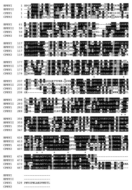
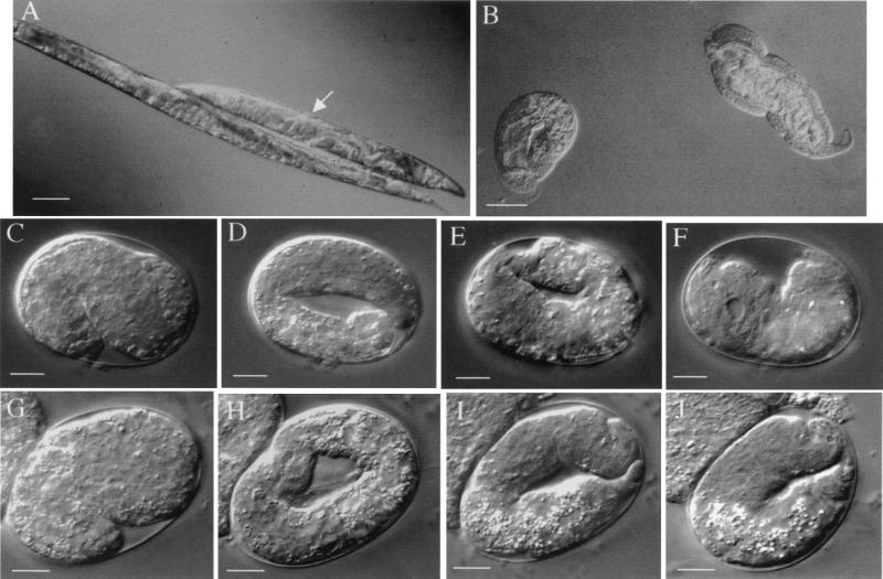
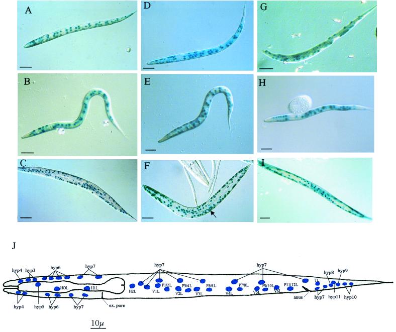
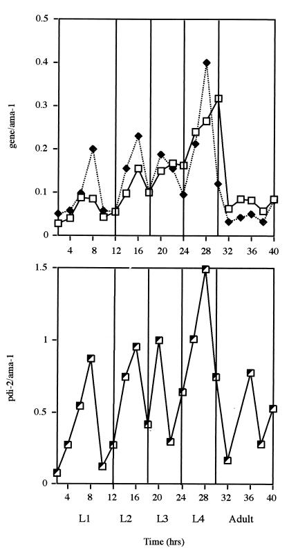
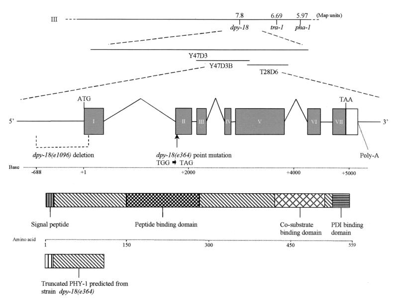
The multienzyme complex prolyl 4-hydroxylase catalyzes the hydroxylation of proline residues and acts as a chaperone during collagen synthesis in multicellular organisms. The beta subunit of this complex is identical to protein disulfide isomerase (PDI). The free-living nematode Caenorhabditis elegans is encased in a collagenous exoskeleton and represents an excellent model for the study of collagen biosynthesis and extracellular matrix formation. In this study, we examined prolyl 4-hydroxylase alpha-subunit (PHY; EC 1.14.11.2)- and beta-subunit (PDI; EC 5.3.4.1)-encoding genes with respect to their role in collagen modification and formation of the C. elegans exoskeleton. We identified genes encoding two PHYs and a single associated PDI and showed that all three are expressed in collagen-synthesizing ectodermal cells at times of maximal collagen synthesis. Disruption of the pdi gene via RNA interference resulted in embryonic lethality. Similarly, the combined phy genes are required for embryonic development. Interference with phy-1 resulted in a morphologically dumpy phenotype, which we determined to be identical to the uncharacterized dpy-18 locus. Two dpy-18 mutant strains were shown to have null alleles for phy-1 and to have a reduced hydroxyproline content in their exoskeleton collagens. This study demonstrates in vivo that this enzyme complex plays a central role in extracellular matrix formation and is essential for normal metazoan development.


Figures

FIG. 1
FIG. 1
Alignment of the C. elegans (C prefix) prolyl 4-hydroxylase α subunits PHY-1 (GenBank no. Z81134) and PHY-2 (GenBank no. Z69637) to the human (H prefix) α(I) (M24487) and α(II) (U90441) subunits using ClustalW. Gaps (dashes) were introduced for maximal alignment, and signal peptides were removed. Highly conserved cysteine and active-site histidine, aspartic acid, and lysine residues (23) are indicated by an asterisk. The conserved tryptophan76, converted to a stop codon in the phy-1 gene of the dpy-18 (e364) allele, is indicated by a dollar sign.
FIG. 2
FIG. 2
Double-stranded RNAi of phy-1, phy-2, and pdi-2. (A) Medium dumpy phy-1 RNAi in wild-type N2 background (arrow) compared to wild-type N2. Adults are depicted. Bar, 100 μm. (B) phy-1phy-2 combined RNAi in wild-type N2 background. A range of severe dumpy phenotypes is represented by coiled larva (left) and adult nematode (right). Bar, 100 μm. (C to F) phy-2 RNAi in dpy-18 (e364) background. The same individual embryo is depicted in all images. Bars, 10 μm. (C) Beginning of elongation (1.5-fold, 440 min). (D) Elongated embryo (3-fold, 570 min). Head and tail of coiled embryo are out of the focal plane. (E) Retracting embryo (710 min). Head and tail of uncoiled embryo are now visible in the same focal plane. (F) Terminal phenotype (approximately 1,800 min), a fully retracted dying embryo with visible vacuoles. (G to J) pdi-2 RNAi in wild-type N2 background. The same individual embryo is depicted in all images. Bars, 10 μm. (G) Beginning of elongation (1.5-fold, 430 min). (H) Elongated embryo (3-fold, 560 min). Head and tail of coiled embryo are out of the focal plane. (I) Retracting embryo (700 min). Head and tail of uncoiled embryo are now visible in the same focal plane. (J) Terminal phenotype (approximately 960 min), a retracted dying embryo with evident small vacuoles.
FIG. 3
FIG. 3
Tissue-specific localization of phy-1, phy-2, and pdi-2. L1 staining patterns correspond to the positions of labeled hypodermal nuclei in panel J, which are particularly apparent at the anterior and posterior ends of the nematode. Staining patterns may be complicated by the corresponding nuclei on the opposite lateral focal plane of the depicted nematode. (A) 5-Bromo-4-chloro-3-indolyl-β-d-galactopyranoside (X-Gal) staining of L1 larvae showing phy-1 (pPD95-03, with a nuclear localization signal [NLS])-driven lacZ expression using the rol-6 marker plasmid. The anterior is to the left, dorsal is at the top, and focus is in the lateral plane. All anterior, posterior, and midbody hyp cells are evident. The lateral seam (H, T, and V) and P cells are stained. Bar, 10 μm. (B) X-Gal staining of L1 larvae showing phy-1 (pPD95-03, with an NLS)-driven lacZ expression using the unc-76 marker plasmid. The anterior is to the left, dorsal is at the top, and focus is in the lateral plane. Most anterior hyp cells (hyp5 to hyp7) are evident; only hyp7 nuclei are visible in the posterior. Some of the lateral seam (H, T, and V) and P cells are visible. Bar, 10 μm. (C) X-Gal staining of adult nematode showing phy-1 (pPD95-03, with an NLS)-driven lacZ expression using the rol-6 marker plasmid. The anterior is to the left, and the body is twisted due to expression of the rol-6 phenotype. Many anterior and posterior hyp cells and lateral hypodermal cells are visible. Bar, 100 μm. (D) X-Gal staining of L1 larvae showing phy-2 (pPD95-03, with an NLS)-driven lacZ expression using the rol-6 marker plasmid. The anterior is to the left, ventral is at the top, and focus is in the lateral plane. All anterior hyp cells are evident; only hyp7 nuclei are present in the tail. The midbody lateral seam cells, P cells, and hyp cells are visible. Additional midbody nonhypodermal cells are also evident. Bar, 10 μm. (E) X-Gal staining of L1 larvae showing phy-2 (pPD95-03, with an NLS)-driven lacZ expression using the unc-76 marker plasmid. The anterior is to the left, dorsal is at the top, and focus is in the lateral plane. Most anterior hyp cells are evident, with weak, incomplete staining of posterior hyp cells. Midbody hyp7 cells, lateral seam cells, and P cells are conspicuous. Additional midbody nonhypodermal cells are present. Bar, 10 μm. (F) X-Gal staining of adult nematode revealing phy-2 (pPD95-03, with an NLS)-driven lacZ expression using the rol-6 marker plasmid. The anterior is to the left, and the body is helically twisted due to expression of the rol-6 phenotype. Anterior, posterior, and lateral hypodermal cells are discernible. Vulval cell nuclear staining is indicated by an arrow. Additional nonhypodermal cell nuclei are also conspicuous. Bar, 100 μm. (G) X-Gal staining of L1 larvae showing pdi-2 (pPD21-28, with an NLS)-driven lacZ expression using the rol-6 marker plasmid. The anterior is to the left, dorsal is at the top, and focus is in the lateral plane. Some of the anterior hyp cells are evident; hyp7 nuclei are present in the tail. Some of the midbody lateral seam cells, P cells, and hyp cells are visible. Bar, 10 μm. (H) X-Gal staining of L1 larvae showing pdi-2 (pPD95-03, with an NLS)-driven lacZ expression using the unc-76 marker plasmid. The anterior is to the left, dorsal is at the top, and focus is in the lateral plane. Most anterior hyp cells are evident (hyp3, hyp6, and hyp7); only hyp7 nuclei are evident in the posterior (partially out of focus). Some of the lateral seam (H, T, and V) and P cells are stained. Bar, 10 μm. (I) X-Gal staining of immature adult nematode showing pdi-2 (pPD21-28, with an NLS)-driven lacZ expression using the rol-6 marker plasmid. The anterior is to the left, and the body is twisted due to expression of the rol-6 phenotype. Anterior and posterior hyp cells and lateral hypodermal cells are visible. Bar, 100 μm. (J) Diagrammatic representation of the L1 left lateral aspect depicting the hypodermal cell nuclei. An identical pattern is present on the right lateral view, with additional hyp7 nuclei dorsal to H2R nuclei. ex, excretory. Panel J is based on the original L1 hypodermal cell nucleus designation (31).
FIG. 4
FIG. 4
Temporal expression pattern of the phy-1 (□), phy-2 (⧫), and pdi-2 (┘) transcripts during postembryonic development. A semiquantitative reverse transcriptase PCR approach was applied to examine the ratio of expression (y axes) of the individual enzyme-encoding genes to that of the constitutively expressed gene ama-1. The arbitrary values for phy-1 and phy-2 were plotted together for comparison. All values were obtained from mRNA of synchronously maintained larval and early adult stages at 25°C. L1 to L4, first to fourth larval stages.
FIG. 5
FIG. 5
Rescue of medium dumpy phenotype of a dpy-18 strain by coinjection of a wild-type copy of phy-1 and the selectable marker dpy-7–GFP. (A) Rescued progeny (arrow) and nonrescued progeny of a dpy-18 strain (e364) viewed by Nomarski optics. (B) Corresponding fluorescence in the rescued dpy-18 strain (e364) (arrow) viewed under a UV filter. Adult stages are depicted.
FIG. 6
FIG. 6
Gene structure of phy-1 showing physical and genetic map locations. Exons are shown as filled boxes. Introns and promoter regions are represented by lines, and 3′ untranslated regions are indicated by an open box. ATG and TAA indicate the positions of the translational start and stop signals, respectively. The 776-bp deletion in phy-1 from dpy-18 (e1096), extending from positions −688 to +88 (relative to the ATG), is depicted. The tryptophan76 (TGG)-to-amber stop codon (TAG) point mutation found in phy-1 from dpy-18 (e364) is also indicated. The domain structure for PHY-1 is shown along with the truncated protein predicted for dpy-18 (e364), revealing missing functional domains.

References

    1. Annunen P, Helaakoski T, Myllyharju J, Veijola J, Pihlajaniemi T, Kivirikko K I. Cloning of the human prolyl 4-hydroxylase alpha subunit isoform alpha(II) and characterization of the type II enzyme tetramer—the alpha(I) and alpha(II) subunits do not form a mixed alpha(I)alpha(II)beta(2) tetramer. J Biol Chem. 1997;272:17342–17348. - PubMed
    1. Annunen P, Koivunen P, Kivirikko K I. Cloning of the alpha subunit of prolyl 4-hydroxylase from Drosophila and expression and characterization of the corresponding enzyme tetramer with some unique properties. J Biol Chem. 1999;274:6790–6796. - PubMed
    1. Baird S E, Emmons S W. Properties of a class of genes required for ray morphogenesis in Caenorhabditis elegans. Genetics. 1990;126:335–344. - PMC - PubMed
    1. Bird D M, Riddle D L. Molecular cloning and sequencing of ama-1, the gene encoding the largest subunit of Caenorhabditis elegans RNA polymerase II. Mol Cell Biol. 1989;9:4119–4130. - PMC - PubMed
    1. Blelloch R, Kimble J. Control of organ shape by a secreted metalloprotease in the nematode Caenorhabditis elegans. Nature. 1999;399:586–590. - PubMed

Publication types

LinkOut - more resources