. 2000 Jun;20(11):4094-105.
doi: 10.1128/MCB.20.11.4094-4105.2000.

hnRNP C is required for postimplantation mouse development but Is dispensable for cell viability

Affiliations

hnRNP C is required for postimplantation mouse development but Is dispensable for cell viability

D J Williamson et al. Mol Cell Biol. 2000 Jun.

Abstract

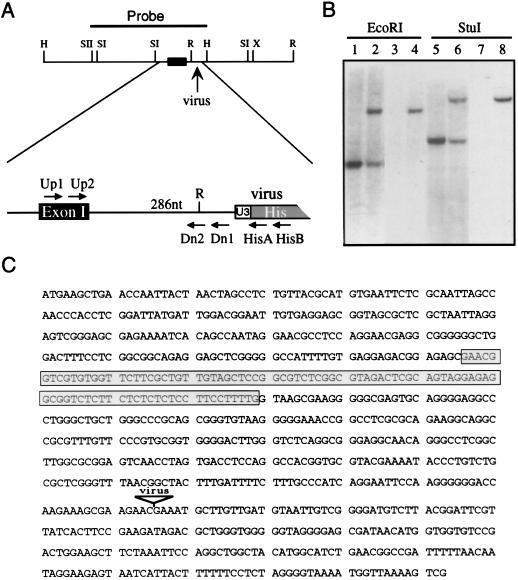
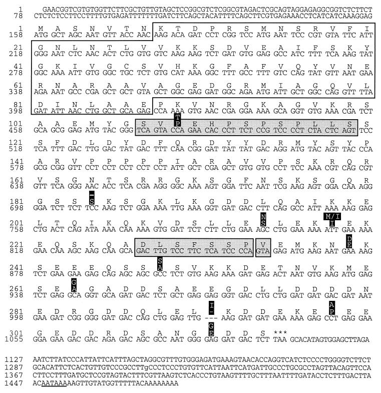
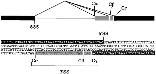
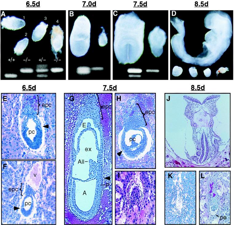
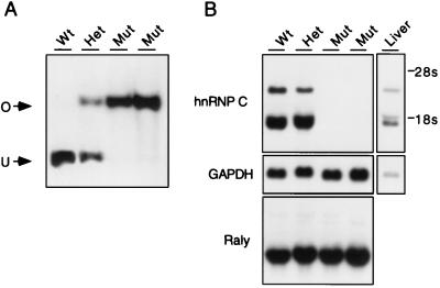
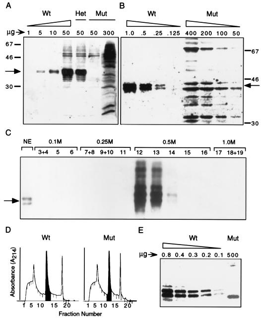
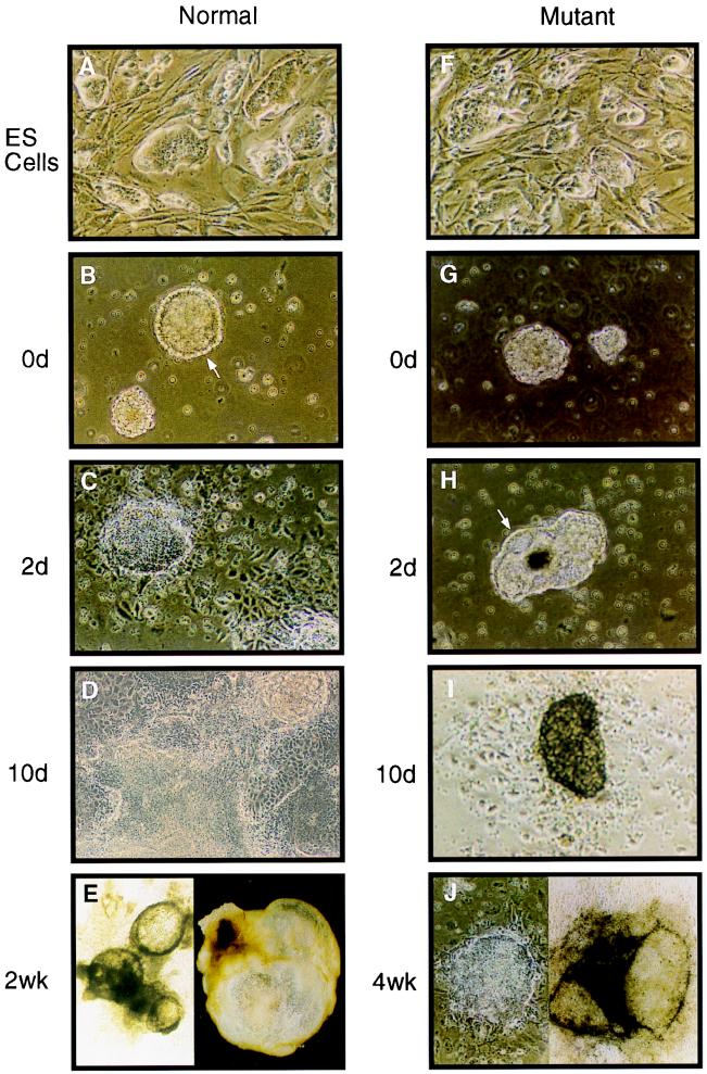
The hnRNP C1 and C2 proteins are among the most abundant proteins in the nucleus, and as ubiquitous components of RNP complexes, they have been implicated in many aspects of mRNA biogenesis. In this report, we have characterized a null mutation induced in embryonic stem cells by insertion of the U3His gene trap retrovirus into the first intron of the hnRNP C1/C2 gene. cDNAs encoding murine hnRNP C1 and C2 were characterized, and the predicted protein sequences were found to be highly conserved among vertebrates. A human consensus sequence, generated from over 400 expressed sequence tags, suggests two revisions to the previously published human sequence. In addition, alternatively spliced transcripts, expressed only by the murine gene, encode four novel proteins: variants of C1 and C2 with either seven additional amino acids or one fewer amino acid in a region between the oligomerization and C-terminal acidic domains. The disrupted gene was transmitted into the germ line and is tightly linked to a recessive, embryonic lethal phenotype. Homozygous mutant embryos fail to develop beyond the egg cylinder stage and are resorbed by 10.5 days of gestation, a phenotype consistent with a fundamental role in cellular metabolism. However, hnRNP C1 and C2 are not required for cell viability. Embryonic stem cell lines established from homozygous mutant blastocysts did not express detectable levels of either protein yet were able to grow and differentiate in vitro, albeit more slowly than wild-type cells. These results indicate that the C1 and C2 hnRNPs are not required for any essential step in mRNA biogenesis; however, the proteins may influence the rate and/or fidelity of one or more steps.


Figures

FIG. 1
FIG. 1
The 4A4 provirus inserts into an intron of the hnRNP C1/C2 gene. (A) Restriction map of a 3.7-kb genomic fragment encompassing the site of viral integration. A SacII-HindIII fragment (solid line) used to probe an ES cell cDNA library and the location of the first hnRNP C1/C2 exon (black box) are indicated. The virus integration site is enlarged, showing the distance (286 nt) to the first exon and the locations of primers used for genotyping embryos (Up1, Up2, Dn1, Dn2, HisA, and HisB). H, HindIII; R, EcoRI; SI, SacI; SII, SacII; X, XbaI. (B) Southern blot analysis of wild-type D3 cells (lanes 1, 3, 5, and 7) and mutant cells heterozygous for the 4A4 provirus (lanes 2, 4, 6, and 8). A flanking sequence probe (lanes 1, 2, 5, and 6) detects a single-copy sequence in wild-type DNA as well as an additional fragment corresponding to the virus-occupied allele in 4A4 cells. A virus-specific probe (lanes 3, 4, 7, and 8) detects only the occupied allele. (C) Sequence surrounding the site of viral integration in 4A4 cells showing the first exon of the mouse hnRNP C1/C2 gene (boxed) and the flanking sequence isolated by iPCR.
FIG. 2
FIG. 2
cDNA sequences of the mouse hnRNP C1/C2 gene. The consensus sequence of a transcript encoding the largest form of murine hnRNP C2 is shown. The alternatively spliced 39-nt exon missing in C1 transcripts is shown (residues 107 to 119, first shaded box). Additional murine sequences that arise by alternative splicing, encoding seven residues (227 to 233) or one residue (234), are also indicated (second and third shaded boxes, respectively). Single amino acid differences between the murine and human hnRNP C proteins are highlighted (white on black), with the human sequence shown above. The RRM is enclosed (residues 8 to 87).
FIG. 3
FIG. 3
Species-specific splicing of murine hnRNP C. A region of genomic DNA containing an intron and flanking exons (white on black and shaded) was amplified by PCR and sequenced as shown. Comparisons of genomic and cDNA sequences reveal that murine hnRNP C transcripts splice from a single 5′ splice site (5′SS) located at nt 835 of the composite cDNA (Fig. 2) to one of three alternative 3′ splice sites (Cα, Cβ, and Cγ). For comparison, human hnRNP C transcripts use the Cβ 3′ splice site exclusively.
FIG. 4
FIG. 4
Phenotype of hnRNP C mutant embryos. (A to D) Whole mount preparations showing failure of hnRNP C null embryos, identified by a single 597-nt PCR product (upper band), to develop beyond E6.5. (A) At E6.5 mutant embryos not yet resorbed were slightly smaller than normal littermates (the rightmost is still surrounded by Reichert's membrane). (B to D) From E7.0 to E8.5, the size difference of the mutant embryos (right and bottom) is clearly appreciated. While normal embryos undergo a rapid cellular proliferation and morphological transformation, surviving mutant embryos appear to grow only slightly, and by E8.5, many were necrotic (rightmost) and difficult to separate from maternal tissue. (E, G, and J) Sagittal sections of normal embryos at egg cylinder (E), late streak (G), and early somite (J) stages. (F) Presumed mutant embryo at the egg cylinder stage. The embryo is smaller than normal littermates, the demarcation between embryonic and extraembryonic regions (arrowhead) is not clear, and the ectoplacental cone is underdeveloped. (H) Mutant embryos at E7.5 failed to undergo gastrulation and appear similar to normal E6.5 embryos. (I) A more severely affected embryo undergoing resorption. This embryo appeared to be misoriented with regard to the mesometrial axis (all embryos are oriented with the mesometrium toward the top of the page). (K) By E8.5, the resorption process for most mutant embryos was nearly complete. (L) A surviving mutant embryo at E8.5 showing that parietal endoderm cells (arrow) had formed although no other structures were apparent. A, amniotic cavity; All, allantois; E, ectoplacental cavity; epc, ectoplacental cone; ex, exocoelom; P, primitive streak; pc, proamniotic cavity; pe, parietal endoderm; v, blood vessel.
FIG. 5
FIG. 5
hnRNP C is not required for cell viability. (A) ES cell lines were derived from 3.5 day blastocysts and genotyped by Southern analysis. Lane 1, wild-type (wt) cells containing a single EcoRI-restricted fragment (U); lane 2, heterozygous (Het) cell line containing one copy of the unoccupied allele and one copy of the virus-occupied (O) allele; lanes 3 and 4, two mutant (Mut) homozygous cell lines containing two copies of the virus-occupied allele. (B) Cell lines shown in panel A were grown without feeder cells for three generations and analyzed by Northern blotting. The murine hnRNP C cDNA detects three transcripts (1.4, 1.9, and ∼3 kb, presumably generated by alternative 3′ polyadenylation) in wild-type and heterozygous cell lines. Expression of all three transcripts is ablated in the two mutant cell lines, indicating that hnRNP C is not essential for cell growth. When the same blot was probed with a Raly cDNA, no compensatory increase in the transcription of this related gene was observed. A glyceraldehyde-3-phosphate dehydrogenase (GAPDH) probe was used to ensure equal loading of RNA for each sample, and mouse liver RNA was included as a control.
FIG. 6
FIG. 6
hnRNP C protein expression is not detectable in homozygous cells. Total cellular lysates (A) and NE (B) were prepared from wild-type (Wt), heterozygous (Het), and mutant (Mut) cells. The indicated amounts of protein were resolved by SDS-PAGE and analyzed by Western blotting using an anti-hnRNP C antibody. The positions of the hnRNP C doublet (C1 and C2, arrow) and protein molecular size standards (in kilodaltons) are indicated. (C) A wild-type ES cell NE was partially purified on a HiTrap Q anion-exchange column developed with a step gradient of 0.25, 0.5, and 1.0 M NaCl. Ten-microliter aliquots of the indicated 1-ml fractions, as well as 1 μg of input NE, were analyzed by Western blotting. (D) Absorbance of fractionated wild-type and mutant NE developed on Mono Q anion-exchange columns. The first two 0.5 M 1-ml fractions (shaded), containing the entire hnRNP C protein fraction, were pooled and analyzed by Western blotting (E). As much as 5,000-fold more partially purified mutant extract than wild-type extract was loaded.
FIG. 7
FIG. 7
hnRNP C null ES cells differentiate in vitro. The hnRNP C null cells (F) grew in tightly packed “nests” when cultured on MEF feeder layers, indistinguishable from wild-type ES cells (A). Both mutant and wild-type ES cells were grown in suspension culture for 4 days to induce simple EB formation and then replated onto culture dishes (counted as day 0) (0d) to determine the differentiation capacity of the two cell lines. (B) Wild-type ES cells gave rise to a large number of simple EBs, many of which had already formed an outer ring of large endoderm cells with an underlying Reichert's membrane (arrow). (G) By contrast, mutant ES cells gave rise to only about 10% of the number of EBs that wild-type cells did. Of those EBs that did form, none exhibited well-differentiated endoderm cells. (C and H) Wild-type EBs quickly attached to the tissue culture plate and by 2 days had elaborated a large outgrowth of endoderm cells. By 2 days in culture, the mutant EBs were just beginning to form a ring of endoderm cells and none had attached to the tissue culture plate. (D and I) By 10 days of tissue culture, wild-type EBs had undergone extensive cellular differentiation whereas the mutant EBs that survived to this point were just beginning to attach to the culture plate. (E) In addition to the wild-type colonies that grew in flattened sheets of differentiated cells, several colonies formed large cystic EBs (right). Many of these cystic EBs coalesced to form interconnected fluid filled chambers with an underlying monolayer of differentiated cells (left). (J) Very few of the mutant EBs survived longer than 2 weeks. However, those EBs that did survive eventually (4 weeks or longer) underwent morphological differentiation similar to that of wild-type EBs, including both well-differentiated monolayers (left) and large cystic EBs. Among the cell types observed for both wild-type and mutant EBs were rhythmically beating myocardial cells.

Similar articles

Cited by

References

    1. Altschul S F, Madden T L, Schaffer A A, Zhang J, Zhang Z, Miller W, Lipman D J. Gapped BLAST and PSI-BLAST: a new generation of protein database search programs. Nucleic Acids Res. 1997;25:3389–3402. - PMC - PubMed
    1. Bachvarova R, De Leon V. Polyadenylated RNA of mouse ova and loss of maternal RNA in early development. Dev Biol. 1980;74:1–8. - PubMed
    1. Barnett S F, LeStourgeon W M, Friedman D L. Rapid purification of native C protein from nuclear ribonucleoprotein particles. J Biochem Biophys Methods. 1988;16:87–97. - PubMed
    1. Bennett M, Michaud S, Kingston J, Reed R. Protein components specifically associated with prespliceosome and spliceosome complexes. Genes Dev. 1992;6:1986–2000. - PubMed
    1. Beyer A L, Christensen M E, Walker B W, LeStourgeon W M. Identification and characterization of the packaging proteins of core 40S hnRNP particles. Cell. 1977;11:127–138. - PubMed

Publication types

MeSH terms

Associated data