. 2000 Jun;20(11):4169-80.
doi: 10.1128/MCB.20.11.4169-4180.2000.

S-Phase progression mediates activation of a silenced gene in synthetic nuclei

Affiliations

S-Phase progression mediates activation of a silenced gene in synthetic nuclei

A J Crowe et al. Mol Cell Biol. 2000 Jun.

Abstract

Aberrant expression of developmentally silenced genes, characteristic of tumor cells and regenerating tissue, is highly correlated with increased cell proliferation. By modeling this process in vitro in synthetic nuclei, we find that DNA replication leads to deregulation of established developmental expression patterns. Chromatin assembly in the presence of adult mouse liver nuclear extract mediates developmental stage-specific silencing of the tumor marker gene alpha-fetoprotein (AFP). Replication of silenced AFP chromatin in synthetic nuclei depletes sequence-specific transcription repressors, thereby disrupting developmentally regulated repression. Hepatoma-derived factors can target partial derepression of AFP, but full transcription activation requires DNA replication. Thus, unscheduled entry into S phase directly mediates activation of a developmentally silenced gene by (i) depleting developmental stage-specific transcription repressors and (ii) facilitating binding of transactivators.


Figures
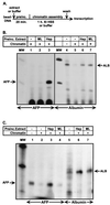
FIG. 1
FIG. 1
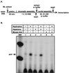
Reconstitution of in vivo expression patterns in vitro. (A) Diagram of coupled chromatin assembly-transcription system. (B) Hepatoma-specific derepression of AFP and albumin chromatin templates. Immobilized AFP (lanes 1 to 3) or albumin (lanes 4 to 7) templates were preincubated (preinc.) in NDB (lanes 1 and 5), adult mouse liver (ML) extract (lanes 2 and 7), HepG2 (lane 3), or Hepa 1-6 extract (lane 6) prior to chromatin assembly in HSS. Chromatin templates were washed and in vitro transcribed in HeLa extract. A nucleosome-free albumin template (lane 4) was in vitro transcribed in HeLa extract (lane 4). (C) Differential regulation of AFP and albumin in simultaneous transcription reactions. Immobilized AFP and albumin templates were coincubated with either NDB (lanes 1, 2, and 5), HepG2 (lanes 3 and 6), or ML (lanes 4 and 7) extract prior to chromatin assembly in HSS (lanes 2 to 4 and 6 to 7) or incubation in Xl buffer in the absence of chromatin assembly (lanes 1 and 5). All templates were washed and in vitro transcribed in HeLa extract. After RNA isolation, each reaction mixture was divided in two, and primer extension analysis was performed with either an AFP-specific primer (lanes 1 to 4) or an albumin-specific primer (lanes 5 to 7). The extension products for AFP (84 bp) and albumin (ALB; 105 bp) are indicated. Radiolabeled φX174 DNA digested with HaeIII was run as a molecular size standard (lane MW).
FIG. 2
FIG. 2
Solid-phase synthetic nuclei. (A) Solid-phase synthetic nuclei support nuclear transport. Immobilized AFP templates were assembled for 1.5 h in LSS to generate solid-phase synthetic nuclei (a to c and e), or in Xl buffer alone (d). Assembly reaction mixtures were further incubated for 30 min with approximately 150 ng of either LEF-1:eGFP (a to d) or GST:eGFP (e). After addition of propidium iodide, samples were visualized by phase-contrast microscopy (a) or by fluorescence microscopy (b to e) with either a GFP wide-band filter (b, d, and e) or a rhodamine filter (c). In the absence of a nuclear membrane (d) or an NLS (e), no concentrated GFP fluorescence was observed. (B) Steady-state DNA replication. Immobilized AFP templates were assembled into synthetic nuclei in the presence of [α-32P]dATP and incubated for 0 (lane 1), 1 (lane 2), 2 (lane 3), or 4 (lane 4 and 5) hours in LSS. Aphidicolin (40 ng/μl) was added to lane 5. Purified DNA was subjected to electrophoresis and visualized by autoradiography of the dried gel. Labeled arrows point to replicated full-length DNA, unresolved replication intermediates or concatemers, and the location of the well. Radiolabeled lambda DNA digested with HindIII was run as molecular size standards (lane MW). (C) DNA replication under transcription conditions. Immobilized AFP templates were assembled into synthetic nuclei by preincubation (preinc.) in HSS plus membranes (MEMB) (lanes 1 to 10) or in LSS (lanes 11 and 12) or by preassembly in HSS followed by nucleus assembly in LSS (lanes 13 to 15). Templates were incubated with either NDB (lanes —) or adult ML (ml), or HepG2 (hep) extract at the indicated times during assembly. Templates in lanes 13 to 15 were preincubated with ML extract prior to assembly in HSS, and these silenced templates were then assembled into nuclei in the presence of the indicated extracts. Incubations and assembly reactions were performed exactly as diagrammed in Fig. 3 and 4. DNA replication was monitored by inclusion of [α-32P]dATP during synthetic nucleus assembly. Purified DNA was subjected to electrophoresis and visualized by autoradiography of the dried gel. Labeled arrows point to replicated full-length DNA and unresolved replication intermediates. Relative replication levels (fold) in comparison to buffer controls are indicated below the lanes. (D) Nucleosome assembly on replicated DNA. Immobilized templates were assembled into synthetic nuclei in LSS in the presence of [α-32P]dATP for 2 h. DNA was then digested with MNase, and aliquots were withdrawn for analysis at 0, 2.5, 5, 10, 20, 40, 60, and 80 min. Nucleosome spacing was estimated by comparison to a 123-bp DNA ladder (Gibco-BRL) and radiolabeled λ HindIII fragments (lane MW). Arrows point to MNase-protected fragments.
FIG. 2
FIG. 2
Solid-phase synthetic nuclei. (A) Solid-phase synthetic nuclei support nuclear transport. Immobilized AFP templates were assembled for 1.5 h in LSS to generate solid-phase synthetic nuclei (a to c and e), or in Xl buffer alone (d). Assembly reaction mixtures were further incubated for 30 min with approximately 150 ng of either LEF-1:eGFP (a to d) or GST:eGFP (e). After addition of propidium iodide, samples were visualized by phase-contrast microscopy (a) or by fluorescence microscopy (b to e) with either a GFP wide-band filter (b, d, and e) or a rhodamine filter (c). In the absence of a nuclear membrane (d) or an NLS (e), no concentrated GFP fluorescence was observed. (B) Steady-state DNA replication. Immobilized AFP templates were assembled into synthetic nuclei in the presence of [α-32P]dATP and incubated for 0 (lane 1), 1 (lane 2), 2 (lane 3), or 4 (lane 4 and 5) hours in LSS. Aphidicolin (40 ng/μl) was added to lane 5. Purified DNA was subjected to electrophoresis and visualized by autoradiography of the dried gel. Labeled arrows point to replicated full-length DNA, unresolved replication intermediates or concatemers, and the location of the well. Radiolabeled lambda DNA digested with HindIII was run as molecular size standards (lane MW). (C) DNA replication under transcription conditions. Immobilized AFP templates were assembled into synthetic nuclei by preincubation (preinc.) in HSS plus membranes (MEMB) (lanes 1 to 10) or in LSS (lanes 11 and 12) or by preassembly in HSS followed by nucleus assembly in LSS (lanes 13 to 15). Templates were incubated with either NDB (lanes —) or adult ML (ml), or HepG2 (hep) extract at the indicated times during assembly. Templates in lanes 13 to 15 were preincubated with ML extract prior to assembly in HSS, and these silenced templates were then assembled into nuclei in the presence of the indicated extracts. Incubations and assembly reactions were performed exactly as diagrammed in Fig. 3 and 4. DNA replication was monitored by inclusion of [α-32P]dATP during synthetic nucleus assembly. Purified DNA was subjected to electrophoresis and visualized by autoradiography of the dried gel. Labeled arrows point to replicated full-length DNA and unresolved replication intermediates. Relative replication levels (fold) in comparison to buffer controls are indicated below the lanes. (D) Nucleosome assembly on replicated DNA. Immobilized templates were assembled into synthetic nuclei in LSS in the presence of [α-32P]dATP for 2 h. DNA was then digested with MNase, and aliquots were withdrawn for analysis at 0, 2.5, 5, 10, 20, 40, 60, and 80 min. Nucleosome spacing was estimated by comparison to a 123-bp DNA ladder (Gibco-BRL) and radiolabeled λ HindIII fragments (lane MW). Arrows point to MNase-protected fragments.
FIG. 3
FIG. 3
Hepatoma-specific activation of AFP during DNA replication. (A) Diagram of solid-phase nucleus assembly and transcription system. (B) In vitro transcription. Immobilized AFP templates were incubated with NDB (lanes 1 and 4 to 9) or HepG2 (lane 2) or ML (lane 3) extract. Templates were assembled into synthetic nuclei by incubation in HSS plus membranes. Synthetic nuclei were further incubated for an additional 30 min in the presence of NDB (lanes 1 to 4 and 7) or HepG2 (lanes 5 and 8) or ML (lanes 6 and 9) extract. Aphidicolin (lanes 7 to 9) was added concomitantly with the HSS plus membranes. All samples were washed and in vitro transcribed in HeLa extract. Radiolabeled φX174 DNA digested with HaeIII was run as molecular size standards (lane MW).
FIG. 4
FIG. 4
DNA replication alone is sufficient to activate a developmentally silenced AFP template. (A) Diagram of two-step solid-phase nucleus replication and transcription system. (B) In vitro transcription. Immobilized AFP templates were incubated with ML extract (lanes 1 to 6) prior to chromatin assembly. Chromatin templates were washed and either incubated in Xl buffer (lanes 1, 3, and 5) or further assembled into synthetic nuclei by incubation with LSS (lanes 2, 4, and 6). NDB (lanes 1 and 2), HepG2 extract (lanes 3 and 4), or ML extract (lanes 5 and 6) was included during the 2-h postchromatin/nucleus assembly period. All samples were washed and in vitro transcribed in HeLa extract. Radiolabeled φX174 DNA digested with HaeIII was run as molecular size standards (lane MW).
FIG. 5
FIG. 5
Sequence specificity and replication dependence of repressor depletion. (A) Diagram of two-step solid-phase nucleus replication and transcription system. (B) In vitro transcription. Immobilized AFP templates were incubated with ML extract prior to chromatin assembly in HSS (lanes 1 to 10). Chromatin templates were washed and further incubated in the presence (lanes 2 to 6 and 8 to 10) or absence (lanes 1 and 7) of LSS to assemble synthetic nuclei. Aphidicolin was added during synthetic nucleus formation to block DNA replication in lane 10. ML extract was added during synthetic nucleus formation (lanes 2 to 6 and 8 to 10). The indicated amount of supercoiled AFP(3.8)-lacZ DNA (lanes 3, 4, 9, and 10) or supercoiled pUC DNA (lanes 5 and 6) was added as a competitor during synthetic nucleus formation. All samples were washed and in vitro transcribed in nuclear HeLa extract. Radiolabeled φX174 DNA digested with HaeIII was run as molecular size standards (lane MW).
FIG. 6
FIG. 6
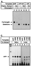
Developmental repression is mediated through sequence-specific binding. (A) Derepression of developmentally silenced AFP chromatin. Immobilized full-length AFP templates (AFP) and AFP templates containing deletions from −1000 to −541 (AFPΔ541) and −1000 to −209 (AFPΔ209) were preincubated (preinc.) in either NDB (lanes 1, 3, 4, and 6) or adult ML extract (lanes 2, 5, and 7). Templates were then either assembled into chromatin in HSS (lanes 2, 4, 5, and 7) or incubated in Xl buffer (lanes 1, 3, and 6). All samples were washed and in vitro transcribed in HeLa extract. The primer extension products obtained from both the full-length template and the deletion templates (13-bp smaller) are indicated by arrows. (B) Developmental repression of AFP is not mediated by chromatin. Immobilized full-length AFP, AFPΔ541, and AFPΔ209 were in vitro transcribed in either HeLa extract (lanes 1 to 3) or adult ML extract (lanes 4 to 6). The primer extension products are indicated by arrows. (C) Deletion of 150 bp is sufficient to derepress AFP. Immobilized full-length AFP (AFP) and AFP templates containing deletions from −1000 to −850 (Δ850), −1000 to −765 (Δ765), and −1000 to −586 (Δ586) were in vitro transcribed in either HeLa extract (lanes 1 to 4) or ML extract (lanes 5 to 8). These deletion mutants were designed to yield a primer extension product identical to that of the full-length AFP transcript, and this product is indicated by a single arrow. Lanes 5 to 8 were overexposed relative to lanes 1 to 4 in order to visualize the ML primer extension products.
FIG. 6
FIG. 6
Developmental repression is mediated through sequence-specific binding. (A) Derepression of developmentally silenced AFP chromatin. Immobilized full-length AFP templates (AFP) and AFP templates containing deletions from −1000 to −541 (AFPΔ541) and −1000 to −209 (AFPΔ209) were preincubated (preinc.) in either NDB (lanes 1, 3, 4, and 6) or adult ML extract (lanes 2, 5, and 7). Templates were then either assembled into chromatin in HSS (lanes 2, 4, 5, and 7) or incubated in Xl buffer (lanes 1, 3, and 6). All samples were washed and in vitro transcribed in HeLa extract. The primer extension products obtained from both the full-length template and the deletion templates (13-bp smaller) are indicated by arrows. (B) Developmental repression of AFP is not mediated by chromatin. Immobilized full-length AFP, AFPΔ541, and AFPΔ209 were in vitro transcribed in either HeLa extract (lanes 1 to 3) or adult ML extract (lanes 4 to 6). The primer extension products are indicated by arrows. (C) Deletion of 150 bp is sufficient to derepress AFP. Immobilized full-length AFP (AFP) and AFP templates containing deletions from −1000 to −850 (Δ850), −1000 to −765 (Δ765), and −1000 to −586 (Δ586) were in vitro transcribed in either HeLa extract (lanes 1 to 4) or ML extract (lanes 5 to 8). These deletion mutants were designed to yield a primer extension product identical to that of the full-length AFP transcript, and this product is indicated by a single arrow. Lanes 5 to 8 were overexposed relative to lanes 1 to 4 in order to visualize the ML primer extension products.
FIG. 7
FIG. 7
Model for DNA replication-mediated activation of a developmentally silenced gene. The developmental repressor region of the AFP gene may be bound by a collection of repressor proteins (symbolized by bricks) during postnatal repression. This brick wall prohibits expression of the gene, perhaps by disrupting an upstream activator-promoter communication of an upstream activator(s) with the promoter. Duplication of repressor-binding sites, through either DNA replication or addition of exogenous AFP DNA, effectively depletes the developmental repressors, resulting in incomplete reassembly of the brick wall on the newly replicated DNA. In the presence of an incomplete complex of repressors, activators can now interact with proximal promoter-bound factors in order to initiate transcription.

Similar articles

Cited by

References

    1. Adams C C, Workman J L. Nucleosome displacement in transcription. Cell. 1993;72:305–308. - PubMed
    1. Almouzni G, Mechali M, Wolffe A P. Competition between transcription complex assembly and chromatin assembly on replicating DNA. EMBO J. 1990;9:573–582. - PMC - PubMed
    1. Aparicio O M, Gottschling D E. Overcoming telomeric silencing: a trans-activator competes to establish gene expression in a cell cycle-dependent way. Genes Dev. 1994;8:1133–1146. - PubMed
    1. Armstrong J A, Bieker J J, Emerson B M. A SWI/SNF-related chromatin remodeling complex, E-RC1, is required for tissue-specific transcriptional regulation by EKLF in vitro. Cell. 1998;95:93–104. - PubMed
    1. Ausubel F M, Brent R, Kingston R E, Moore D D, Seidman J G, Smith J A, Struhl K. Current protocols in molecular biology. Boston, Mass: Current Protocols; 1987.

Publication types

MeSH terms

LinkOut - more resources