. 2000 May 9;97(10):5243-8.
doi: 10.1073/pnas.97.10.5243.

MEK kinase 1 is critically required for c-Jun N-terminal kinase activation by proinflammatory stimuli and growth factor-induced cell migration

Affiliations

MEK kinase 1 is critically required for c-Jun N-terminal kinase activation by proinflammatory stimuli and growth factor-induced cell migration

Y Xia et al. Proc Natl Acad Sci U S A. .

Abstract

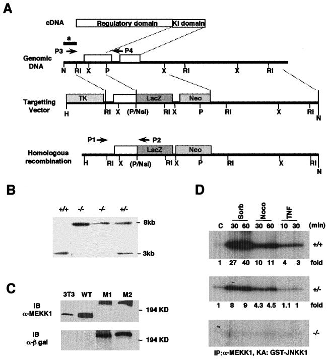
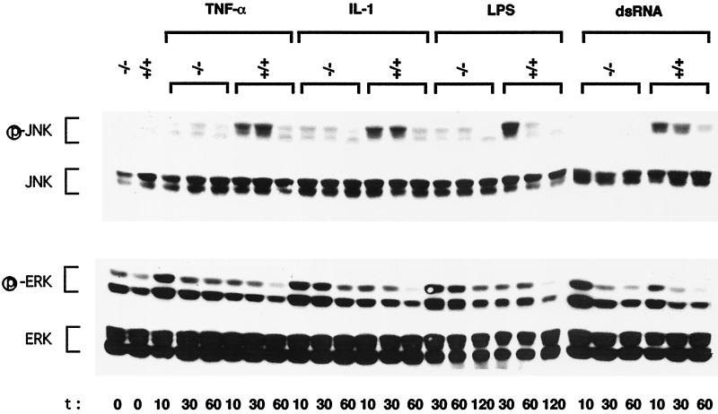
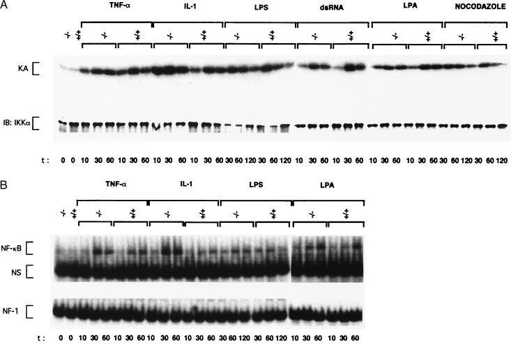
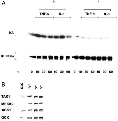
Exposure of eukaryotic cells to extracellular stimuli results in activation of mitogen-activated protein kinase (MAPK) cascades composed of MAPKs, MAPK kinases (MAP2Ks), and MAPK kinase kinases (MAP3Ks). Mammals possess a large number of MAP3Ks, many of which can activate the c-Jun N-terminal kinase (JNK) MAPK cascade when overexpressed, but whose biological function is poorly understood. We examined the function of the MAP3K MEK kinase 1 (MEKK1) in proinflammatory signaling. Using MEKK1-deficient embryonic stem cells prepared by gene targeting, we find that, in addition to its function in JNK activation by growth factors, MEKK1 is required for JNK activation by diverse proinflammatory stimuli, including tumor necrosis factor alpha, IL-1, double-stranded RNA, and lipopolysaccharide. MEKK1 is also essential for induction of embryonic stem cell migration by serum factors, but is not required for activation of other MAPKs or the IkappaB kinase signaling cascade.


Figures

Figure 1
Figure 1
Generation of Mekk1−/− ES cells. (A) Diagram of the targeting strategy. Schematic structures of Mekk1 cDNA, the relevant portion of the Mekk1 locus, the targeting vector, and the homologous recombinant. Indicated are locations of the N-terminal regulatory region and the C-terminal catalytic (Ki) domain of MEKK1, restriction sites (H, HindIII; RI, EcoRI; P, PstI; N, NotI; X, XbaI; B, BamHI), and the position of a 5′ external probe (a). Arrowheads labeled P1, P2, P3, and P4 indicate positions of primers used in genotyping. (B) Southern blot demonstrating homologous recombination within the Mekk1 locus. The 5′ external probe detects a 3-kb HindIII/PstI fragment for the wild-type locus and an 8-kb HindIII/PstI fragment for the targeted locus. (C) Immunoblot (IB) using MEKK1 and β-galactosidase antibodies revealing expression of 180-kDa mouse MEKK1 polypeptide in mouse fibroblasts (3T3) and ES cells (WT) and a 242-kDa MEKK1-β-galactosidase fusion protein in two homozygous recombinant clones. (D) MEKK1 kinase activity in Mekk1 wild-type (+/+), heterozygous (+/−), and homozygous (−/−) recombinant ES cells. Lysates were prepared from cells stimulated with sorbitol (300 mM), nocodazole (2 μM), or TNFα (10 ng/ml) for the indicated times. After immunoprecipitation (IP) with anti-MEKK1, immunocomplex kinase assays (KA) were performed by using GST-JNKK1 as a substrate. Fold-activation was calculated based on phosphorimager analysis.
Figure 2
Figure 2
MEKK1 is required for JNK activation by proinflammatory stimuli. Wild-type (+/+) and MEKK1-deficient (−/−) ES cells were left untreated or incubated with TNFα (10 ng/ml), IL-1 (10 ng/ml), LPS (15 μg/ml), or dsRNA (5 μg/ml) for the indicated times (min) after which the cells were lysed. Equal amounts (100 μg) of lysates were resolved by SDS/PAGE, transferred to a nitrocellulose membrane, and sequentially probed with antibodies to activated (phosphorylated) JNK (p-JNK) and activated (phosphorylated) ERK (p-ERK). The membranes were stripped and reprobed with antibodies to all JNK and ERK isoforms. Shown are the results of one typical experiment of at least three similar and separate experiments.
Figure 3
Figure 3
MEKK1 is not involved in IKK and NF-κB activation in ES cells. (A) IKK activation. Wild-type (+/+) and MEKK1-deficient (−/−) ES cells were left untreated or stimulated with TNFα, IL-1, LPS, dsRNA, as described above, LPA (20 μM), or nocodazole (2 μM). IKK complexes were isolated from equal amounts of cell lysates by immunoprecipitation with anti-IKKγ monoclonal antibody, and immunocomplex kinase assays (KA) were performed by using GST-IκBα (1–54) as a substrate. Recovery of IKK was determined by immunoblotting (IB) with anti-IKKα. (B) NF-κB activation. Wild-type and MEKK1-deficient ES cells were treated as described above with TNFα, IL-1, LPS, and LPA. At the indicated times, cell extracts were prepared, and NF-κB and NF1 DNA binding activities were measured. NS, nonspecific protein–DNA complex.
Figure 4
Figure 4
Dependence on IKKγ and expression of MAP3Ks and GCK in wild-type (+/+) and mutant (−/−) ES cells. (A) Wild-type and Ikkγ- (only a single Ikkγ allele was disrupted, but as Ikkγ is an x-linked gene and the ES cells we used were XO, this resulted in complete loss of IKKγ expression) ES cells were left untreated (0 time point) or treated with TNFα or IL-1 as described above. At the indicated times, cell lysates were prepared and IKK activity was determined by using an IKKα monoclonal antibody and immunocomplex kinase assay. (B) Exponentially growing ES cells or 3T3 fibroblasts were lysed, and equal amounts (50 μg) of total cell lysates were resolved by SDS/PAGE, transferred to a nitrocellulose membrane, and probed with antibodies to ASK1, TAK1, MEKK2, or GCK (Santa Cruz Biotechnology). All of these antibodies were found to specifically recognize their cognate antigens based on immunoblot analysis of cells transiently transfected with expression vectors for the different kinases (not shown).
Figure 5
Figure 5
MEKK1 is involved in serum- and Ras-induced JNK activation and induction of ES cell migration. (A) Serum- and LPA-induced MAPK activation. Exponentially growing wild-type (+/+) and MEKK1-deficient (−/−) ES cells were serum-starved for 2 h. The cells were either left untreated or incubated with serum (15%) or LPA (20 μM) for the indicated times (min). The cells were lysed and JNK, p38, and ERK activation and expression were determined as described in Fig. 2 by immunoblotting. (B) MEKK1 is involved in Ras-induced JNK activation. Wild-type or MEKK1-deficient ES cells were transfected with HA-JNK2 vector along with either empty vector or expression vectors for Ha-Ras(V12), Rac(L61) or full-length human MEKK1. JNK activation was determined by immunocomplex kinase assay (KA) by using GST-c-Jun (1–79) as a substrate. Fold-activation was determined after phosphorimager analysis. HA-JNK2 expression was examined by immunoblotting (IB) using anti-HA antibody. (C) MEKK1 is required for serum-stimulated ES cell migration on fibronectin. Wild-type and MEKK1-deficient ES cells were serum-starved for 2 h. Cells were allowed to migrate on fibronectin (10 μg/ml)-coated transwells (modified Boyden chambers) in the presence of BSA (10 μg/ml, control), serum (2%), TNFα (10 ng/ml), or LPA (20 μM). The numbers of migrated cells were determined after fixing and staining the cells with crystal violet. The results represent the average of three independent experiments.

References

    1. Lange-Carter C A, Pleiman C M, Gardner A M, Blumer K J, Johnson G L. Science. 1993;260:315–319. - PubMed
    1. Minden A, Lin A, McMahon M, Lange-Carter C, Dérijard B, Davis R J, Johnson G L, Karin M. Science. 1994;266:1719–1723. - PubMed
    1. Xia Y, Wu Z, Su B, Murray B, Karin M. Genes Dev. 1998;12:3369–3381. - PMC - PubMed
    1. Hibi M, Lin A, Smeal T, Minden A, Karin M. Genes Dev. 1993;7:2135–2148. - PubMed
    1. Dérijard B, Hibi M, Wu I-H, Barrett T, Su B, Deng T, Karin M, Davis R J. Cell. 1994;76:1025–1037. - PubMed

Publication types

MeSH terms