. 2000 May 9;97(10):5322-7.
doi: 10.1073/pnas.97.10.5322.

Rapid evolution in plant chitinases: molecular targets of selection in plant-pathogen coevolution

Affiliations

Rapid evolution in plant chitinases: molecular targets of selection in plant-pathogen coevolution

J G Bishop et al. Proc Natl Acad Sci U S A. .

Abstract

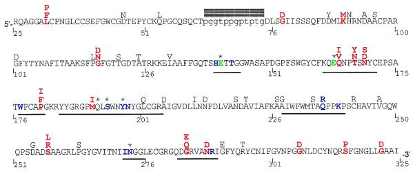
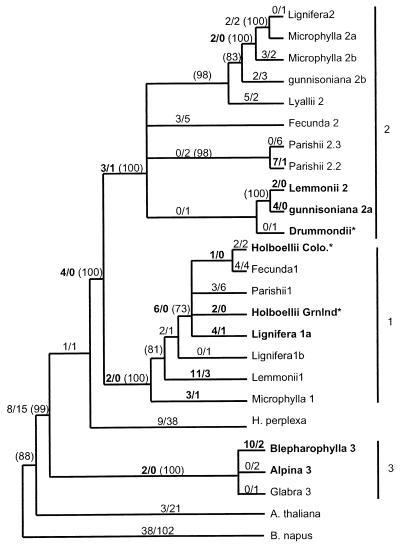
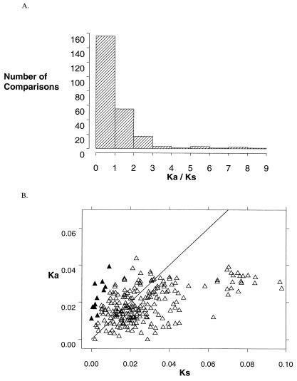
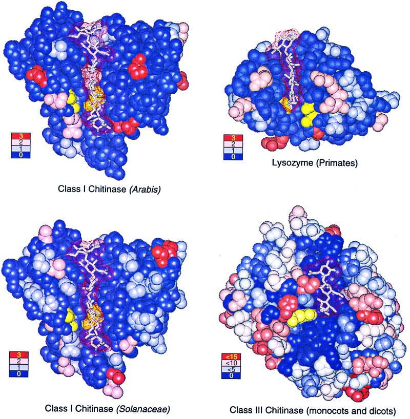
Many pathogen recognition genes, such as plant R-genes, undergo rapid adaptive evolution, providing evidence that these genes play a critical role in plant-pathogen coevolution. Surprisingly, whether rapid adaptive evolution also occurs in genes encoding other kinds of plant defense proteins is unknown. Unlike recognition proteins, plant chitinases attack pathogens directly, conferring disease resistance by degrading chitin, a component of fungal cell walls. Here, we show that nonsynonymous substitution rates in plant class I chitinase often exceed synonymous rates in the plant genus Arabis (Cruciferae) and in other dicots, indicating a succession of adaptively driven amino acid replacements. We identify individual residues that are likely subject to positive selection by using codon substitution models and determine the location of these residues on the three-dimensional structure of class I chitinase. In contrast to primate lysozymes and plant class III chitinases, structural and functional relatives of class I chitinase, the adaptive replacements of class I chitinase occur disproportionately in the active site cleft. This highly unusual pattern of replacements suggests that fungi directly defend against chitinolytic activity through enzymatic inhibition or other forms of chemical resistance and identifies target residues for manipulating chitinolytic activity. These data also provide empirical evidence that plant defense proteins not involved in pathogen recognition also evolve in a manner consistent with rapid coevolutionary interactions.


Figures

Figure 1
Figure 1
A. parishii class I chitinase amino acid sequence. Residue number 1 corresponds to the start codon, but only residues 25–325 are included in this study. The mature protein is ≈298 residues long, consisting of a cysteine-rich chitin binding domain (5′) and a chitinolytic domain (3′), connected by a hypervariable proline-glycine rich hinge (lowercase). Residues 1–22 and 319–325 form the signal peptide and vacuolar targeting peptide, respectively, and are cleaved from the mature peptide. Positively selected residues are red, catalytic residues Glu-141 and Glu-163 are green, putative substrate-binding residues are shown in blue (40), active site residues (defined as residues within 0.6 nm of bound substrate) are underlined, blocks denote indels, and * denotes importance for enzyme function confirmed by directed mutagenesis (51, 52). Alternative residues found among the 19 sequences are shown above the A. parishii sequence. Positively selected residues are identified as sites having a posterior probability > 0.95 of being in the positive category, for a majority of phylogenies tested.
Figure 2
Figure 2
Combined phylogeny of several parsimony phylogenies rooted with B. napus. See Materials and Methods for details of construction. Numbers along each branch show the number of nonsynonymous substitutions/number of synonymous substitutions for that branch, estimated by maximum likelihood for an unrooted tree, and numbers in parentheses show the percent support among 1,000 bootstrap data sets, calculated for each of the component parsimony trees. Branches showing only bootstrap support have estimated branch length = 0, but bootstrap support > 70%, whereas those with no bootstrap values have support < 70%. Numbers shown in bold highlight Ka > Ks (but do not denote significance), and gene names in bold indicate genes involved in significant pairwise comparisons. * denotes phylogenetic position based on coding sequence only. Substitution counts differ from direct pairwise comparisons because the former are estimated by likelihood methods and the latter by approximate methods. Branch lengths are drawn so as to make clades easily discernable.
Figure 3
Figure 3
(A) Distribution of 231 Ka/Ks ratios into intervals, where Ka and Ks are the number of nucleotide substitutions that cause amino acid replacements or are silent, respectively, divided by the number of possible substitutions of each type. Ka/Ks > 1 indicates comparisons where amino acid substitutions are more frequent than predicted by neutral substitution rates. (B) Ka vs. Ks. Ratios > 1 occur above the line Ka = Ks. ▴ denote significant comparisons.
Figure 4
Figure 4
Crystal structures with bound polysaccharide ligands (dot surfaces) showing the location and frequency of amino acid replacements for Arabis class I chitinase (Upper Left), Solanaceae class I chitinase (Lower Left), primate lysozyme (Upper Right), and plant class III chitinase (Lower Right). Ligands are hexa-N-acetyl-d-glucosamine (class I chitinases), tetra-acetyl-chitotetraose (lysozyme), and allosamadin inhibitor (class III chitinase). Catalytic residues are colored yellow. Color-coded legends show the number of replacements occurring at positions illustrated in the corresponding color.

Similar articles

Cited by

References

    1. Thompson J N, Burdon J J. Nature (London) 1992;360:121–125.
    1. Simms E L. BioScience. 1996;46:136–145.
    1. Caicedo A L, Schaal B A, Kunkel B N. Proc Natl Acad Sci USA. 1999;96:302–306. - PMC - PubMed
    1. Stahl E A, Dwyer G, Mauricio R, Kreitman M, Bergelson J. Nature (London) 1999;400:667–671. - PubMed
    1. Meyers B C, Shen K A, Rohani P, Gaut B S, Michelmore R W. Plant Cell. 1998;11:1833–1846. - PMC - PubMed

Publication types

Associated data

LinkOut - more resources