

LexA chimeras reveal the function of Drosophila Fos as a context-dependent transcriptional activator
- PMID: 10805795
- PMCID: PMC25832
- DOI: 10.1073/pnas.97.10.5351
LexA chimeras reveal the function of Drosophila Fos as a context-dependent transcriptional activator
Abstract
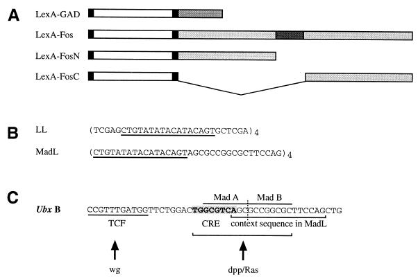
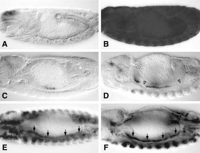
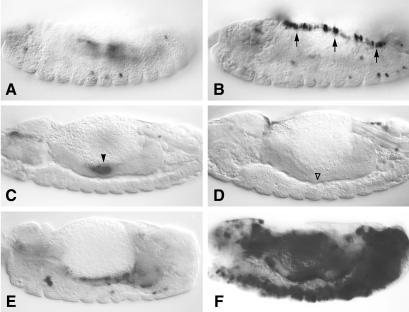
The transcriptional activation potential of proteins can be assayed in chimeras containing a heterologous DNA-binding domain that mediates their recruitment to reporter genes. This approach has been widely used in yeast and in transient mammalian cell assays. Here, we applied it to assay the transactivation potential of proteins in transgenic Drosophila embryos. We found that a chimera between the DNA-binding bacterial LexA protein and the transactivation domain from yeast GAL4 behaved as a potent synthetic activator in all embryonic tissues. In contrast, a LexA chimera containing Drosophila Fos (Dfos) required an unexpected degree of context to function as a transcriptional activator. We provide evidence to suggest that this context is provided by Djun and Mad (a Drosophila Smad), and that these partner factors need to be activated by signaling from Jun N-terminal kinase and decapentaplegic, respectively. Because Dfos behaves as an autonomous transcriptional activator in more artificial assays systems, our data suggest that context-dependence of transcription factors may be more prevalent than previously thought.
Figures

Similar articles
-
Yeast SNF2/SWI2, SNF5, and SNF6 proteins function coordinately with the gene-specific transcriptional activators GAL4 and Bicoid.Genes Dev. 1992 Sep;6(9):1707-15. doi: 10.1101/gad.6.9.1707. Genes Dev. 1992. PMID: 1516829
-
Characterization of a serum response factor-like protein in Saccharomyces cerevisiae, Rlm1, which has transcriptional activity regulated by the Mpk1 (Slt2) mitogen-activated protein kinase pathway.Mol Cell Biol. 1997 May;17(5):2615-23. doi: 10.1128/MCB.17.5.2615. Mol Cell Biol. 1997. PMID: 9111331 Free PMC article.
-
Teashirt is required for transcriptional repression mediated by high Wingless levels.EMBO J. 2001 Jan 15;20(1-2):137-45. doi: 10.1093/emboj/20.1.137. EMBO J. 2001. PMID: 11226164 Free PMC article.
-
Homeodomain interactions.Curr Opin Struct Biol. 1996 Feb;6(1):62-8. doi: 10.1016/s0959-440x(96)80096-0. Curr Opin Struct Biol. 1996. PMID: 8696974 Review.
-
[The molecular mechanism of dorsoventral polarity formation in Drosophila embryos].Tanpakushitsu Kakusan Koso. 1993 Nov;38(15):2480-91. Tanpakushitsu Kakusan Koso. 1993. PMID: 8284442 Review. Japanese. No abstract available.
Cited by
-
The Drosophila ERG channel seizure plays a role in the neuronal homeostatic stress response.PLoS Genet. 2019 Aug 8;15(8):e1008288. doi: 10.1371/journal.pgen.1008288. eCollection 2019 Aug. PLoS Genet. 2019. PMID: 31393878 Free PMC article.
-
Spatial and temporal control of expression with light-gated LOV-LexA.G3 (Bethesda). 2022 Sep 30;12(10):jkac178. doi: 10.1093/g3journal/jkac178. G3 (Bethesda). 2022. PMID: 35876796 Free PMC article.
-
An Interscholastic Network To Generate LexA Enhancer Trap Lines in Drosophila.G3 (Bethesda). 2019 Jul 9;9(7):2097-2106. doi: 10.1534/g3.119.400105. G3 (Bethesda). 2019. PMID: 31040111 Free PMC article.
-
The Drosophila Epidermal Growth Factor Receptor does not act in the nucleus.J Cell Sci. 2018 Sep 20;131(18):jcs220251. doi: 10.1242/jcs.220251. J Cell Sci. 2018. PMID: 30158176 Free PMC article.
-
The fight to understand fighting: neurogenetic approaches to the study of aggression in insects.Curr Opin Insect Sci. 2019 Dec;36:18-24. doi: 10.1016/j.cois.2019.06.004. Epub 2019 Jun 27. Curr Opin Insect Sci. 2019. PMID: 31302354 Free PMC article. Review.
References
-
- Brent R, Ptashne M. Cell. 1985;43:729–736. - PubMed
-
- Ptashne M, Gann A. Nature (London) 1997;386:569–577. - PubMed
-
- Hope I A, Mahadevan S, Struhl K. Nature (London) 1988;333:635–640. - PubMed
-
- Lin Y S, Carey M, Ptashne M, Green M R. Nature (London) 1990;345:359–361. - PubMed
-
- Carey M, Lin Y S, Green M R, Ptashne M. Nature (London) 1990;345:361–364. - PubMed
Publication types
MeSH terms
Substances
LinkOut - more resources
Full Text Sources
Other Literature Sources
Molecular Biology Databases
Research Materials
Miscellaneous
