

doi: 10.1073/pnas.080520697.
Staphylococcus aureus sortase mutants defective in the display of surface proteins and in the pathogenesis of animal infections
Affiliations
- PMID: 10805806
- PMCID: PMC25859
- DOI: 10.1073/pnas.080520697
Item in Clipboard
Staphylococcus aureus sortase mutants defective in the display of surface proteins and in the pathogenesis of animal infections
Proc Natl Acad Sci U S A.
.
Abstract
Many gram-positive bacteria covalently tether their surface adhesins to the cell wall peptidoglycan. We find that surface proteins of Staphylococcus aureus are linked to the cell wall by sortase, an enzyme that cleaves polypeptides at a conserved LPXTG motif. S. aureus mutants lacking sortase fail to process and display surface proteins and are defective in the establishment of infections. Thus, the cell wall envelope of gram-positive bacteria represents a surface organelle responsible for interactions with the host environment during the pathogenesis of bacterial infections.
Figures
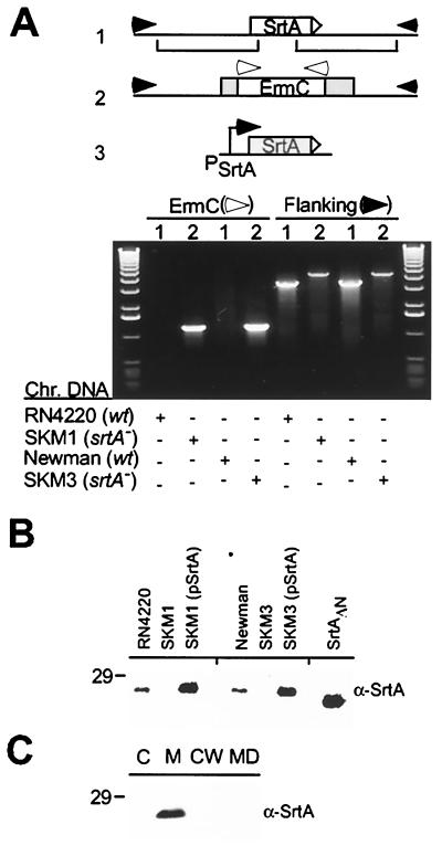
S. aureus sortase (srtA) mutants. (A) Drawing depicts the wild-type sortase (srtA) gene (1) and the srtA−:ermC allele (2). Plasmid-encoded wild-type srtA (pSrtA) is expressed from its own promoter and was used for complementation studies (3). Oligonucleotide primers binding to sequences flanking the srtA gene (filled arrows) or the ermC gene (open arrows) were used to amplify DNA fragments from the chromosomal DNA of strains RN4220 (srtA, 1), SKM1 (srtA−, 2), Newman (srtA, 1), and SKM3 (srtA−, 2). DNA fragments were separated on ethidium bromide-stained agarose gel, flanked by the 1-kb DNA ladder. (B) Immunoblotting with anti-SrtA (α-SrtA) revealed the presence of sortase (26 kDa) in extracts of wild-type strains RN4220 and Newman and the absence of sortase in the mutant strains SKM1 and SKM3 (srtA−). SrtAΔN lacking the N-terminal membrane anchor was expressed in E. coli and purified. (C) RN4220 cultures were fractionated into medium (MD), cell wall (CW), membrane (M), and cytosolic (C) compartments and immunoblotted with α-SrtA.
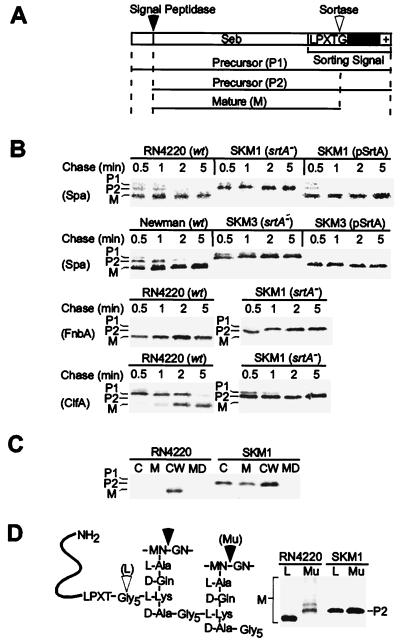
Anchoring surface proteins to the cell wall of staphylococci. (A) Drawing depicts the structure of the Seb-Cws surface protein, which is comprised of enterotoxin B (Seb) with an N-terminal signal peptide and a C-terminal cell wall sorting signal. The cell wall sorting signal contains an LPXTG motif, a hydrophobic domain (black box), and a positively charged tail. (B) The sorting signals of protein A (Spa), fibronectin-binding protein (FnbA), and clumping factor (ClfA) were fused to Seb, and cell wall sorting was followed in a pulse–chase experiment. (C) Pulse-labeled staphylococci cultures were fractionated into medium (MD), cell wall (CW), membrane (M), and cytosolic (C) compartments, and Seb-Spa490–524 was immunoprecipitated with anti-Seb (α-Seb). (D) The peptidoglycan of staphylococci was digested with lysostaphin (L) or mutanolysin (Mu), and radiolabeled surface proteins were analyzed by SDS/PAGE.
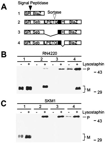
Protein secretion and sorting pathways of staphylococci. (A) Drawing depicts the structure of protein fusions with the mature domain of staphylococcal β-lactamase (BlaZ). 1, (SebSP-BlaZ) fusion of the enterotoxin B signal peptide (SP). 2, (Seb-Cws-BlaZ) fusion of enterotoxin B (Seb) and the protein A sorting signal to BlaZ. 3, (Seb-CwsΔLPXTG-BlaZ) same fusion as in 2 but lacking the LPXTG motif. 4, (Seb-CwsΔR-BlaZ) same fusion as in 2 but lacking the retention signal (+). (B) Pulse-labeled staphylococcal cultures (strain RN4220) were divided into two aliquots and precipitated with TCA. One sample was directly boiled in SDS, whereas the other was first subjected to peptidoglycan hydrolysis with lysostaphin and then boiled in SDS. Samples were subjected to immunoprecipitation with anti-BlaZ (α-BlaZ) and analyzed by SDS/PAGE and PhosphorImager. (C) Same experiment as in B, but using the sortase mutant strain SKM1.
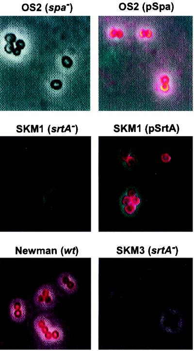
Display of protein A on the staphylococcal surface. Binding of CY3-labeled Ig to protein A was measured by capturing dark-field and fluorescent microscopy images with a charge-coupled device camera and superimposing the data. S. aureus OS2 (spa−) cannot express protein A; however, Ig binding was restored by transformation with plasmid encoding wild-type spa (pSpa). S. aureus SKM1 (srtA:ermC) failed to bind CY3-labeled Ig, a defect that was complemented by transformation with plasmids encoding wild-type srtA (pSrtA). S. aureus Newman (wild-type, wt) displayed protein A on the staphylococcal surface; however, the isogenic sortase knockout mutant SKM3 failed to bind CY3-labeled Ig.
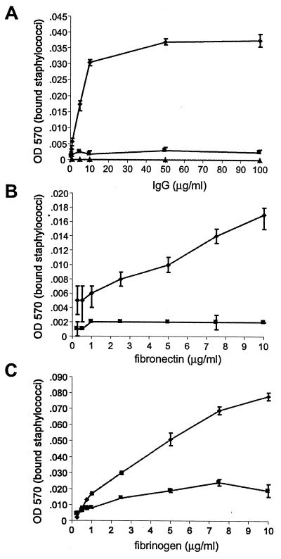
Display of surface proteins by staphylococci. Microtiter dishes were coated with increasing amounts of Ig, fibronectin, or fibrinogen. Binding of staphylococci to mammalian proteins was detected by staining with crystal violet and measuring the absorbance at 570 nm in a spectrophotometer. (A) Binding of S. aureus RN4220 (♦), OS2 (spa−, ▴), and SKM1 (srtA−, ■) to Ig. (B) Binding of S. aureus RN4220 (♦), and SKM1 (srtA−, ■) to fibronectin. (C) Binding of S. aureus Newman (♦) and SKM3 (srtA−, ■) to fibrinogen.
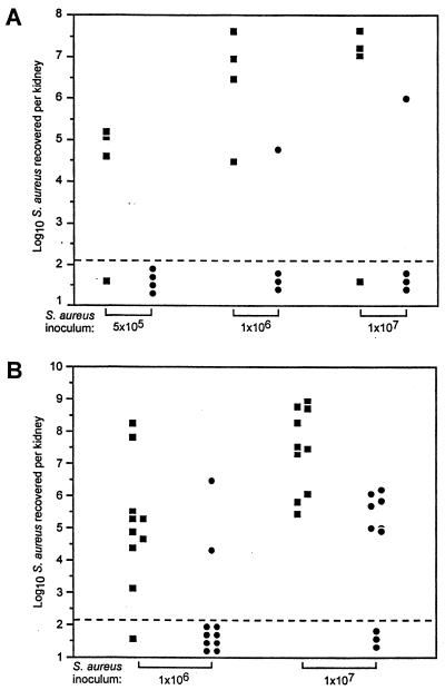
Surface proteins and the pathogenesis of S. aureus infections. S. aureus Newman (human clinical isolate) and the isogenic sortase mutant SKM3 were injected into the tail vein of C57BL/6 (A) or Swiss–Webster mice (B) as indicated. Five days after infection, animals were killed, kidneys excised, homogenized, and plated. Symbols indicate cfu of S. aureus Newman (■) and the sortase mutant strain SKM3 (●). The dashed line represents the limit of detection of staphylococci in renal tissues. P values were calculated after log transformation of the data using the Student t test or the Mann–Whitney U test (parenthesis): C57BL/6: 5 × 10⁵ cfu, P = 0.03 (0.08); 1 × 10⁶ cfu, P = 0.01 (0.04); 1 × 10⁷ cfu, P = 0.12 (0.11). Swiss–Webster: 1 × 10⁶ cfu, P = 0.049 (0.006); 1 × 10⁷ cfu, P = 0.0004 (0.001).
Comment in
-
Sortase, a universal target for therapeutic agents against gram-positive bacteria?Proc Natl Acad Sci U S A. 2000 May 9;97(10):5013-5. doi: 10.1073/pnas.97.10.5013. Proc Natl Acad Sci U S A. 2000. PMID: 10805759 Free PMC article. Review. No abstract available.
References
Publication types
MeSH terms
Substances
Grants and funding
LinkOut - more resources
Full Text Sources
Other Literature Sources
Medical
