

Production of infectious bovine papillomavirus from cloned viral DNA by using an organotypic raft/xenograft technique
- PMID: 10805809
- PMCID: PMC25863
- DOI: 10.1073/pnas.97.10.5534
Production of infectious bovine papillomavirus from cloned viral DNA by using an organotypic raft/xenograft technique
Abstract
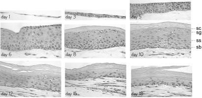
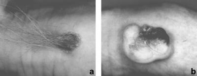
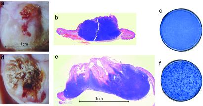
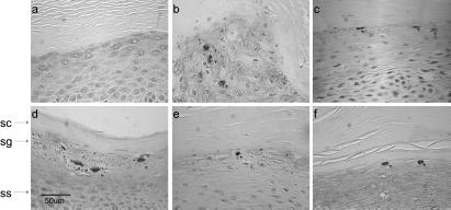
Bovine papillomavirus type 1 (BPV-1) induces fibropapillomas in its natural host and can transform fibroblasts in culture. The viral genome is maintained as an episome within fibroblasts, which has allowed extensive genetic analyses of the viral functions required for DNA replication, gene expression, and transformation. Much less is known about BPV-1 gene expression and replication in bovine epithelial cells because the study of the complete viral life cycle requires an experimental system capable of generating a fully differentiated stratified bovine epithelium. Using a combination of organotypic raft cultures and xenografts on nude mice, we have developed a system in which BPV-1 can replicate and produce infectious viral particles. Organotypic cultures were established with bovine keratinocytes plated on a collagen raft containing BPV-1-transformed fibroblasts. These keratinocytes were infected with virus particles isolated from a bovine wart or were transfected with cloned BPV-1 DNA. Several days after the rafts were lifted to the air interface, they were grafted on nude mice. After 6-8 weeks, large xenografts were produced that exhibited a hyperplastic and hyperkeratotic epithelium overlying a large dermal fibroma. These lesions were strikingly similar to a fibropapilloma caused by BPV-1 in the natural host. Amplified viral DNA and capsid antigens were detected in the suprabasal cells of the epithelium. Moreover, infectious virus particles could be isolated from these lesions and quantitated by a focus formation assay on mouse cells in culture. Interestingly, analysis of grafts produced with infected and uninfected fibroblasts indicated that the fibroma component was not required for productive infection or morphological changes characteristic of papillomavirus-infected epithelium. This system will be a powerful tool for the genetic analysis of the roles of the viral gene products in the complete viral life cycle.
Figures


Similar articles
-
DNA from bovine papillomavirus type 2 induces warts in a xenograft model.Virus Res. 2002 Dec;90(1-2):365-70. doi: 10.1016/s0168-1702(02)00246-0. Virus Res. 2002. PMID: 12457989
-
Differentiation-specific expression from the bovine papillomavirus type 1 P2443 and late promoters.J Virol. 1993 Sep;67(9):5605-16. doi: 10.1128/JVI.67.9.5605-5616.1993. J Virol. 1993. PMID: 8394463 Free PMC article.
-
Bovine papillomavirus DNA can be detected in keratinocytes of equine sarcoid tumors.Vet Microbiol. 2010 Dec 15;146(3-4):269-75. doi: 10.1016/j.vetmic.2010.05.032. Epub 2010 Jun 1. Vet Microbiol. 2010. PMID: 21095508
-
Bovine Papillomavirus: New Insights into an Old Disease.Transbound Emerg Dis. 2016 Feb;63(1):14-23. doi: 10.1111/tbed.12222. Epub 2014 Mar 24. Transbound Emerg Dis. 2016. PMID: 24661978 Review.
-
Association of bovine papillomavirus with the equine sarcoid.J Gen Virol. 2003 May;84(Pt 5):1055-1062. doi: 10.1099/vir.0.18947-0. J Gen Virol. 2003. PMID: 12692268 Review.
Cited by
-
Life cycle heterogeneity in animal models of human papillomavirus-associated disease.J Virol. 2002 Oct;76(20):10401-16. doi: 10.1128/jvi.76.20.10401-10416.2002. J Virol. 2002. PMID: 12239317 Free PMC article.
-
Pseudotyped Virus for Papillomavirus.Adv Exp Med Biol. 2023;1407:85-103. doi: 10.1007/978-981-99-0113-5_5. Adv Exp Med Biol. 2023. PMID: 36920693
-
Human papillomavirus type 16 E6 induces cell competition.PLoS Pathog. 2022 Mar 23;18(3):e1010431. doi: 10.1371/journal.ppat.1010431. eCollection 2022 Mar. PLoS Pathog. 2022. PMID: 35320322 Free PMC article.
-
Suppression of Stromal Interferon Signaling by Human Papillomavirus 16.J Virol. 2019 Sep 12;93(19):e00458-19. doi: 10.1128/JVI.00458-19. Print 2019 Oct 1. J Virol. 2019. PMID: 31292244 Free PMC article.
-
Specific inactivation of inhibitory sequences in the 5' end of the human papillomavirus type 16 L1 open reading frame results in production of high levels of L1 protein in human epithelial cells.J Virol. 2002 Mar;76(6):2739-52. doi: 10.1128/jvi.76.6.2739-2752.2002. J Virol. 2002. PMID: 11861841 Free PMC article.
References
-
- Howley P M. In: Virology. Fields B N, Knipe D M, Howley P M, editors. Philadelphia: Lippincott; 1995. pp. 2045–2076.
-
- Howett M K, Christensen N D, Kreider J W. Clin Dermatol. 1997;15:229–236. - PubMed
-
- Meyers C, Laimins L A. Curr Top Microbiol Immunol. 1994;186:199–215. - PubMed
-
- Chow L T, Broker T R. Clin Dermatol. 1997;15:217–227. - PubMed
-
- McMillan N A, Payne E, Frazer I H, Evander M. Virology. 1999;261:271–279. - PubMed
MeSH terms
Substances
LinkOut - more resources
Full Text Sources
