. 2000 May 9;97(10):5574-9.
doi: 10.1073/pnas.97.10.5574.

Accumulation of protease-resistant prion protein (PrP) and apoptosis of cerebellar granule cells in transgenic mice expressing a PrP insertional mutation

Affiliations

Accumulation of protease-resistant prion protein (PrP) and apoptosis of cerebellar granule cells in transgenic mice expressing a PrP insertional mutation

R Chiesa et al. Proc Natl Acad Sci U S A. .

Abstract

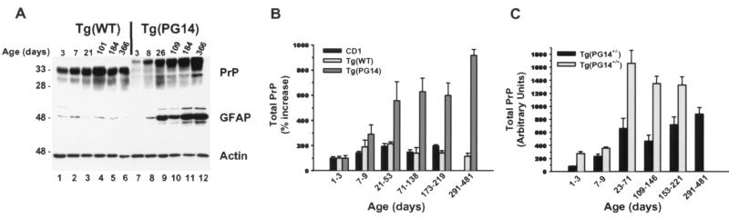
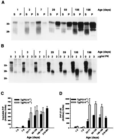
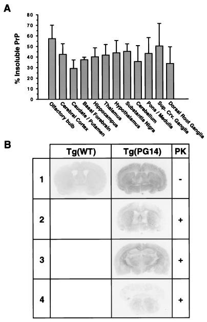
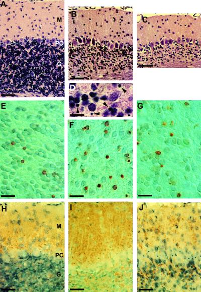
We have generated lines of transgenic mice that express a mutant prion protein (PrP) containing 14 octapeptide repeats whose human homologue is associated with an inherited prion dementia. These mice develop a neurological illness with prominent ataxia at 65 or 240 days of age, depending on whether the transgene array is, respectively, homozygous or hemizygous. Starting from birth, mutant PrP is converted into a protease-resistant and detergent-insoluble form that resembles the scrapie isoform of PrP, and this form accumulates dramatically in many brain regions throughout the lifetime of the mice. As PrP accumulates, there is massive apoptosis of granule cells in the cerebellum. Our analysis provides important insights into the molecular pathogenesis of inherited prion disorders in humans.


Figures

Figure 1
Figure 1
PG14 PrP accumulates with age in the brains of Tg(PG14) mice. (A) Brain extracts of Tg(WT+/+) and Tg(PG14+/−) mice of the indicated ages were separated by SDS-PAGE, and immunoblotted with antibodies specific for PrP, GFAP, or actin. Each lane represents 10 μg of total protein. (B) The amount of PrP in the brains of Tg(WT+/+), Tg(PG14+/−), and nontransgenic CD1 mice in each age group was quantitated by densitometric analysis of Western blots and was expressed as a percentage of the amount present in 1- to 3-day-old mice. Each bar represents the mean ± SEM of values from three to seven animals. (C) The amount of PrP in the brains of Tg(PG14+/−) and Tg(PG14+/+) mice in each age group was quantitated by densitometric analysis of Western blots and was expressed in arbitrary units. Each bar represents the mean ± SEM of values from three to seven animals. Size markers are given in kDa.
Figure 2
Figure 2
The amount of detergent-insoluble and protease-resistant PG14 PrP increases with age in the brains of Tg(PG14) mice. (A) Brain lysates from Tg(PG14+/+) mice of the indicated ages were subjected to ultracentrifugation, and PrP in the supernatants (S lanes) and pellets (P lanes) was analyzed by Western blotting. (B) Brain lysates from Tg(PG14+/+) mice of the indicated ages were incubated with 0–3 μg of proteinase K (PK) for 30 min at 37°C, and PrP was visualized by Western blotting. The undigested samples (0 μg/ml PK) represent 50 μg of protein, and the other samples represent 200 μg of protein. The protease-resistant fragment (PrP 27-30) migrates between 27 and 30 kDa. (C) The amount of PG14 PrP in the pellet fraction after ultracentrifugation (see A) was quantitated by densitometric analysis of Western blots of samples from Tg(PG14+/−) and Tg(PG14+/+) mice. Each bar represents the mean ± SEM of four to eight replicate analyses of samples from four to seven brains. (D) The amount of PrP 27-30 that was produced by digestion with 2 μg/ml of proteinase K (see B) was quantitated by densitometric analysis of Western blots of samples from Tg(PG14+/−) and Tg(PG14+/+) mice. Each bar represents the mean ± SEM of four to eight replicate analyses of samples from four to seven brains.
Figure 3
Figure 3
Protease-resistant and detergent-insoluble PrP are widely distributed in the brains of Tg(PG14) mice. (A) Detergent insolubility of PrP from dissected brain regions was analyzed and quantitated as in Fig. 2 A and C. Each bar represents the mean ± SEM of three to five replicates from four Tg(PG14+/−) mice, ranging in age from 79 to 239 days. (B) Coronal cryostat sections from the brain of a Tg(WT+/+) mouse (182 days old) and a Tg(PG14+/+) mouse (192 days old) at the level of the caudate-putamen (rows 1 and 2), thalamus (row 3), and cerebellum (row 4) were subjected to histoblotting, either before (row 1) or after (rows 2–4) treatment with proteinase K.
Figure 4
Figure 4
Neuropathological changes in the cerebella of Tg(PG14+/+) mice. Hematoxylin and eosin-stained sections showing the cerebellar cortex of mice of 22 days (A), 100 days (B and D), and 183 days (C) of age. M, molecular layer; PC, Purkinje cell layer; G, granule cell layer. Note the dramatic decrease in the number of granule cells with age. Arrowheads in D indicate pyknotic nuclei. ISEL-stained sections showing positively stained cells (brown) in the granule cell layer from mice of 31 days (E), 53 days (F), and 181 days (G) of age. PrP immunostaining of cerebellar cortex from mice of 22 days (H), 100 days (I), and 181 days (J) of age. Note the small punctate deposits of PrP. Scale bars are: 50 μm (A–C), 10 μm (D), 13 μm (E–G), 32 μm (H–J).
Figure 5
Figure 5
DNA extracted from the cerebella of Tg(PG14) mice displays a 200-bp ladder. Detergent extracts of cerebella were centrifuged at 16,000 × g, and DNA extracted from pellet fraction (lanes 1, 3, 5, and 7) and supernatant fraction (lanes 2, 4, 6, and 8) was subjected to Southern blotting using restriction-digested mouse genomic DNA as a probe. Samples were from mice of the following ages: 182 days (lanes 1 and 2), 187 days (lanes 3 and 4), 178 days (lanes 5 and 6), and 176 days (lanes 7 and 8). One-thirtieth of the DNA extracted from the pellet fractions, and the whole amount of DNA extracted from the supernatant fractions were analyzed. Size markers are given in base-pairs.

References

    1. Prusiner S B. Proc Natl Acad Sci USA. 1998;95:13363–13383. - PMC - PubMed
    1. Young K, Piccardo P, Dlouhy S, Bugiani O, Tagliavini F, Ghetti B. In: Prions: Molecular and Cellular Biology. Harris D A, editor. Wymondham: Horizon Scientific Press; 1999. pp. 139–175.
    1. Gambetti P, Petersen R B, Parchi P, Chen S G, Capellari S, Goldfarb L, Gabizon R, Montagna P, Lugaresi E, Piccardo P, Ghetti B. In: Prion Biology and Diseases. Prusiner S B, editor. Cold Spring Harbor, NY: Cold Spring Harbor Lab. Press; 1999. pp. 509–583.
    1. Chiesa R, Piccardo P, Ghetti B, Harris D A. Neuron. 1998;21:1339–1351. - PubMed
    1. Owen F, Poulter M, Collinge J, Leach M, Lofthouse R, Crow T J, Harding A E. Mol Brain Res. 1992;13:155–157. - PubMed

Publication types

LinkOut - more resources