. 2000 May 9;97(10):5633-8.
doi: 10.1073/pnas.97.10.5633.

Potent and nontoxic antisense oligonucleotides containing locked nucleic acids

Affiliations

Potent and nontoxic antisense oligonucleotides containing locked nucleic acids

C Wahlestedt et al. Proc Natl Acad Sci U S A. .

Abstract

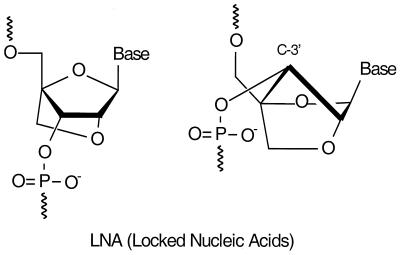
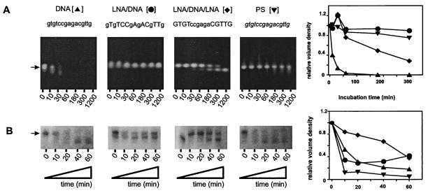
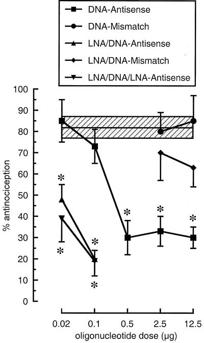
Insufficient efficacy and/or specificity of antisense oligonucleotides limit their in vivo usefulness. We demonstrate here that a high-affinity DNA analog, locked nucleic acid (LNA), confers several desired properties to antisense agents. Unlike DNA, LNA/DNA copolymers were not degraded readily in blood serum and cell extracts. However, like DNA, the LNA/DNA copolymers were capable of activating RNase H, an important antisense mechanism of action. In contrast to phosphorothioate-containing oligonucleotides, isosequential LNA analogs did not cause detectable toxic reactions in rat brain. LNA/DNA copolymers exhibited potent antisense activity on assay systems as disparate as a G-protein-coupled receptor in living rat brain and an Escherichia coli reporter gene. LNA-containing oligonucleotides will likely be useful for many antisense applications.


Figures

Figure 1
Figure 1
Two representations of the chemical structure of LNA (one nucleotide monomer shown). We have defined LNA as an oligonucleotide containing one or more 2′-C,4′-C-oxy-methylene-linked bicyclic ribonucleotide monomers (LNA monomers). The representation shown to the left highlights the chemical connectivities of an LNA monomer. The representation shown to the right highlights the locked 3′-endo (3E) conformation of the furanose ring of an LNA monomer.
Figure 2
Figure 2
Uptake of FITC-labeled all-LNA 15-mer oligonucleotide into human MCF-7 breast cancer cells. (A–C) Low-magnification pictures of living cells. A, light microscopy; C, fluorescence microscopy; B, superimposed view of A and C. (D–F) High magnification of fixed cells. D is phase contrast; F is fluorescence; E is a superimposed view of F and E.
Figure 3
Figure 3
Stability in rat blood serum (A) and RNase H activation (B) of DOR-AS-1 oligonucleotides. LNA monomers are represented by uppercase letters, DNA monomers by lowercase letters, and phosphorothioate (PS) monomers by italic lowercase letters. The location of the full-length RNA is indicated (arrow), and the relative volume densities for single measurements are shown as line graphs.
Figure 4
Figure 4
RNase H cleavage of a DOR-2 RNA target sequence with DNA and DNA/LNA copolymers. (Upper) RNase H cleavage of a 15-mer synthetic RNA (5′UGCCUUCUGCCCGUG) lowercase letters corresponding to the DOR-2 target. (Right) Control incubations lacking added antisense DNA or RNase H, thus demonstrating that the degradation visible (Upper) is mediated by added RNase H and antisense DNA. LNA monomers are represented by uppercase letters, DNA monomers by lowercase letter, and phosphorothioate (PS) monomers by italic lowercase letters. The location of the full-length RNA is indicated (arrow), and the relative volume densities for single measurements are shown as line graphs.
Figure 5
Figure 5
Rat core temperature and immunofluorescence micrographs from rat caudate-putamen. Rats were injected into the caudate-putamen with phosphorothioate (PS) oligodeoxynucleotides (A), gap-mer DNA/phosphorothioate oligodeoxynucleotides (B), or LNA oligodeoxynucleotides (C), on day 1 and day 3 (10 nmol/day), and core temperature was measured 2 h after last injection. Temperature data are presented as means ± SEM based on three animals per group. A pooled group of DNA-injected animals served as controls (n = 24). Statistical analysis was done by means of an independent Student's t test (nsP > 0.05, *P < 0.05, **P < 0.01). Fluorescence micrographs from sections stained for tyrosine hydroxylase from caudate-putamen (same rats as used in temperature experiments). Arrow indicates lesion with absence of tyrosine hydroxylase staining. (Scale bar, as shown in A, = 100 μm) (CPu, caudate-putamen; cc, corpus callosum)
Figure 6
Figure 6
Blockade by DOR-AS-1 antisense oligonucleotides of [D-Ala2]Deltorphin-(Deltorphin II)-induced antinociception in the warm water tail-flick test in rats. The oligonucleotides were administered in a 5-μl volume twice daily (intrathecal) (0800 and 1700 h) for 3 days. The day after the last injection, the rats were injected with [D-Ala2]deltorphin (60 μg, intrathecal) and tested in the warm water (52°C) tail-flick test for DOR-mediated antinociception. Data are presented as means ± SEM based on six to eight animals per group. The shaded area shows means ± SEM for deltorphin-treated controls. Student's t test was used for comparisons with deltorphin-treated controls, and all differences were considered as statistically significant at P < 0.05 (as indicated by the asterisk).

Similar articles

Cited by

References

    1. Wahlestedt C, Good L. Curr Opin Drug Disc Dev. 1999;2:142–146. - PubMed
    1. Stein C A. Nat Biotechnol. 1999;17:209. - PubMed
    1. Branch A D. Trends Biochem Sci. 1998;23:45–50. - PubMed
    1. Crooke S T. Antisense Nucleic Acid Drug Dev. 1998;8:vii–viii. - PubMed
    1. Perry C M, Balfour J A. Drugs. 1999;57:375–380. - PubMed

Publication types

MeSH terms

LinkOut - more resources