. 2000 May 1;14(9):1072-84.

MEI-1/MEI-2 katanin-like microtubule severing activity is required for Caenorhabditis elegans meiosis

Affiliations

MEI-1/MEI-2 katanin-like microtubule severing activity is required for Caenorhabditis elegans meiosis

M Srayko et al. Genes Dev. .

Abstract

The Caenorhabditis elegans meiotic spindle is morphologically distinct from the first mitotic spindle, yet both structures form in the same cytoplasm approximately 20 minutes apart. The mei-1 and mei-2 genes of C. elegans are required for the establishment of the oocyte meiotic spindle but are not required for mitotic spindle function. mei-1 encodes an AAA ATPase family member with similarity to the p60 catalytic subunit of the heterodimeric sea urchin microtubule-severing protein, katanin. We report that mei-2 encodes a 280-amino acid protein containing a region similar to the p80-targeting subunit of katanin. MEI-1 and MEI-2 antibodies decorate the polar ends of meiotic spindle microtubules and meiotic chromatin. We find that the subcellular location of MEI-2 depends on wild-type mei-1 activity and vice versa. These experiments, combined with MEI-1 and MEI-2's similarity to p60 and p80 katanin, suggest that the C. elegans proteins function as a complex. In support of this idea, MEI-1 and MEI-2 physically associate in HeLa cells. Furthermore, co-expression of MEI-1 and MEI-2 in HeLa cells results in the disassembly of microtubules. These data lead us to conclude that MEI-1/MEI-2 microtubule-severing activity is required for meiotic spindle organization in C. elegans.


Figures
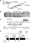
Figure 1
Figure 1
Mapping, RNAi, and sequencing of molecular lesions identifies mei-2. (A) mei-2 was genetically mapped to LGI, 0.016 cm (∼1/2 the length of a cosmid) to the left of let-607, a gene which we rescued with cosmids F57B10 and F48A9. The duplications hDp39 and hDp54 did not rescue mei-2 mutations. The endpoint of hDp54 was mapped to cosmid C01B6, thus placing mei-2 to the right. Cosmids that rescued the mei-2(ct102) mutation when coinjected are shown in bold. (B) Embryos from hermaphrodites injected with RNA transcribed from a 4.8 kb XbaI subclone of cosmid F57B10 (shown in C) exhibited the mei-2-like phenotype. Note the large polar body in the anterior, compared with wild type (WT) (arrows). (C) To identify the mei-2 gene, RNA was transcribed from three genomic subclones of cosmid F57B10 indicated. RNA transcribed from a SacI–EcoRI subclone containing F57B10.4 (white boxes) did not produce any obvious phenotype. A SpeI–XbaI subclone containing mei-2 (F57B10.12, black boxes) did produce the mei-2 phenotype, identical to the entire XbaI 4.8-kb subclone. (Xb) XbaI; (Sp) SpeI; (E) EcoRI; (S) SacI. (D) The mei-2 structure, as predicted by Genefinder and confirmed by RT–PCR, is shown. mei-2 is trans-spliced to the leader sequence SL1(white box) and encodes four exons (black boxes). The 3′ UTR is 122 nucleotides long (grey box). The DNA sequence alterations responsible for each of the four mei-2 mutations are also shown. mei-2(sb31) contains two lesions, one point mutation and a 35-bp deletion (▵) that results in an in-frame insertion of four amino acids. The sequence of mei-2 is available from GenBank/EMBL/DDBJ under accession number AF248052.
Figure 2
Figure 2
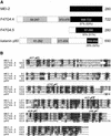
Comparison of MEI-2 and related proteins. (A) Schematic representations of full-length C. elegans MEI-2, F47G4.4, F47G4.5, and S. purpuratus katanin p80 proteins are shown. The region of similarity as determined by BLAST searching with MEI-2 sequence is represented by black boxes containing amino acid coordinates. Percent identity (and similarity in brackets) to MEI-2 are shown below each protein. Two regions of homology between F47G4.4 and katanin p80, as determined from a BLAST search with F47G4.4 are also depicted (28% identity/51% similarity for the amino-terminal region, and 23% identity/37% similarity for the middle region, grey boxes). (B) Pileup results for MEI-2, F47G4.4, F47G4.5, and katanin p80 are shown. Full-length MEI-2 protein sequence is aligned with the carboxy-terminal portion of F47G4.4, most of F47G4.5, and the carboxyl terminus of katanin p80. Identical amino acids (█) and similar amino acids (grey boxes) to MEI-2 are indicated. Also shown are the positions and nature of molecular lesions found in each mei-2 mutant. ▿ Insertion of four amino acids (FCNS) caused by the 35-bp deletion in mei-2(sb31) (see Fig. 1D). Alignment of sequences was performed with the Genetic Corporation Group (Madison, WI) Pileup program and the freeware program, Boxshade.
Figure 3
Figure 3
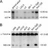
Expression of mei-2 mRNA and protein. (A) Northern blot analysis of mei-2 mRNA. Poly A+ RNA was loaded into each lane and probed with a genomic clone containing mei-2 (see Materials and Methods). RNA was harvested from gravid wild-type hermaphrodites, larval stages L1, L2/L3, L4, as well as glp-1 hermaphrodites (lacking a germ line), fem-3 adults (which produce sperm, but not oocytes), and fem-1 adults (which do not produce sperm, and tend to bloat with unfertilized oocytes). As a loading control, the blot was probed simultaneously with rp21, a ribosomal protein gene. fem-1(lf) lane exposure was adjusted until the rp21 signal was approximately equal to the signal in other lanes. (B) Western blot analysis of MEI-2. Homogenates prepared from mei-2 mutant hermaphrodites, wild type, fem-3, and fem-1 were probed with anti-MEI-2 serum. Note the slightly retarded migration of MEI-2(sb31) protein, consistent with the net insertion of four amino acids. As a loading control, the blot was probed simultaneously with anti-α-tubulin.
Figure 4
Figure 4
Immunolocation of MEI-2. (A) MEI-2 expression was first apparent in the proximal gonad of adult hermaphrodites. Typically, the last 2–3 oocytes in this region stained with anti-MEI-2. MEI-2 was not observed in the distal portion of the gonad (*), where mitotic proliferation of germ cells occurs. A dotted line indicates the outline of one gonad arm (as determined by Nomarski microscopy and DAPI immunofluoresence, not shown). (B) An embryo undergoing meiosis II is shown. MEI-2 antiserum decorated the meiotic spindle and condensed meiotic chromatin (clear arrowhead), the polar body from meiosis I (arrow) and the condensed sperm pronucleus (white arrowhead). Anti-MEI-2 also stained the meiosis-I spindle and polar body from meiosis II (not shown). With the exception of the polar bodies, all MEI-2 staining disappeared after the completion of meiosis (see Fig 6C). (C–E) Deconvolution imaging of a meiotic spindle in metaphase is shown, stained with (C) DAPI, to visualize chromatin, (D) anti-α-tubulin, and (E) anti-MEI-2. The MEI-2 staining patterns were also documented in the absence of DAPI and α-tubulin staining (data not shown), to ensure that the observed patterns did not result from artifacts of multichannel fluorescence microscopy. Bars: A and B 10 μm; E 1 μm.
Figure 5
Figure 5
MEI-2 immunolocation in mei-2 mutants. Meiotic spindles from wild-type (WT; A–C) and mei-2 mutant embryos (D–I) were fixed and stained with DAPI (A,D,G) anti-α-tubulin (B,E,H) and anti-MEI-2 (C,F,I). ct98 and ct102 are mei-2 alleles (top-down is in order of increasing genetic severity). The ts allele mei-2(ct98) was grown at the restrictive temperature of 25°C. Bar; 2 μm.
Figure 6
Figure 6
Mislocalization of MEI-2 to mitotic chromatin and centrosomes in mei-1(ct46gf) mutant embryos. Wild-type (WT; A–C) and mei-1(ct46gf) (D–I) mutant embryos were fixed and stained with DAPI (A,D,G), anti-α-tubulin (B,E,H), and anti-MEI-2 (C,F,I). All meiotic staining of MEI-2 appeared normal in both wild-type and mei-1(ct46gf) embryos (not shown). (A–C) Wild-type embryo in first mitosis showed no MEI-2 staining other than the polar bodies (arrow). (D–I) mei-1(ct46gf) embryos exhibited centrosomal staining during prometaphase (F), and centrosomal and chromatin staining during metaphase (see Fig. 7D) and anaphase (I). The centrosomal and mitotic chromatin staining patterns reiterated for several divisions. We also observed the same staining pattern in mel-26(ct61) mutant embryos (not shown). Bar; 10 μm.
Figure 7
Figure 7
MEI-1 and MEI-2 immunolocation. Spindles from wild-type (WT) and mei-1(ct46gf) mutant embryos were fixed and immunostained with anti-MEI-1 (A and C) and anti-MEI-2 (B and D). In wild type (A and B), MEI-1 and MEI-2 were found at meiotic spindle poles (out of focal plane, white arrowheads) and chromatin (clear arrowheads). In mei-1(ct46gf) (C and D), MEI-1 and MEI-2 were found at centrosomes (white arrowheads) and chromatin (clear arrowheads). Bar; 2 μm. Embryos in C and D have approximately 10 cells; a close-up of one metaphase spindle from each embryo is shown.
Figure 8
Figure 8
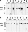
In vivo binding of GFP–MEI-1 with cotransfected GST fusions of MEI-2 and homologs. HeLa cells were cotransfected with GFP–MEI-1 and various GST fusion proteins and were subsequently lysed. GST fusion proteins, along with associated proteins, were isolated by glutathione–sepharose chromatography. The presence of transfected proteins in the glutathione–sepharose bound fraction (B) and unbound fraction (U) was monitored by immunoblotting. (A) Anti-GST-probed immunoblots. HeLa cells were cotransfected with GFP–MEI-1 and GST–MEI-2 (lane 1), GFP–MEI-1 and GST (lane 2), GFP–MEI-1 and GST-con80 (lane 3), GFP-p60 and GST–MEI-2 (lane 4), GFP-VPS4 and GST–MEI-2 (lane 5), GFP-p60 and GST-con80 (lane 6), GFP–MEI-1 and GST–F47G4.4 (lane 7), or GFP–MEI-1 and GST-F47G4.5 (lane 8). (B) Anti-GFP probed immunoblots loaded identically as those in A. Positions of standards and their molecular weights (in kD) are indicated to the left of each panel.
Figure 9
Figure 9
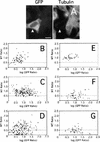
Reduction in the intensity of anti-tubulin immunofluorescence in HeLa cells cotransfected with GFP–MEI-1 and GST fusions of MEI-2 and homologs. (A) Fluorescence micrographs of a HeLa cell coexpressing GFP–MEI-1 and GST–MEI-2 (arrowhead). The intensity of antitubulin staining of the MEI-1-expressing cell is reduced relative to that of adjacent untransfected cells, indicating that MT disassembly has occurred. Bar; 10 μm. (B–G) The ratio of the antitubulin fluorescence intensity of individual transfected cells to that of neighboring untransfected cells (MT Ratio) was measured and plotted as a function of the GFP-fluorescence ratio of the same cells (GFP Ratio). HeLa cells were cotransfected with (B) GFP–MEI-1 and GST, (C) GFP–MEI-1 and GST-con80, (D) GFP–MEI-1 and GST–MEI-2, (E) GFP-VPS4 and GST–MEI-2, (F) GFP–MEI-1 and GST-F47G4.4, or (G) GFP–MEI-1 and GST-F47G4.5.

Similar articles

Cited by

References

    1. Afshar K, Barton NR, Hawley RS, Goldstein LSB. DNA binding and meiotic chromosomal localization of the Drosophila nod kinesin-like protein. Cell. 1995;81:129–138. - PubMed
    1. Ahmad FJ, Yu W, McNally FJ, Baas PW. An essential role for katanin in severing microtubules in the neuron. J Cell Biol. 1999;145:305–315. - PMC - PubMed
    1. Albertson DG. Formation of the first cleavage spindle in nematode embryos. Dev Biol. 1984;101:61–72. - PubMed
    1. Albertson DG, Thomson JN. Segregation of holocentric chromosomes at meiosis in the nematode, Caenorhabditis elegans. Chromosome Research. 1993;1:15–26. - PubMed
    1. Altschul SF, Gish W, Miller W, Myers EW, Lipman DJ. Basic local alignment search tool. J Mol Biol. 1990;215:403–410. - PubMed

Publication types

MeSH terms

LinkOut - more resources