. 2000 May 1;14(9):1098-108.

Pre-mRNA splicing alters mRNP composition: evidence for stable association of proteins at exon-exon junctions

Affiliations

Pre-mRNA splicing alters mRNP composition: evidence for stable association of proteins at exon-exon junctions

H Le Hir et al. Genes Dev. .

Abstract

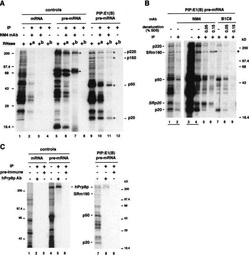
We provide direct evidence that pre-mRNA splicing alters mRNP protein composition. Using a novel in vitro cross-linking approach, we detected several proteins that associate with mRNA exon-exon junctions only as a consequence of splicing. Immunoprecipitation experiments suggested that these proteins are part of a tight complex around the junction. Two were identified as SRm160, a nuclear matrix-associated splicing coactivator, and hPrp8p, a core component of U5 snRNP and spliceosomes. Glycerol gradient fractionation showed that a subset of these proteins remain associated with mRNA after its release from the spliceosome. These results demonstrate that the spliceosome can leave behind signature proteins at exon-exon junctions. Such proteins could influence downstream metabolic events in vivo such as mRNA transport, translation, and nonsense-mediated decay.


Figures

Figure 1
Figure 1
Schematic structures of site-specifically modified PIP:E1(B) RNAs. The experimental pre-mRNA (right) contained a benzophenone (B) at the penultimate nucleotide of exon 1 (E1, open box) and a single 32P (star) at the beginning of exon 2 (shaded box). Control mRNA (left) was structurally identical to the spliced product of experimental pre-mRNA. Control pre-mRNA (center) was identical to experimental pre-mRNA except that the 32P was positioned at the 5′ splice site.
Figure 2
Figure 2
Splicing and cross-linking of benzophenone-containing PIP:E1(B) RNAs. (A) Splicing time courses. RNAs were incubated under splicing conditions for times indicated and separated by denaturing PAGE. Substrate and product structures are indicated above and to the right of each panel. Because all constructs contained only one 32P label (star), only a subset of splicing products are detectable. The small amount of RNA of similar mobility to mRNA in lanes 8–10 is debranched intron; this species contains label only when generated from control pre-mRNA. Species likely generated by exonucleolytic digestion of the lariat intron–exon 2 (Moore and Sharp 1992) (•) and four-way RNA ligation intermediates that copurified with full-length pre-mRNA (••) are indicated. (B) Protein cross-linking. After incubation under splicing conditions for times indicated, samples were irradiated on ice at 302 nm and digested with RNase A. Reactions in lanes 4, 9, and 14 were treated with proteinase K after cross-linking; RNAs in lanes 5, 10, and 15 were subjected to splicing and cross-linking but contained no benzophenone. Lanes 16–18 are longer exposures of lanes 11–13. Molecular mass standards (kD) and apparent molecular masses of proteins that cross-linked in a splicing-dependent manner (*) are indicated to the left and right, respectively. (•) indicates position of the ∼70-kD protein discussed in the text. (C) Side-by-side comparisons of protein cross-linking patterns. RNAs [(ctl) control; (exp) experimental] were incubated under splicing conditions for 90 min, UV irradiated, and RNase A digested. Cross-linked proteins were separated in 7.5% (lanes 1–3), 10% (lanes 4–6), or 16% (lanes 7–9) denaturating polyacrylamide gels. Proteins that cross-link in a splicing dependent manner are indicated. (D,E) Dependence of cross-links on splicing. The indicated RNAs were incubated under splicing conditions for times shown and irradiated as in B. As specified, splicing was inhibited by ATP omission (−ATP) or blocking U2 snRNA with a complementary 2′-O-methyl oligonucleotide (+α-U2 oligonucleotide). (D) RNAs; (E) cross-linked proteins. • and * are as in A and B, respectively.
Figure 3
Figure 3
IP of proteins cross-linked to PIP:E1(B) RNAs. (A) IP of native complexes with mAb NM4. RNAs were incubated under splicing conditions for 90 min, irradiated, and treated with RNase without IP (+) (lanes 1, 5, and 9), treated with RNase after (+a) IP (lanes 2,6,10), or treated with RNase before (+b) IP (lanes 3,4,7,8,11,12). IPs in lanes 4, 8, and 12 contained no antibody. (•) Indicates position of the ∼70-kD protein discussed in text. (B) IP of native and denatured proteins with mAb NM4 or B1C8. Experimental pre-mRNA was spliced for 90 min, irradiated, and treated with RNase. Lanes 1 and 2 are shorter exposures of lanes 3 and 4. Samples in lanes 1 and 3 were not further purified. IPs with either mAb NM4 (lanes 2,4–6) or mAb B1C8 (lanes 7–9) were performed without prior protein denaturation (−; lanes 2,4,7) or after boiling in either 0.05% SDS (lanes 5,8) or 0.15% SDS (lanes 6,9). (C) IP of denatured proteins with hPrp8p antiserum or preimmune serum. After incubation of RNAs under splicing conditions for 90 min, irradiation, and RNase treatment, proteins were denatured by boiling in 0.25% SDS. Samples in lanes 1, 4, and 7 were not further purified. IPs were performed with either hPrp8p antiserum (lanes 2,5,8) or preimmune serum (lanes 3,6,9).
Figure 4
Figure 4
Protein cross-linking to 4-thio-dU-containing PIP RNAs. (A) Schematic structures of the three experimental PIP pre-mRNAs and sequences around the 5′ and 3′ splice sites. (Star) Positions of benzophenone (B), 4-thio-dU (S) and 32P. Underlined nucleotides correspond to those differing between constructs. (B) RNAs as indicated were incubated under splicing conditions for 90 min, UV irradiated, and treated with either RNase A or T1 (see Materials and Methods). Proteins that cross-linked in a splicing-dependent manner are specified to the right of each panel.
Figure 5
Figure 5
Glycerol gradient fractionation. (A) Profile of proteins cross-linked to experimental PIP:E1(B) pre-mRNA after 2 hr of splicing, UV irradiation, and fractionation in a 10%–30% glycerol gradient (top panel). RNAs extracted from a portion of each fraction were analyzed in a denaturing RNA gel (bottom panel) to reveal the sedimentation profiles of mRNP (mRNA) and spliceosomes (lariat–exon 2). (Bars) Positions of mRNP and spliceosomes. Lane T (total) is an aliquot of the same reaction mixture before fractionation. Extracted RNAs were also analyzed by Northern blotting using a U5 snRNA specific probe (middle panel). (Arrow) Peak position of U5 snRNA. (B) Same as A, except that UV irradiation was performed after density sedimentation and U5 snRNA was not analyzed. Species likely generated by exonucleolytic degradation of the lariat intron–exon 2 (Moore and Sharp 1992) are indicated (•) in the lower panels of both A and B.

Similar articles

Cited by

References

    1. Abmayr SM, Workman JL, Roeder RG. The pseudorabies immediate early protein stimulates in vitro transcription by facilitating TFIID: Promoter interactions. Genes & Dev. 1988;2:542–553. - PubMed
    1. Achsel T, Ahrens K, Brahms H, Teigelkamp S, Luhrmann R. The human U5-220kD protein (hPrp8) forms a stable RNA-free complex with several U5-specific proteins, including an RNA unwindase, a homologue of ribosomal elongation factor EF-2, and a novel WD-40 protein. Mol Cell Biol. 1998;18:6756–6766. - PMC - PubMed
    1. Blencowe BJ, Issner R, Kim J, McCaw P, Sharp PA. New proteins related to the Ser-Arg family of splicing factors. RNA. 1995;1:852–865. - PMC - PubMed
    1. Blencowe BJ, Issner R, Nickerson JA, Sharp PA. A coactivator of pre-mRNA splicing. Genes & Dev. 1998;12:996–1009. - PMC - PubMed
    1. Cáceres JF, Screaton GR, Krainer AR. A specific subset of SR proteins shuttles continuously between the nucleus and the cytoplasm. Genes & Dev. 1998;12:55–66. - PMC - PubMed

Publication types

MeSH terms

LinkOut - more resources