. 2000 May 1;14(9):1119-31.

A subtilisin-like serine protease involved in the regulation of stomatal density and distribution in Arabidopsis thaliana

Affiliations

A subtilisin-like serine protease involved in the regulation of stomatal density and distribution in Arabidopsis thaliana

D Berger et al. Genes Dev. .

Abstract

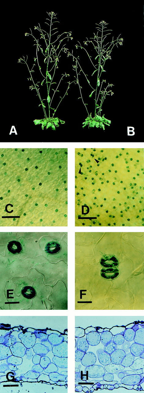
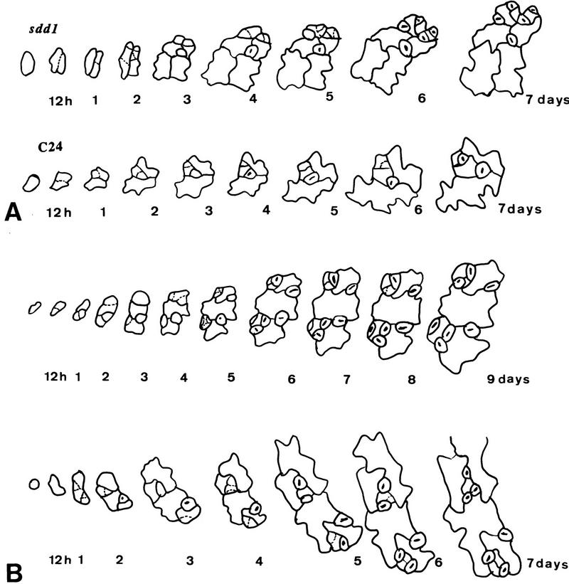
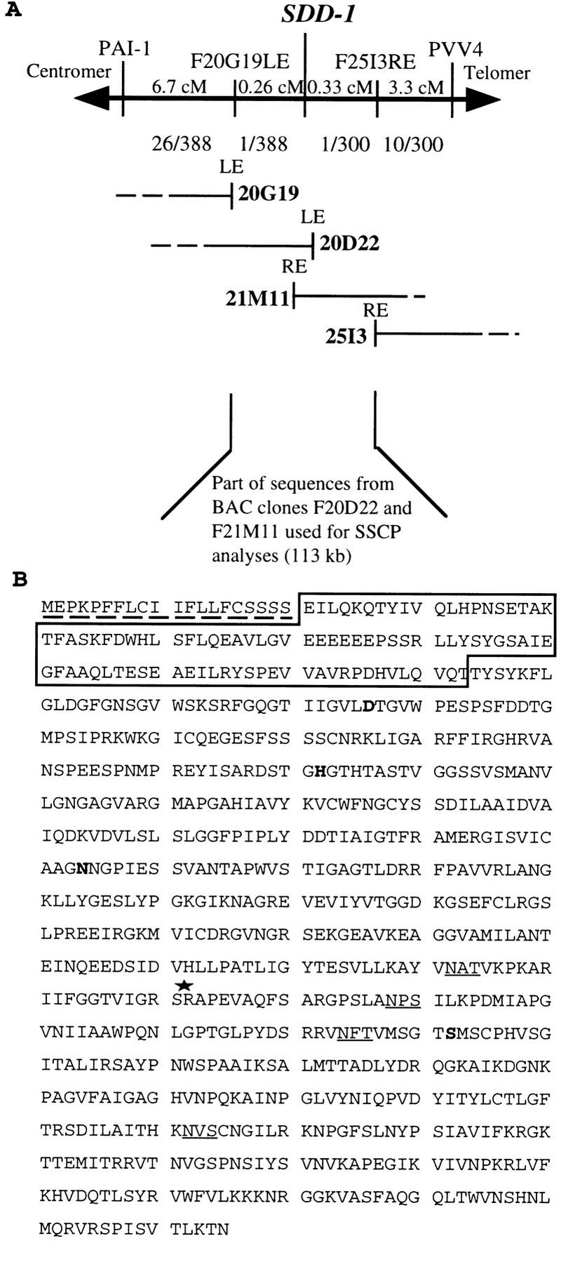
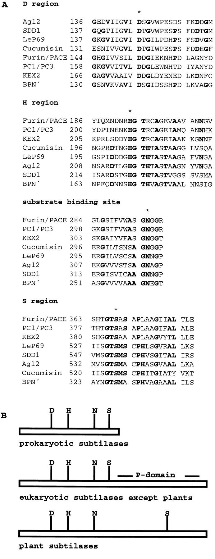
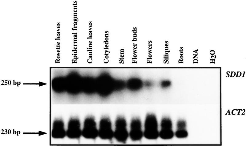
Stomata are specialized cellular structures in the epidermis of aerial plant organs that control gas exchange (H(2)O release and CO(2) uptake) between leaves and the atmosphere by modulating the aperture of a pore flanked by two guard cells. Stomata are nonrandomly distributed, and their density is controlled by endogenous and environmental factors. To gain insight into the molecular mechanisms regulating stomatal distribution, Arabidopsis thaliana mutants with altered stomatal characteristics were isolated and examined. The sdd1-1 mutant exhibits a two- to fourfold increase of stomatal density and formation of clustered stomata (i.e., stomata that are not separated by intervening pavement cells), whereas the internal leaf architecture is not altered. The SDD1 gene was identified by map-based cloning. It encodes a subtilisin-like serine protease related to prokaryotic and eukaryotic proteins. We propose that SDD1 acts as a processing protease involved in the mediation of a signal that controls the development of cell lineages that lead to guard cell formation.


Figures

Figure 1
Figure 1
Habitus of wild-type and sdd1-1, increase of stomatal density, formation of paired stomata in leaves of sdd1-1, and cross sections of rosette leaves of wild-type and mutant. Macroscopically, wild type (A) and sdd1-1 (B) show no differences in morphology. Alteration in stomatal density is shown in images of the abaxial surfaces of rosette leaves of the transgenic wild-type control (C) and of the sdd1-1 mutant (D). Bars, 200 μm. Paired stomata in sdd1-1 are indicated in D by arrowheads and shown in higher magnification in F in comparison to single stomata in the wild type (E). Bars, 10 μm. For better visibility, guard cells expressing β-glucuronidase were histochemically stained with X-gluc. Cross sections of rosette leaves of wild type (G) and sdd1-1 (H) demonstrate that the structure or arrangement of internal leaf tissues is not affected by the sdd1-1 mutation. Large substomatal cavities are present at some but not all stomata present in sdd1-1 leaves. Bars, 25 μm.
Figure 2
Figure 2
Stomatal complex formation from protodermal cells in the wild-type (C24) and the sdd1-1 mutant (A) and formation of stomatal clusters in sdd1-1 (B). The development of individual stomatal complexes was monitored through serial dental resin imprints repeatedly taken from the abaxial surface of developing (maturing) primary leaves. In sdd1-1, cell lineages are extended leading to the enhanced formation of higher order satellite stomata (A). The first imprint was taken from a leaf ∼0.2 mm in length; the second imprint, 12 hr later. Further imprints were taken at daily intervals. Cluster formation in sdd1-1 occurred exclusively within individual cell lineages (B). The formation of a secondary double cluster with simultaneously maturing guard cells is shown in the top panel, and a nonsynchronous formation of a tertiary double cluster in the bottom panel. Dotted lines indicating the positions of new cell walls formed during the intervals between impressions were introduced for better clarity.
Figure 3
Figure 3
Map-based cloning of the SDD1 gene in the top region of chromosome 1 (A) and the deduced amino acid sequence of SDD1 (B). Two CAPS markers (PAI1 and PVV4) and two RFLP markers (F20G19LE and F25I3RE) used for mapping of SDD1 are shown. The genetic distance in cM between SDD1 and these markers is indicated by numbers above the horizontal line. The numbers below the line represent the fraction of chromosomes that showed recombination between SDD1 and the various markers. Four different, overlapping IGF–BAC clones (20G19, 20D22, 21M11, and 25I3) forming a contig covering the SDD1 locus are shown. The region within the BAC contig, which was subjected to SSCP analysis for the identification of the sdd1-1 mutation, is marked below the BAC contig. At the very bottom, the SDD1 amino acid sequence is shown (B). The putative (amino-terminal) signal sequence for ER uptake is marked by a broken line. The sequence within the box represents the predicted prodomain of the protein, cleavage of which is probably required for activation of the proteolytic activity of the protein. The invariant amino acids in the catalytic triade (D, H, and S domains) and the core region of the substrate binding site (N domain), which are present in all known subtilases, are printed in boldface type. Putative glycosylation sites are underlined. The site of the premature stop caused by the sdd1-1 mutation is marked with an asterisk.
Figure 4
Figure 4
(A) Alignment of the sequences of the characteristic domains of various subtilisin-like serine proteases and SDD1; (B) schematic representation of the overall structure of subtilases. The D, H, and S regions, which together form the catalytic triad, and the substrate-binding site of different subtilisin-like serine proteases—Ag12 from A. glutinosa (Ribeiro et al. 1995), LeP69 from tomato (Jorda et al. 1999), cucumisin from melon (Yamagata et al. 1994), FURIN/PACE (Wise et al. 1990) and PC1/PC3 (Smeekens and Steiner 1990) from human, KEX2 from Sacharomyces cerevisiae (Mizuno et al. 1988), and subtilisin BPN′ from B. amyloliquefaciens (Wells et al. 1983) are shown. Consensus sequences are printed in boldface type, invariant amino acids are marked with an asterisk. (B) The substrate binding sites are marked by an N. The P-domain is exclusively found in nonplant eukaryotic subtilases; the plant representatives are characterized by an extension in the central region of the protein causing a shift of the S-domain toward the amino terminus.
Figure 5
Figure 5
Expression analysis of SDD1 (top) by NASBA. SDD1-specific sequences were amplified by NASBA from RNA isolated from various organs, separated by gel electrophoresis, and hybridized with a labeled SDD1 DNA fragment. As control for the amount and the quality of the RNA preparations, mRNA levels of the constitutively expressed ACT2 gene were determined in the same way (bottom). As further control for the specificity of the method for the detection of mRNA, total genomic DNA and a water (mock) control were subjected to the same treatments.

Similar articles

Cited by

References

    1. Abak K, Yanmaz R. Investigation on the stomatal density in certain pepper lines and their F1 hybrids. Capsicum Newslett. 1985;4:22.
    1. An Y-Q, McDowell JM, Huang S, McKinney EC, Chambliss S, Meagher RB. Strong, constitutive expression of the Arabidopsis ACT2/ACT8 actin subclass in vegetative tissues. Plant j. 1996;10:107–121. - PubMed
    1. Barr PJ. Mammalian subtilisins: The long-sought dibasic processing endoproteases. Cell. 1991;66:1–3. - PubMed
    1. Bechthold N, Ellis J, Pelletier G. In planta Agrobacterium mediated gene transfer by infiltration of adult Arabidopsis thaliana plants. CR Acad Sci. 1993;316:1194–1199.
    1. Becker D. Binary vectors which allow the exchange of selectable markers and reporter genes. Nucleic Acids Res. 1990;18:203. - PMC - PubMed

Publication types