. 2000 May 1;14(9):1146-55.

SPC4/PACE4 regulates a TGFbeta signaling network during axis formation

Affiliations

SPC4/PACE4 regulates a TGFbeta signaling network during axis formation

D B Constam et al. Genes Dev. .

Abstract

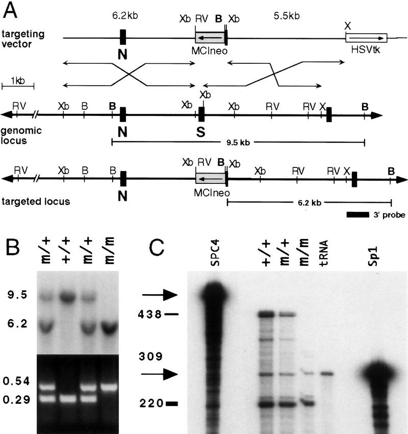
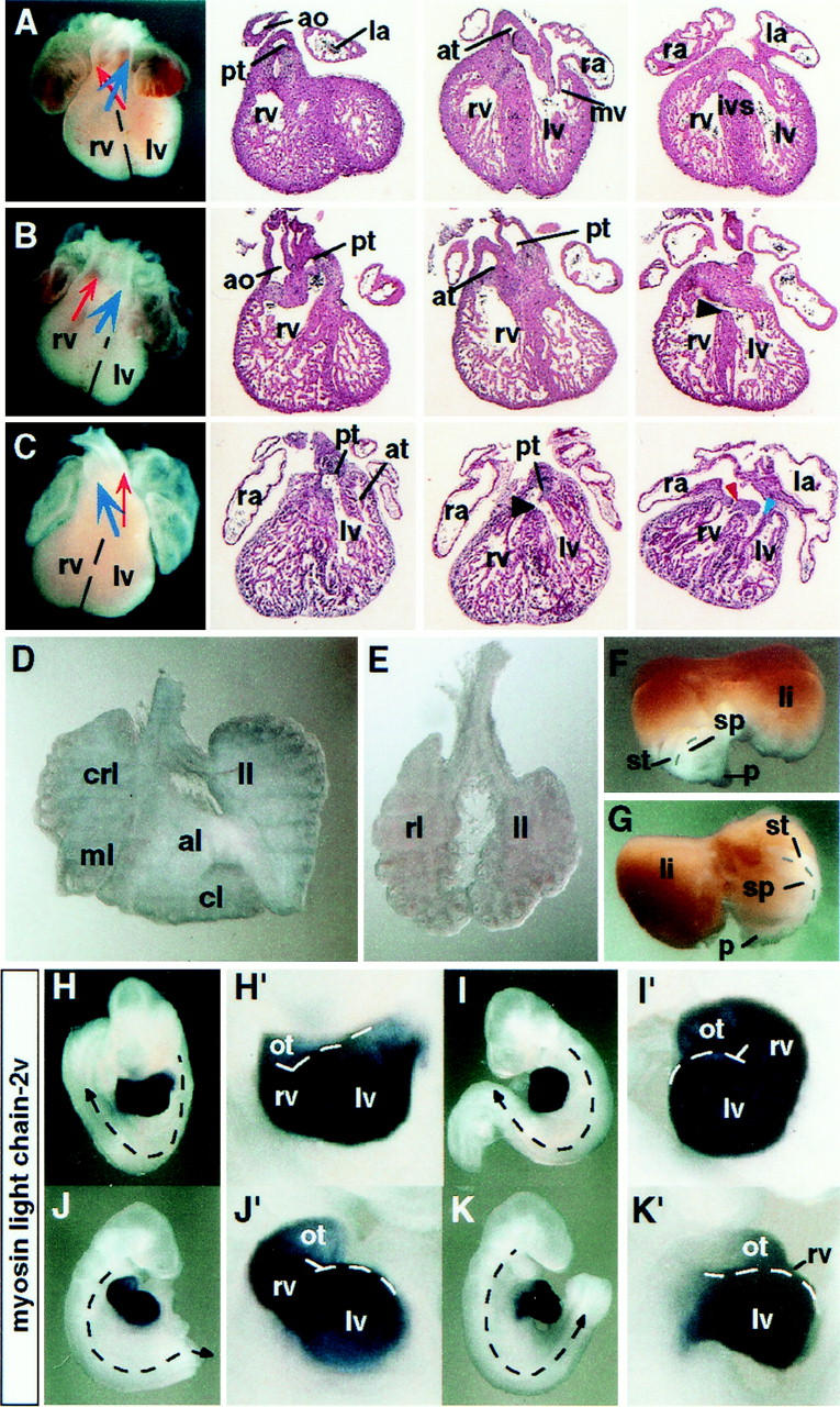
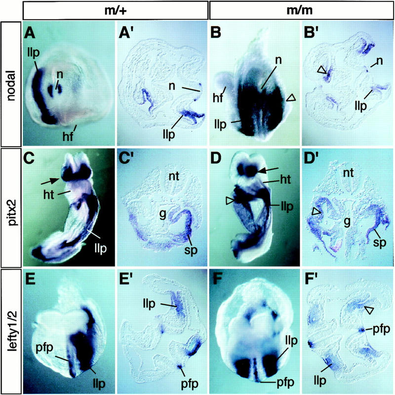
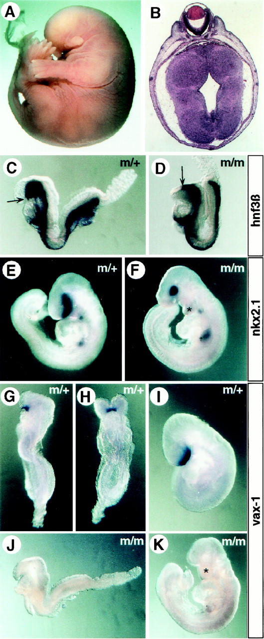
In vertebrates, specification of anteroposterior (A/P) and left-right (L/R) axes depends on TGFbeta-related signals, including Nodal, Lefty, and BMPs. Endoproteolytic maturation of these proteins is probably mediated by the proprotein convertase SPC1/Furin. In addition, precursor processing may be regulated by related activities such as SPC4 (also known as PACE4). Here, we show that a proportion of embryos lacking SPC4 develop situs ambiguus combined with left pulmonary isomerism or complex craniofacial malformations including cyclopia, or both. Gene expression analysis during early somite stages indicates that spc4 is genetically upstream of nodal, pitx2, lefty1, and lefty2 and perhaps maintains the balance between Nodal and BMP signaling in the lateral plate that is critical for L/R axis formation. Furthermore, genetic interactions between nodal and spc4, together with our analysis of chimeric embryos, strongly suggest that during A/P axis formation, SPC4 acts primarily in the foregut. These findings establish an important role for SPC4 in patterning the early mouse embryo.


Figures

Figure 1
Figure 1
Generation of a loss-of-function allele at the spc4 locus. (A) Schematic representation of the wild-type and mutant alleles and the targeting vector. (B) BamHI; (RV) EcoRV; (Xb) XbaI; (X) XhoI. (B) Southern blot and PCR analysis of pups obtained from heterozygous intercrosses. Genomic tail DNA digested with BamHI was hybridized with an external 3′-flanking probe to detect the wild-type (+) or mutant (m) allele, respectively (top). Embryos were genotyped by PCR analysis of yolk sac DNA, amplifying 0.54-kb and 0.29-kb fragments of the mutant or wild-type allele, respectively (bottom). (C) RNase protection analysis. An antisense RNA probe complementary to spc4 sequences comprising the exons designated N and S and a control probe specific for mouse Sp-1 (arrows) were hybridized to total RNA obtained from adult brains. Solid bars indicate the position and size of the full-length protected fragments. A partially protected 309-basepair fragment (shaded bar) weakly detectable in homozygous mutants (m/m) indicates low-level expression of a transcript lacking the coding region for the catalytic serine.
Figure 2
Figure 2
Situs defects of spc4 mutants. (A–C) Whole-mount view and frontal sections of hearts collected from E13.5 embryos. Cardiac abnormalities of spc4 mutants include double outlet right ventricle formation (B) and dextrocardia, associated with ventricular septal defects (black arrowheads), and common right atrium (C). (Red arrowhead) mitral valve; (blue arrowhead) tricuspid valve. (D,E) Left pulmonary isomerism. Whereas normal lungs form four lobes on the right side and only one on the left (D), the lungs of spc4 mutants are often bilaterally symmetric, consisting of one lobe on each side (E). (F,G) Dorsal view of visceral organs dissected from E13.5 embryos showing wild-type positioning of the stomach, spleen, and pancreatic primordium on the left side (F). The mirror image configuration of an spc4 mutant embryo is shown in G. (H–K) Reversal of the direction of heart looping and/or turning in embryos stained for the cardiac marker mlc-2v mRNA. Stippled arrows indicate the direction of turning that is reversed in the embryos shown in J and K. Close-up magnifications (H′–K′) show abnormal heart looping to the left side (I′,K′). (al) Anterior lobe; (ao) aorta; (at) aortic trunk; (cl) caudal lobe; (crl) cranial lobe; (ivs) interventricular septum; (la) left atrium; (li) liver; (ll) left lobe; (lv) left ventricle; (ml) medial lobe; (ot) outflow tract; (pt) pulmonary trunk; (ra) right atrium; (rl) right lobe; (rv) right ventricle; (sp) spleen; (st) stomach; (p) pancreatic primordium.
Figure 3
Figure 3
Ectopic expression of nodal, pitx2, and lefty presage laterality defects. (A,A′) Whole-mount in situ hybridization showing wild-type nodal mRNA expression pattern in the node (n) and left lateral plate (llp). (B,B′) Caudal view (B) and transverse section (B′) through the trunk of SPC4-deficient embryos showing ectopic nodal expression on the right side (open arrowheads). (C,C′) Ventral view (C) and transverse section (C′) of control embryo stained to visualize pitx2 mRNA expression in head mesenchyme (arrow) and in the left lateral plate. (D,D′) Ventral–lateral view (D) and section (D′) of mutant embryos showing ectopic pitx2 expression in the splanchnic component of the right lateral plate (open arrowheads). (E,F) Expression of lefty1 and lefty2 mRNAs in the ventral neural tube and lateral plate mesoderm of control (E,E′) or homozygous mutant embryos (F,F′). (g) Gut; (hf) headfold; (hg) hindgut; (ht) heart; (llp/rlp) left and right lateral plate; (n) node; (nt) neural tube; (pfp) prospective floorplate; (so) somite; (sp) splanchnic mesoderm.
Figure 4
Figure 4
A/P axis defects of spc4−/− embryos. (A) Whole mount view of an E15.5 homozygous mutant showing truncation of the anterior head structures. (B) Coronal section obtained from this embryo showing cyclopia. (C,D) The anterior extension of the axial midline (arrows) marked by the expression of hnf3β mRNA is truncated in a proportion of homozygous mutant E8.5 embryos (D) compared with heterozygous litter mates (C). (E,F) Compared with normal E9.5 embryos (E), mutants with craniofacial abnormalities (F) form an abnormally shaped first branchial arch that prematurely fuses at the ventral midline (asterisk; see also in K), and the number of nkx2.1-expressing cells in the ventral forebrain overlying the prechordal plate mesoderm is significantly reduced (F). (G–K) Expression of vax1 marking the ventral forebrain between E8.5 (G,H) and E9.5 (I) is abolished in anteriorly truncated, stage-matched mutant embryos (J,K).
Figure 5
Figure 5
Expression of spc4 mRNA between 6.5 dpc and 8.5 dpc. (A) Whole-mount view and (B) sagittal section of a 6.5-dpc embryo showing expression in the extraembryonic ectoderm lining the exocoelomic cavity. (C) Lateral view of a headfold-stage (8.0 dpc) embryo showing spc4 expression in the chorion. (D) Ventral whole-mount view and (E) transverse section of an 8.5-dpc embryo showing high expression levels in the definitive endoderm of the foregut and adjacent splanchnic mesoderm. Arrow in D indicates where the section in E was obtained. (aip) anterior intestinal portal; (al) allantois; (ch) chorion; (cm) cranial mesenchyme; (ec) ectoplacental cone; (en) definitive endoderm; (ep) epiblast; (hf) headfold; (hg) hindgut; (ht) heart; (np) neural plate; (xe) extraembryonic ectoderm.
Figure 6
Figure 6
Genetic interaction between spc4 and nodal. (A,E) Whole-mount view and (B–D, F–I) transverse sections of (A–D) wild-type and (E–I) nodallacZ/+; spc4−/− embryos stained for shh mRNA expression. (E–H) The foregut and anterior notochord are severely reduced in the mutant (G). Asterisk, arrow, and arrowhead in F and H indicate the absence of notochord, neural tube, and endoderm expression of shh, respectively. (aip) Anterior intestinal portal; (anf) anterior neural fold; (en) endoderm; (fg) foregut; (fp) floor plate; (hg) hindgut; (ht) heart; (nc) notochord; (np) neural plate; (so) somite; (spl) splanchnic mesoderm.
Figure 7
Figure 7
Schematic representation of the proposed mechanism by which SPC4 controls TGFβ-related signals and their target genes during L/R axis formation. According to this model, the transcriptional status of Nodal and BMP target genes (solid bars), including nodal, pitx2, and lefties is either OFF (darkly shaded area), ON (lightly shaded area), or ambivalent (intermediately shaded area in between), depending on the net sum of positive and negative regulatory input of Nodal (N) and BMP (B) signals (red hatched bars and blue crosshatched bars, respectively). (A) In wild-type (wt) embryos, BMP signals on the right side (R) effectively repress Nodal target genes but are sequestered on the left side (L) by antagonistic activities (Rodriguez Esteban et al. 1999; Yokouchi et al. 1999; Zhu et al. 1999). Consequently, the threshold concentration of mature Nodal (red broken horizontal line) that is required to reliably induce Nodal target genes is considerably reduced on the left side (arbitrary units for the concentration-dependent regulatory input of mature Nodal and BMP proteins are indicated by the scale bar). (B) According to this model, the threshold concentration of Nodal protein required for activation of its target genes in spc4 mutants is reduced by an arbitrary factor of twofold because of impaired BMP processing. Because of a similar reduction in the efficiency of Nodal processing, however, mature Nodal still cannot reach the threshold concentrations necessary to reliably overcome the residual inhibitory BMP signals on the right side. Consequently, Nodal targets may fail to become ectopically induced on the right side, even if Nodal concentrations are symmetric with respect to the midline. (C) In spc4 mutants lacking one copy of nodal, the net sum of positive and negative signals is further decreased so that Nodal targets may fail to be reliably induced even on the left side.

Similar articles

Cited by

References

    1. Beddington RSP, Robertson EJ. An assessment of the developmental potential of embryonic stem cells in the midgestation mouse embryo. Development. 1989;105:733–737. - PubMed
    1. Benjannet S, Savaria D, Laslop A, Munzer JS, Chretien M, Marcinkiewicz M, Seidah NG. Alpha1-antitrypsin Portland inhibits processing of precursors mediated by proprotein convertases primarily within the constitutive secretory pathway. J Biol Chem. 1997;272:26210–26218. - PubMed
    1. Bisgrove BW, Essner JJ, Yost HJ. Regulation of midline development by antagonism of lefty and nodal signaling. Development. 1999;126:3253–3262. - PubMed
    1. Chang H, Huylebroeck D, Verschueren K, Guo Q, Matzuk MM, Zwijsen A. Smad5 knockout mice die at mid-gestation due to multiple embryonic and extraembryonic defects. Development. 1999;126:1631–1642. - PubMed
    1. Chang H, Zwijsen A, Vogel H, Huylebroeck D, Matzuk MM. Smad5 is essential for left-right asymmetry in mice. Dev Biol. 2000;219:71–78. - PubMed

Publication types

MeSH terms

LinkOut - more resources