. 2000 Jun;182(11):3183-90.
doi: 10.1128/JB.182.11.3183-3190.2000.

Yersinia enterocolitica TyeA, an intracellular regulator of the type III machinery, is required for specific targeting of YopE, YopH, YopM, and YopN into the cytosol of eukaryotic cells

Affiliations

Yersinia enterocolitica TyeA, an intracellular regulator of the type III machinery, is required for specific targeting of YopE, YopH, YopM, and YopN into the cytosol of eukaryotic cells

L W Cheng et al. J Bacteriol. 2000 Jun.

Abstract

Pathogenic Yersinia species employ type III machines to target effector Yops into the cytosol of eukaryotic cells. Yersinia tyeA mutants are thought to be defective in the targeting of YopE and YopH without affecting the injection of YopM, YopN, YopO, YopP, and YopT into the cytosol of eukaryotic cells. One model suggests that TyeA may form a tether between YopN (LcrE) and YopD on the bacterial surface, a structure that may translocate YopE and YopH across the plasma membrane of eukaryotic cells (M. Iriarte, M. P. Sory, A. Boland, A. P. Boyd, S. D. Mills, I. Lambermont, and G. R. Cornelis, EMBO J. 17:1907-1918, 1998). We have examined the injection of Yop proteins by tyeA mutant yersiniae with the digitonin fractionation technique. We find that tyeA mutant yersiniae not only secreted YopE, YopH, YopM, and YopN into the extracellular medium but also targeted these polypeptides into the cytosol of HeLa cells. Protease protection, cell fractionation, and affinity purification experiments suggest that TyeA is located intracellularly and binds to YopN or YopD. We propose a model whereby TyeA functions as a negative regulator of the type III targeting pathway in the cytoplasm of yersiniae, presumably by preventing the export of YopN.


Figures
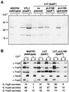
FIG. 1
FIG. 1
tyeA mutant yersiniae secrete Yops in the presence of calcium. The wild-type Y. enterocolitica strain W22703, yopN (VTL1) and tyeA (LC7 [tyeA2]) isogenic mutant strains, and LC7 transformed with either pLC186 (wild-type tyeA) or pLC199 (gst-tyeA) were grown at 37°C in TSB in the presence or absence of calcium. Cultures were centrifuged, and the supernatant (S) was separated from the cell pellet (P). Protein in each sample was precipitated with TCA, solubilized in sample buffer, and analyzed by SDS-PAGE. (A) Coomassie blue staining of culture supernatants. (B) Immunoblotting of culture supernatants and bacterial extracts with antisera raised against YopE, YopN, YopD, SycE, and YopQ. The wild-type Y. enterocolitica strain W22703 secretes Yops in the absence but not in the presence of calcium. In contrast, yopN and tyeA mutant strains display a calcium-blind phenotype and secrete Yops in the presence and absence of calcium. Transformation of tyeA mutant cells with either pLC186 or pLC199 restored the wild-type phenotype. Secretion is indicated as the percentage of polypeptide that is present in the culture medium divided by the total amount of polypeptide.
FIG. 2
FIG. 2
tyeA mutant yersiniae display a Los phenotype and secrete effector Yops into the extracellular medium during the infection of HeLa cells. HeLa cells were infected with wild-type Y. enterocolitica W22703, the tyeA isogenic mutant strain LC7 (tyeA2), LC7(pLC186) (expressing wild-type tyeA), or LC7(pLC199) (expressing gst-tyeA). After incubation for 3 h at 37°C, the tissue culture medium (M) was decanted and centrifuged to separate secreted proteins from those present within nonadherent bacteria. HeLa cells as well as adherent yersiniae were extracted with digitonin (D), a detergent that solubilizes the eukaryotic plasma membrane but not the bacterial envelope. Extracts were centrifuged to separate proteins solubilized from the HeLa cytoplasm from those that sediment with the bacteria. Proteins in each fraction were precipitated with chloroform-methanol and analyzed by SDS-PAGE and immunoblotting with antibodies directed against YopB, YopD, YopE, YopH, YopM, YopN, YopQ, YopR, SycE, and Fpt. The wild-type Y. enterocolitica strain W22703 targeted YopE, YopH, YopM, and YopN into the cytosol of HeLa cells (digitonin supernatant). In contrast, the tyeA2 mutant strain LC7 displayed a loss of targeting specificity phenotype (Los) and secreted large amounts of YopE, YopH, YopM, and YopN into the culture medium. The Los phenotype was complemented by transforming LC7 cells with plasmid pLC186 or pLC199.
FIG. 3
FIG. 3
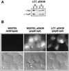
Y. enterocolitica LC7 (tyeA2) targets YopE-Npt into the cytosol of HeLa cells. (A) HeLa cell cultures were infected with Y. enterocolitica LC7 (tyeA2) carrying pDA36, expressing the YopE-Npt fusion protein (23), fractionated by the digitonin technique (see the legend to Fig. 2), and analyzed by immunoblotting. α-Npt measures the distribution of YopE-Npt in various fractions, whereas α-YopE measures the distribution of YopE. (B) Immunofluorescence microscopy of HeLa cells infected with Y. enterocolitica W22703, W22703(pDA36), or LC7(pDA36). Samples were fixed with formaldehyde and stained with α-Npt antibodies followed with an α-rabbit IgG-FITC conjugate. YopE-Npt staining was detected in the cytosol of HeLa cells infected with W22703(pDA36) and LC7(pDA36) but not in HeLa cells that were infected with strain W22703.
FIG. 4
FIG. 4
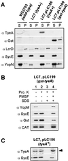
Subcellular location of TyeA. (A) Cell fractionation of Yersinia strains. Y. enterocolitica W22703 (wild type), the tyeA2 mutant strain LC7 (tyeA2), LC7(pLC186), and LC7(pLC199) were grown at 37°C and induced for type III secretion by the chelation of calcium ions. Bacteria were lysed in a French pressure cell. Unbroken cells were removed by low-speed centrifugation, and crude cell extracts were subjected to ultracentrifugation at 100,000 × g. The supernatant (S), containing soluble cytoplasmic contents, was separated from the membrane pellet (P). Samples were analyzed by separating proteins on SDS-PAGE gels and immunoblotting with antibodies raised against TyeA, Gst, LcrD, SycE, and YopN. α-TyeA recognized both wild-type TyeA and Gst-TyeA. The α-TyeA immunoblot of Gst-TyeA samples did not reveal an immunoreactive signal at the same mobility as wild-type TyeA, consistent with the notion that Gst-TyeA is not cleaved to generate native TyeA. The amount of YopN present in the supernatant fraction was quantified and calculated as the YopN/SycE ratio: we observed ratios of 2.9 for Y. enterocolitica W22703 (wild type) and 0.28 for strain LC7 (tyeA2). Small amounts of YopN were found in the pellet fraction, consistent with the notion that these species may represent transport intermediates. (B) Protease protection assay. Y. enterocolitica LC7(pLC199) were grown at 37°C and induced for type III secretion by the chelation of calcium ions. Four 6-ml culture aliquots (108 CFU/ml) were incubated at 37°C for 30 min with or without 10 μg of proteinase K/ml, alone or with 1% SDS or 1 mM PMSF. Lane 1, control reaction; lane 2, sensitivity to extracellular protease; lane 3, protease sensitivity of cytoplasmic proteins; lane 4, control for the inhibition of proteinase K by PMSF. Following incubation at 37°C, all samples were placed on ice and 1 mM PMSF was added to quench all proteolysis. Proteinase K (reactions 1 and 2) and SDS (reaction 2) were added to control for any interference of reagents during precipitation and solubilization. Samples were precipitated with chloroform-methanol, dried, and solubilized in sample buffer. Proteins were analyzed by separation on SDS-PAGE gels followed by immunoblotting. (C) Experiments similar to that for which results are shown in panel B, using cultures of Y. enterocolitica strain LC7(pLC186). TyeA and Gst-TyeA were protected from extracellular proteinase K unless the double membrane envelope of yersinae was dissolved with SDS.
FIG. 5
FIG. 5
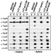
Xylene extraction of yersiniae. Y. enterocolitica W22703 was grown at 37°C and induced for type III secretion by the chelation of calcium ions. Culture aliquots were centrifuged, and the bacterial sediment was washed and suspended in TSB. After extraction with xylene and centrifugation, the extract supernatant (S) and pellet (P) were separated, precipitated with acetone, and analyzed by immunoblotting. Small amounts of YopD, YopE, and LcrV, known type III secretion substrates, were extracted with xylene. The cytoplasmic protein SycE, the cytoplasmic membrane protein LcrD, and the outer membrane protein YscC were used as controls. YopN, another type III secretion substrate, Gst-TyeA, and TyeA were not released from yersiniae by treatment with xylene.
FIG. 6
FIG. 6
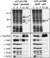
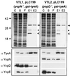
Binding of Gst-TyeA to YopN and YopD. Y. enterocolitica LC7(pLC199) and Y. enterocolitica LC7(pLC100) were grown at 37°C and induced for type III secretion by the chelation of calcium ions. Expression of the Gst and the Gst-TyeA proteins was induced by the addition of 1 mM IPTG to the culture medium. Cells (1012 CFU) were harvested by centrifugation, suspended in buffer, and lysed in a French pressure cell to generate the crude extract (C). Unbroken cells were removed by centrifugation at 6,000 × g. Membranes were sedimented by ultracentrifugation at 100,000 × g, and the supernatant (S), containing soluble cytosolic contents, was subjected to affinity chromatography on glutathione-Sepharose. Flowthrough (F) and eluate (E) fractions after the addition of 10 mM glutathione were collected and analyzed by SDS-PAGE and Coomassie staining. The migrations of full-length Gst-TyeA and Gst are marked by filled arrowheads. Gst-TyeA species within the bracket marked with a star represent degradation products that were observed in large-scale purifications but not during immunoblotting of TCA-precipitated cultures. The open arrow represents an unknown glutathione-Sepharose binding protein of Y. enterocolitica. Samples were analyzed by immunoblotting with antisera raised against Gst-TyeA, YopN, YopD, YopB, YscB, and SycN. YopN and YopD copurified with Gst-TyeA but not with Gst. Small star, migration of YscB and SycN on an SDS-PAGE gel.
FIG. 7
FIG. 7
YopN and YopD bind independently to Gst-TyeA. The experimental protocol described in Fig. 6 was used with Y. enterocolitica VTL1 (YopN) carrying pLC199 and Y. enterocolitica VTL2 (YopD) carrying pLC199. YopN copurified with Gst-TyeA in the absence of YopD, and YopD copurified with Gst-TyeA in the absence of YopN. See the legend to Fig. 6 for details.

Similar articles

Cited by

References

    1. Anderson D M, Schneewind O. A mRNA signal for the type III secretion of Yop proteins by Yersinia enterocolitica. Science. 1997;278:1140–1143. - PubMed
    1. Anderson D M, Schneewind O. Type III machines of Gram-negative pathogens: injecting virulence factors into host cells and more. Curr Opin Microbiol. 1999;2:18–24. - PubMed
    1. Anderson D M, Schneewind O. Yersinia enterocolitica type III secretion: an mRNA signal that couples translation and secretion of YopQ. Mol Microbiol. 1999;31:1139–1148. - PubMed
    1. Bergman T, Håkansson S, Forsberg A, Norlander L, Macellaro A, Bäckman A, Bölin I, Wolf-Watz H. Analysis of the V antigen lcrGVH-yopBD operon of Yersinia pseudotuberculosis: evidence for a regulatory role of LcrH and LcrV. J Bacteriol. 1991;173:1607–1616. - PMC - PubMed
    1. Boland A, Sory M-P, Iriarte M, Kerbourch C, Wattiau P, Cornelis G R. Status of YopM and YopN in the Yersinia yop virulon: YopM of Y. enterocolitica is internalized inside the cytosol of PU5-1.8 macrophages by the YopB, D, N delivery apparatus. EMBO J. 1996;15:5191–5201. - PMC - PubMed

Publication types

MeSH terms

LinkOut - more resources